. 2023 Oct-Dec;64(4):493-500.
doi: 10.47162/RJME.64.4.05.

In vitro effects of heparin-binding epidermal growth factor on adhesion stage of implantation

Affiliations

In vitro effects of heparin-binding epidermal growth factor on adhesion stage of implantation

Burcu Biltekin et al. Rom J Morphol Embryol. 2023 Oct-Dec.

Abstract

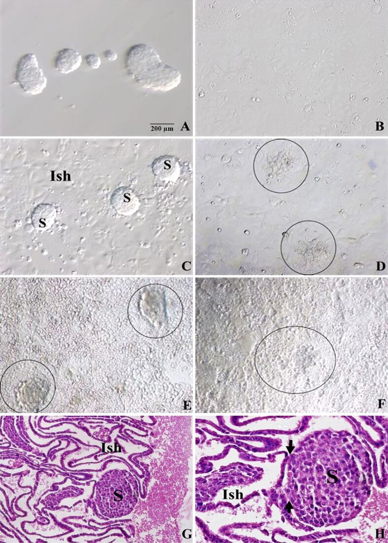
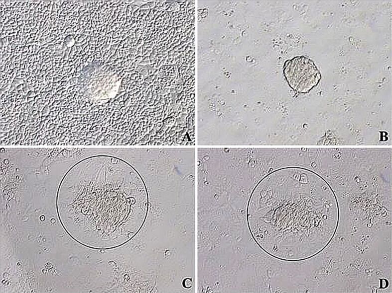
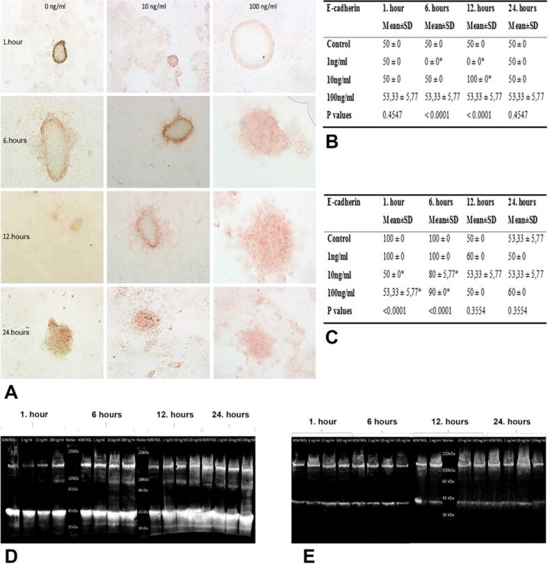
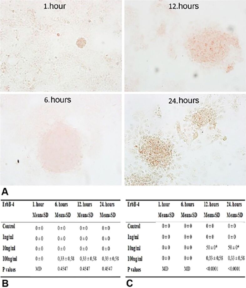
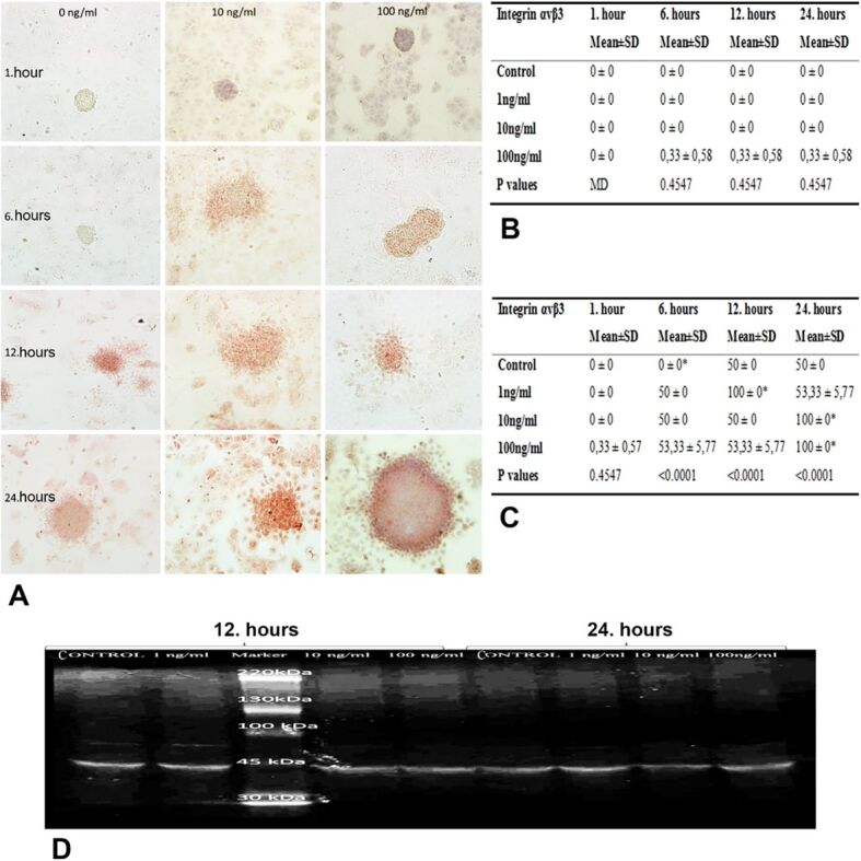
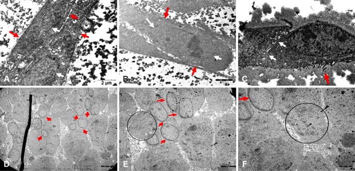
A member of the epidermal growth factor (EGF) family, the heparin-binding EGF (HB-EGF) is expressed in the uteri of both humans and mice during the implantation process. To study the effects of HB-EGF on adhesion stage, we developed an in vitro implantation model employing Ishikawa cell line and JAR cell line, which may attach to Ishikawa cells. For 1, 6, 12, and 24 hours, co-cultures of JAR spheroids grown on Ishikawa monolayers were treated with 1, 10, and 100 ng∕mL doses of HB-EGF. Using immunocytochemistry and Western blot analysis, the effects of HB-EGF on the protein expressions of E-cadherin, Erb-B2 receptor tyrosine kinase 4 (ErbB4), and integrin ανβ3 in Ishikawa and JAR cells were examined semi-quantitatively and quantitatively. Ultrastructural changes of in vitro implantation model were investigated by transmission electron microscopy. We revealed that HB-EGF influenced trophoblast cell adhesion to endometrial cells by upregulating the expression of the proteins ErbB4 and trophoblastic integrin ανβ3. Decrease in trophoblastic E-cadherin expression and increase in endometrial E-cadherin expression were demonstrated accompanying morphological variations in cells required for the invasion. We discovered ultrastructurally that Ishikawa cells acquired uterodome-like appearance, including the organelles, when 10 and 100 ng∕mL dosages of HB-EGF were administered for 12 and 24 hours. However, following additional hours of adhesion and invasion, their intercellular spaces enlarged. The trafficking of vesicular transport was enhanced by JAR spheroids. We therefore discovered that in this implantation paradigm, HB-EGF may enhance the receptivity of Ishikawa cells and the adherence of JAR cells.


Conflict of interest statement

The authors declare that they have no conflict of interests.

Figures

Figure 1
Figure 1
Inverted microscopic images of co-cultures of S on the Ish monolayers: (A) S incubated for 24 hours; (B) Ish monolayers incubated for 24 hours before co-cultures; (C–F) Co-cultures incubated for given hours, showing S on the Ish monolayers: (C) 1-hour incubation, (D) 6-hour incubated S (circle) adhered partially onto Ish cells, (E) 12-hour incubated S (circle) attached totally onto Ish cells, (F) 24-hour incubated, barely definable S (circle) started to invade through Ish cells; (G and H) HE-stained images of co-cultures of S on the Ish monolayers; successful focal attachment of S was observed on the Ish cells (arrow). (A–G) ×200; (H) ×400. HE: Hematoxylin–Eosin; Ish: Ishikawa; S: JAR spheroids
Figure 2
Figure 2
Inverted microscopic images of co-cultures of JAR spheroids (circle) on the Ishikawa monolayers incubated with given doses of HB-EGF for six hours: (A) Control group; (B) 1 ng/mL HB-EGF group showed attached JAR spheroids onto Ishikawa monolayers; (C) 10 ng/mL HB-EGF group showed tightly adhered spheroids on monolayers; (D) 100 ng/mL HB-EGF group showed tightly adhered spheroids on monolayers. (A–D) ×200. HB-EGF: Heparin-binding epidermal growth factor
Figure 3
Figure 3
(A) E-cadherin immunoreactivities of Ishikawa and JAR cells from co-cultures incubated with 1, 10 and 100 ng/mL HB-EGF for 1, 6, 12 and 24 hours; (B) H-score table of E-cadherin immunopositive Ishikawa cells; (C) H-score table of E-cadherin immunopositive JAR cells (*p<0.001 vs control group); (D and E) Western blot images of E-cadherin expression in Ishikawa (D) and JAR (E) cells applied 1, 10 and 100 ng/mL HB-EGF for 1, 6, 12 and 24 hours. H-score: Histoscore; HB-EGF: Heparin-binding epidermal growth factor; SD: Standard deviation
Figure 4
Figure 4
(A) ErbB4 immunoreactivities of JAR cells from co-cultures incubated with 1, 10 and 100 ng/mL HB-EGF for 1, 6, 12 and 24 hours; (B) H-score table of ErbB4 immunopositive Ishikawa cells; (C) H-score table of ErbB4 immunopositive JAR cells (*p<0.001 vs control group). ErbB4: Erb-B2 receptor tyrosine kinase 4; H-score: Histoscore; HB-EGF: Heparin-binding epidermal growth factor; SD: Standard deviation
Figure 5
Figure 5
(A) Integrin ανβ3 immunoreactivities of Ishikawa and JAR cells from co-cultures incubated with 1, 10 and 100 ng/mL HB-EGF for 1, 6, 12 and 24 hours; (B) H-score table of integrin ανβ3 immunopositive Ishikawa cells; (C) H-score table of integrin ανβ3 immunopositive JAR cells (*p<0.001 vs control group); (D) Western blot image of integrin ανβ3 expression in JAR cells applied 1, 10 and 100 ng/mL HB-EGF
Figure 6
Figure 6
Electron micrographs of Ishikawa cells: (A) 10 ng/mL HB-EGF for six hours, showing regular nuclei (red star) with loose chromatin and compact nucleoli (white star); (B) 10 ng/mL HB-EGF for 12 hours, showing locally shortened microvilli (red arrow) and enlarged intercellular spaces; (C) 100 ng/mL HB-EGF for one hour, showing mitotic cells (red arrow); (D) 100 ng/mL HB-EGF for six hours, showing normal structure of cell membrane (red arrow) and nuclei (red star); (E) JAR cells incubated with 10 ng/mL HB-EGF group for one hour, showing local thickening of nuclear membrane (red arrow) and intracytoplasmic vesicles peripherally aligned in chain-like series (circle); (F) JAR cells incubated with 10 ng/mL HB-EGF group for six hours, showing local thickening of nuclear membrane (red arrow) and intracytoplasmic vesicles peripherally aligned in chain-like series (circle). Scale bars: (A–C and F) 2 μm; (D and E) 5 μm. HB-EGF: Heparin-binding epidermal growth factor

Similar articles

References

    1. Ai Z, Jing W, Fang L. Cytokine-like protein 1 (Cytl1): a potential molecular mediator in embryo implantation. PLoS One. 2016;11(1):e0147424–e0147424. - PMC - PubMed
    1. Weimar CHE, Uiterweer EDP, Teklenburg G, Heijnen CJ, Macklon NS. In-vitro model systems for the study of human embryo-endometrium interactions. Reprod Biomed Online. 2013;27(5):461–476. - PubMed
    1. Mardon H, Grewal S, Mills K. Experimental models for investigating implantation of the human embryo. Semin Reprod Med. 2007;25(6):410–417. - PubMed
    1. Zhao HB, Wang C, Li RX, Tang CL, Li MQ, Du MR, Hou XF, Li DJ. E-cadherin, as a negative regulator of invasive behavior of human trophoblast cells, is down-regulated by cyclosporin A via epidermal growth factor/extracellular signal-regulated protein kinase signaling pathway. Biol Reprod. 2010;83(3):370–376. - PubMed
    1. Johnson GA, Burghardt RC, Bazer FW, Seo H, Cain JW. Integrins and their potential roles in mammalian pregnancy. J Anim Sci Biotechnol. 2023;14(1):115–115. - PMC - PubMed