Case Reports
. 2023 Oct-Dec;64(4):579-585.
doi: 10.47162/RJME.64.4.15.

Intracardiac mass presenting as acute myocardial infarction

Affiliations
Case Reports

Intracardiac mass presenting as acute myocardial infarction

Diana Ruxandra Hădăreanu et al. Rom J Morphol Embryol. 2023 Oct-Dec.

Abstract

Cardiac tumors, although rare, present intricate diagnostic and therapeutic challenges, necessitating timely intervention for optimal patient outcomes. This case report focuses on a 65-year-old woman admitted with chest pain and loss of consciousness, ultimately diagnosed with a left ventricular cardiac myxoma. The patient's presentation mimicked acute coronary syndrome, highlighting the diagnostic complexity associated with cardiac tumors. Advanced imaging modalities, including transthoracic echocardiography, computed tomography, and invasive coronary angiography, played a pivotal role in characterizing the intracardiac mass. Histopathological (HP) examination, utilizing immunohistochemistry, confirmed the tumor as a cardiac myxoma. The patient management involved a multidisciplinary approach, leading to surgical resection of the mass and mitral valve replacement. The case underscores the importance of the HP confirmation in patients with cardiac masses, especially when multimodality cardiac imaging suggests various tumor types, simultaneously emphasizing the need for a comprehensive diagnostic approach that includes advanced imaging and histopathology to ensure an accurate diagnosis and tailored management of cardiac tumors.


Conflict of interest statement

The authors declare that they have no conflict of interests.

Figures

Figure 1
Figure 1
Echocardiographic evaluation showing a large, hyperechoic cardiac mass (white arrows) with irregular borders, attached at the level of the left ventricular posterior wall and posterior mitral valve leaflet in transthoracic parasternal long axis view (A), and apical 4-chamber view (B), and transesophageal 2-chamber view (C). Three-dimensional rendering of the cardiac mass by transthoracic (D) and transesophageal echocardiography (E). AS: Left atrium; VS: Left ventricle
Figure 2
Figure 2
Invasive coronary angiography showing no significant coronary lesions at the level of the right coronary artery (A), and left anterior descending artery (C), and with an embolic occlusion of a distal branch of the obtuse marginal branch of the left circumflex coronary artery (B, white arrow
Figure 3
Figure 3
Cardiac computed tomography. Arterial phase in short axis (A) and long axis (B) showing the cardiac mass with irregular borders localized at the base of the left ventricle, on the lateral wall, and posterior mitral valve leaflet. Venous phase in short axis (D) and long axis (E) showing the cardiac mass with low initial contrast uptake. Three-dimensional rendering of the cardiac mass (C and F).
Figure 4
Figure 4
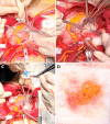
Intraoperative images of the cardiac mass showing its relationship with the mitral valve (A and B), and the mitral valve replacement with a biological prosthesis (C). The macroscopic appearance of the tumor (D) with large dimensions, irregular borders, yellowish color, and areas of hemorrhage was suggestive of cardiac myxoma.
Figure 5
Figure 5
Overview of the tumor structure consisting of fusiform cells with multiple extensions, collagen fibers arranged in all directions, and amorphous conjunctival matrix. Hematoxylin–Eosin (HE) staining, ×100
Figure 6
Figure 6
Detail image from the previous figure. Among the fusiform connective cells, there are rare round cells of inflammatory type. HE staining, ×200
Figure 7
Figure 7
The tumor area formed by connective cells and collagen fibers with a plexiform arrangement. Goldner–Szekely (GS) trichrome staining, ×100
Figure 8
Figure 8
The tumor area formed mainly of amorphous conjunctival matrix. HE staining, ×100
Figure 9
Figure 9
Positive reaction of tumor cells to the anti-vimentin antibody. Immunostaining with anti-vimentin antibody, ×100
Figure 10
Figure 10
Tumor cells positive for the anti-alpha-smooth muscle actin (α-SMA) antibody. Immunostaining with anti-α-SMA antibody, ×100
Figure 11
Figure 11
Tumor cells intensely positive for the anti-cluster of differentiation (CD)34 antibody. Immunostaining with anti-CD34 antibody, ×200
Figure 12
Figure 12
Numerous macrophages unevenly distributed in the tumor stroma. Immunostaining with anti-CD68 antibody, ×200.
Figure 13
Figure 13
The area of the tumor infiltrated with numerous mast cells arranged mainly perivascularly. Immunostaining with anti-tryptase antibody, ×200
Figure 14
Figure 14
Imaging features of the cardiac mass suggestive of several tumor types

References

    1. Tyebally S, Chen D, Bhattacharyya S, Mughrabi A, Hussain Z, Manisty C, Westwood M, Ghosh AK, Guha A. Cardiac tumors: JACC CardioOncology state-of-the-art review. JACC CardioOncol. 2020;2(2):293–311. - PMC - PubMed
    1. Reynen K. Frequency of primary tumors of the heart. Am J Cardiol. 1996;77(1):107–107. - PubMed
    1. Patel J, Sheppard MN. Pathological study of primary cardiac and pericardial tumours in a specialist UK Centre: surgical and autopsy series. Cardiovasc Pathol. 2010;19(6):343–352. - PubMed
    1. Butany J, Nair V, Naseemuddin A, Nair GM, Catton C, Yau T. Cardiac tumours: diagnosis and management. Lancet Oncol. 2005;6(4):219–228. - PubMed
    1. Grebenc ML, Rosado de, Burke AP, Green CE, Galvin JR. Primary cardiac and pericardial neoplasms: radiologic-pathologic correlation. Radiographics. 2000;20(4):1073–1103. - PubMed

Publication types