Review
. 2024 Jan 8;9(1):12.
doi: 10.1038/s41392-023-01688-x.

Ischemia-reperfusion injury: molecular mechanisms and therapeutic targets

Affiliations
Review

Ischemia-reperfusion injury: molecular mechanisms and therapeutic targets

Meng Zhang et al. Signal Transduct Target Ther. .

Abstract

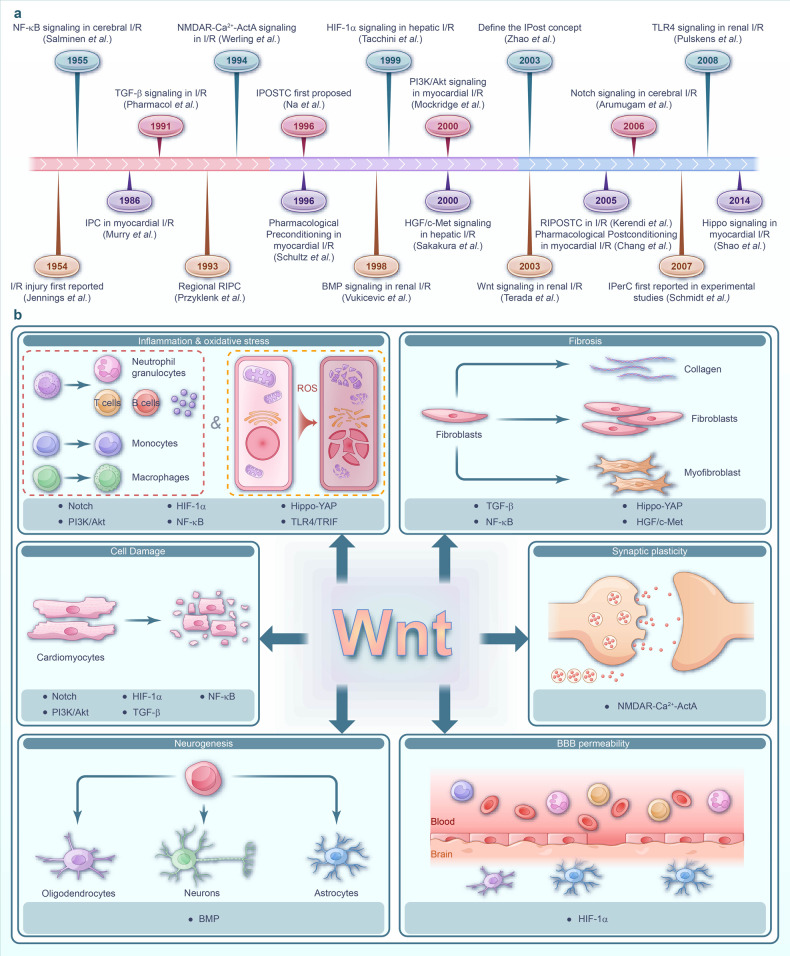
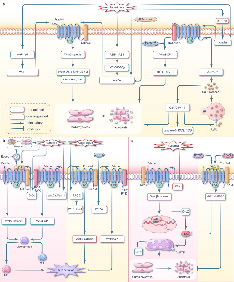
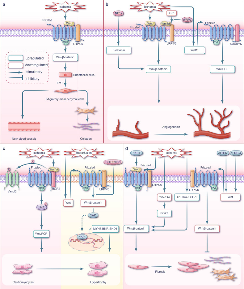
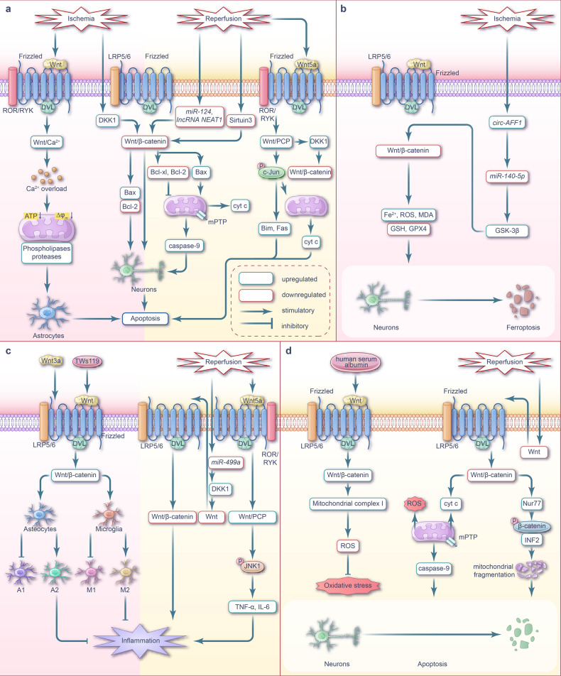
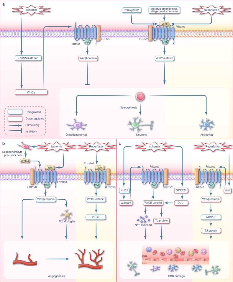
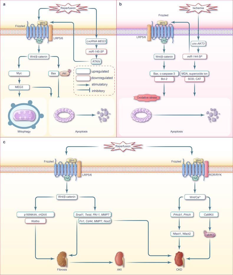
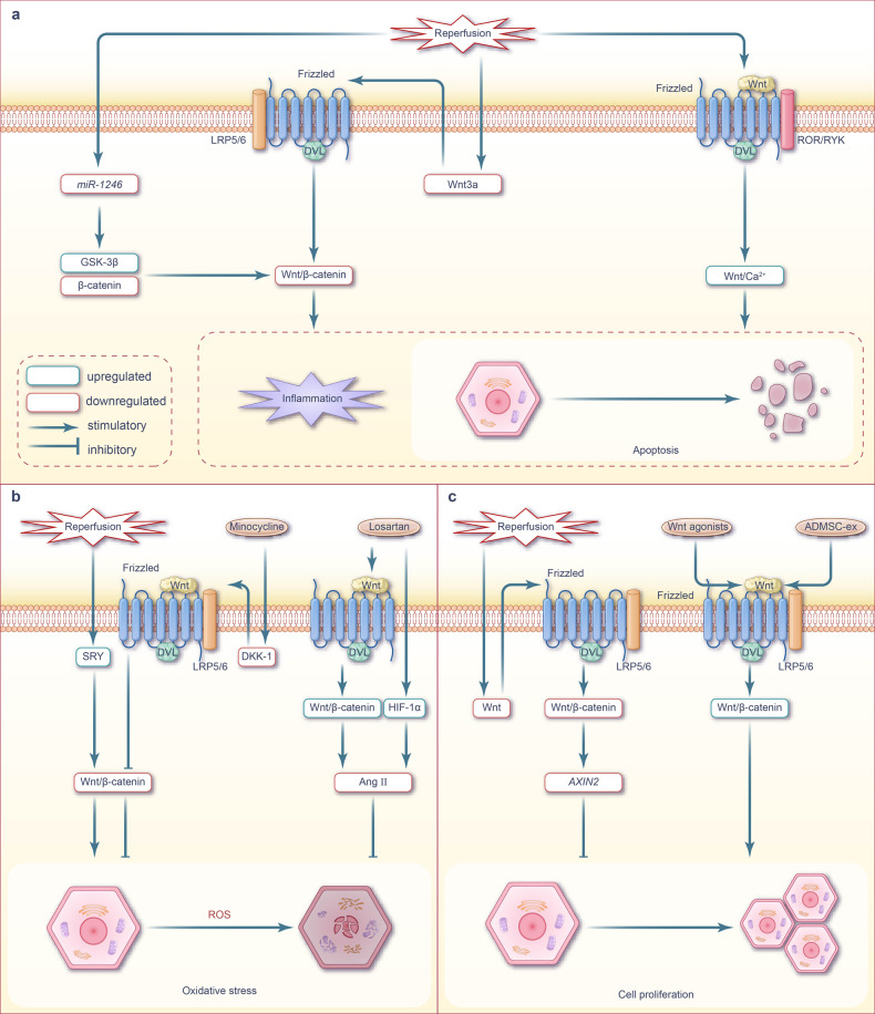
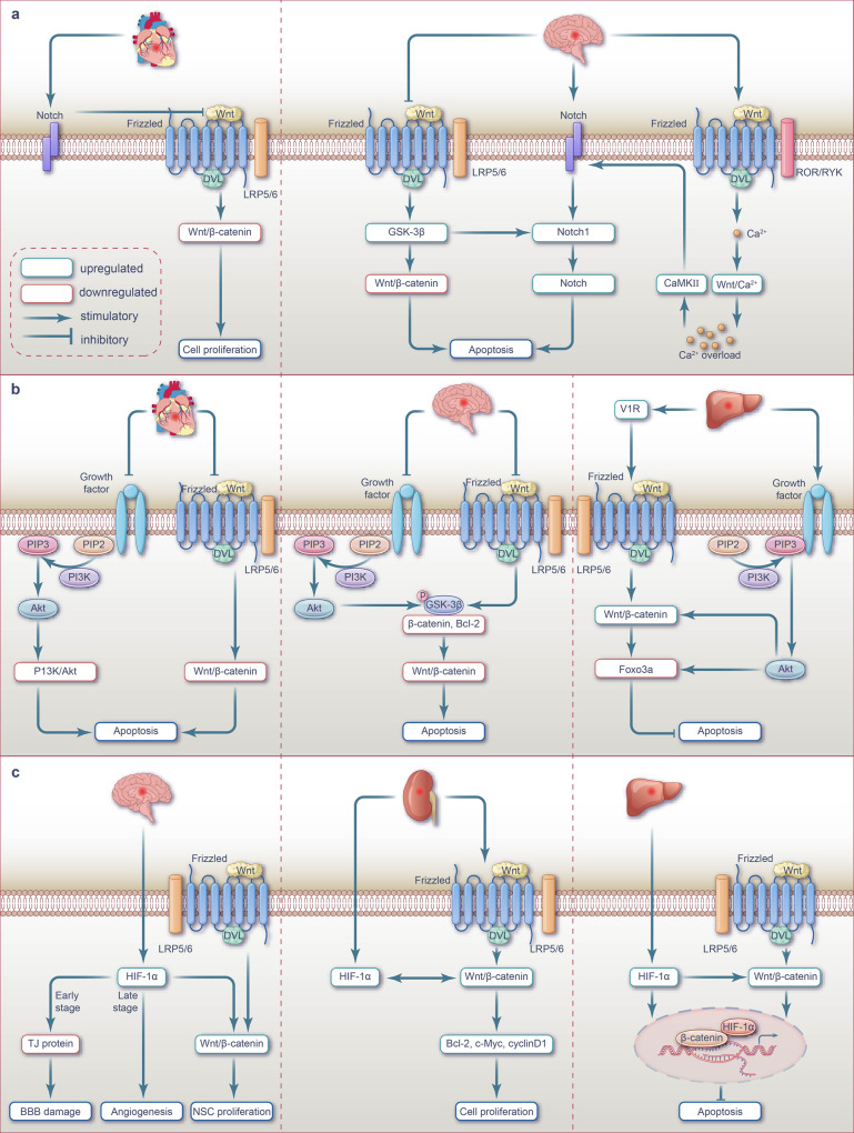
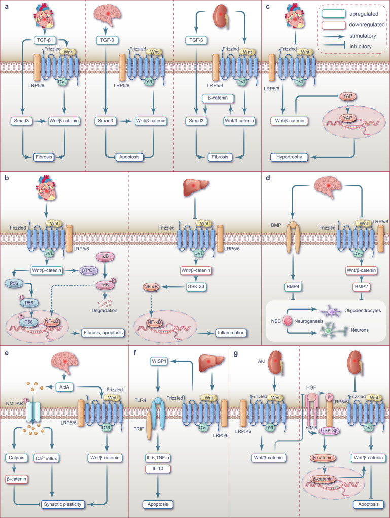
Ischemia-reperfusion (I/R) injury paradoxically occurs during reperfusion following ischemia, exacerbating the initial tissue damage. The limited understanding of the intricate mechanisms underlying I/R injury hinders the development of effective therapeutic interventions. The Wnt signaling pathway exhibits extensive crosstalk with various other pathways, forming a network system of signaling pathways involved in I/R injury. This review article elucidates the underlying mechanisms involved in Wnt signaling, as well as the complex interplay between Wnt and other pathways, including Notch, phosphatidylinositol 3-kinase/protein kinase B, transforming growth factor-β, nuclear factor kappa, bone morphogenetic protein, N-methyl-D-aspartic acid receptor-Ca2+-Activin A, Hippo-Yes-associated protein, toll-like receptor 4/toll-interleukine-1 receptor domain-containing adapter-inducing interferon-β, and hepatocyte growth factor/mesenchymal-epithelial transition factor. In particular, we delve into their respective contributions to key pathological processes, including apoptosis, the inflammatory response, oxidative stress, extracellular matrix remodeling, angiogenesis, cell hypertrophy, fibrosis, ferroptosis, neurogenesis, and blood-brain barrier damage during I/R injury. Our comprehensive analysis of the mechanisms involved in Wnt signaling during I/R reveals that activation of the canonical Wnt pathway promotes organ recovery, while activation of the non-canonical Wnt pathways exacerbates injury. Moreover, we explore novel therapeutic approaches based on these mechanistic findings, incorporating evidence from animal experiments, current standards, and clinical trials. The objective of this review is to provide deeper insights into the roles of Wnt and its crosstalk signaling pathways in I/R-mediated processes and organ dysfunction, to facilitate the development of innovative therapeutic agents for I/R injury.


Conflict of interest statement

The authors declare no competing interests.

Figures

Fig. 1
Fig. 1
The intricate signaling network in I/R injury pathogenesis. a Timeline diagram of key milestones in I/R Injury research: revealing crucial discoveries and emphasizing complex signaling pathways. b The complexity of signaling pathways in I/R injury Pathology. Involvement of Wnt signaling and crosstalk with diverse signaling pathways in the pathological process of I/R injury: impact on cellular injury, inflammation, oxidation, fibrosis, neurogenesis, synaptic plasticity, and BBB permeability across organs I/R events. I/R ischemia-reperfusion, NF-κB nuclear factor-κB, IPC ischemic preconditioning, TGF-β transforming growth factor-β, RIPC remote ischemic preconditioning, NMDAR N-Methyl-D-Aspartate Receptor, ActA Activin A, IPOSTC ischemic postconditioning, BMP bone morphogenetic protein, HIF-1α hypoxia-inducible factor-1α, PI3K/Akt phosphoinositide-3 kinase/protein kinase B, HGF/c-Met hepatocyte growth factor receptor/mesenchymal-epithelial transition factor, RIPOSTC remote ischemic postconditioning, YAP Yes-associated protein, TLR4/TRIF toll-like receptor 4/toll-interleukine-1 receptor domain-containing adapter-inducing interferon-β, BBB blood-brain barrier
Fig. 2
Fig. 2
Wnt signaling pathway and targeted therapy for apoptosis, inflammation, and oxidative stress during myocardial I/R injury. a Wnt signaling pathway-mediated apoptosis during myocardial I/R injury. During myocardial I/R injury, the inhibition of Wnt/β-catenin signaling promotes cardiomyocytes apoptosis. However, the roles of non-canonical Wnt/PCP and Wnt/Ca2+ signaling pathways in myocardial I/R injury are the opposite. The activation of these two Wnt signaling pathways may exacerbate cardiomyocyte apoptosis through the activation of the JNK pathway or the induction of calcium overload. b Wnt signaling-mediated inflammation during myocardial I/R injury. Cardiomyocyte ferroptosis occurs during the ischemia phase, and the exosomes derived from the ferroptotic cells induce M1 macrophage transformation by activating the Wnt/β-catenin pathway. During myocardial ischemic phase, the activation of Wnt/β-catenin signaling promotes the polarization of macrophages towards the M1 phenotype while suppressing the M2 phenotype, ultimately exacerbating the inflammatory response. Upregulation of Wnt ligands and DKK family members in macrophages stimulates inflammation by activating the Wnt/β-catenin signaling pathway. During the process I/R injury, there is an upregulation of RAGE expression in the infarct border zone of rat cardiomyocytes, accompanied by downregulation of Wnt1 and Dvl3 expression. By inhibiting the Wnt/β-catenin signaling pathway, this leads to the promotion of inflammatory response, exacerbating cardiomyocyte apoptosis. Additionally, activation of the Wnt/PCP pathway in macrophages during the ischemic phase increases the expression of inflammatory cytokines, which aggravates cardiac inflammation. c Wnt signaling-mediated oxidative stress during myocardial I/R injury. During myocardial ischemia phase, the downregulation of Wnt protein inhibits Wnt/β-catenin signaling transduction, resulting in increased transcription of intracellular CysC. The elevated expression of CysC exacerbates intracellular oxidative stress and promotes the generation of ROS leading to cardiomyocyte apoptosis. Note: The pink background represents the ischemic phase, while the cream background represents the reperfusion phase. ADMSCs-ex exosomes isolated from adipose-derived mesenchymal stem cells, sFRP-5 secretory frizzled-related protein 5, LRP low-density lipoprotein receptor-related protein, ROR recombinant receptor tyrosine kinase like orphan receptor, RYK receptor tyrosine kinase, CaMKII calmodulin-dependent protein kinase II, ROS reactive oxygen species, NOS nitric oxide synthase, RyR ryanodine receptors, TNF-α tumor necrosis factor-α, MCP-1 monocyte chemoattractant protein-1, APTBP a peptide from tuna backbone protein, CysC cystatin C, mPTP mitochondrial permeability transition pore, cyt c cytochrome c, iPS-CM induced plenipotentiary stem cell-derived conditioned medium
Fig. 3
Fig. 3
Wnt signaling pathway and targeted therapy in ECM remodeling, angiogenesis, cardiac hypertrophy, and fibrosis during myocardial I/R injury. a Wnt signaling-mediated ECM remodeling during myocardial I/R injury. During myocardial ischemic phase, there is a substantial activation of Wnt/β-catenin in endothelial cells, which leads to the promotion of EMT. b Wnt signaling-mediated angiogenesis during myocardial I/R injury. During myocardial ischemic phase, Wnt/β-catenin signaling is activated in vascular endothelial cells, which affects the proliferation and migration of these cells during the process of neovascularization. Conversely, in the infarct region of ischemic mice, the activation of the canonical Wnt pathway functions to suppress cardiac angiogenesis. NP12, an allosteric inhibitor of GSK-3β promotes cardiac angiogenesis by activating Wnt/β-catenin signaling. Additionally, the deficiency of GR activates Wnt/β-catenin signaling, leading to the upregulation of cyclin D1 and ultimately promoting cardiac angiogenesis. Moreover, Wnt/PCP activation contributes to angiogenesis during myocardial ischemia injury. c Wnt signaling pathway-mediated cardiac hypertrophy during myocardial I/R injury. During myocardial ischemic phase, Wnt5a triggers the activation of Wnt/PCP signaling through JNK phosphorylation, which subsequently induces induces cardiomyocyte hypertrophy. In the AC16 human cardiomyocyte cell line model of I/R injury, Wnt/β-catenin signaling is inhibited, and this inhibition synergistically exacerbates myocardial hypertrophy in cooperates with YAP signaling. d Wnt signaling-mediated fibrosis during myocardial I/R injury. During myocardial ischemic phase, the expression of Wnt1 is increased, resulting in the activation of Wnt/β-catenin signaling in cardiac fibroblasts. This activation leads to the proliferation of fibroblasts and ultimately contributes to cardiac repair. MiR‐145 expression was lower in myocardial ischemic phase. The down-regulation of miR-145 directly targets SOX9 in fibroblasts, leading to cardiac fibrosis by activating Wnt/β-catenin signaling. Both PRELP and S100A4/FSP1 promote myocardial fibrosis by activating Wnt/β-catenin signaling. On the other hand, the Wnt antagonist sFRP-4 reduces myocardial fibrosis by inhibiting Wnt/β-catenin signaling. Additionally, ALDH2 suppresses myocardial fibrosis by inhibiting Wnt/β-catenin signaling. Note: The pink background represents the ischemic phase, while the cream background represents the reperfusion phase. EMT endothelial-mesenchymal transition, GR glucocorticoid receptor, sFRP secretory frizzled-related protein, LRP low-density lipoprotein receptor-related protein, ROR recombinant receptor tyrosine kinase like orphan receptor, RYK receptor tyrosine kinase, YAP Yes-associated protein, MYH7 myosin heavy chain 7, BNP brain natriuretic peptide, END1 endothelin 1, SOX9 sex‐determining region Y box 9, FSP1 fibroblast-specific protein 1, PRELP proline/arginine-terminal leucine-rich repeat protein
Fig. 4
Fig. 4
Wnt signaling pathway and targeted therapy for apoptosis, ferroptosis, inflammation, and oxidative stress during cerebral I/R injury. a Wnt signaling-mediated apoptosis during cerebral I/R injury. During cerebral ischemic phase, Wnt/Ca2+ signaling is activated, leading to intracellular calcium overload and and subsequent astrocyte apoptosis. The upregulation of DKK1 inhibits Wnt/β-catenin pathway, leading to neuronal apoptosis. During cerebral I/R phase, Wnt5a-mediated Wnt/PCP signaling is activated, promoting c-Jun phosphorylation, inducing cyt c release from mitochondria, inhibiting Wnt/β-catenin signaling, and ultimately leads to neuronal apoptosis. Downregulation of Sirtuin3, miR-124 and lncRNA NEAT1 also inhibit Wnt/β-catenin signaling. b Wnt signaling-mediated ferroptosis during cerebral I/R injury. During cerebral ischemia phase, circ-AFF1 is highly expressed and directly targets miR-140-5p to upregulate GSK-3β. The highly expressed GSK-3β inhibits Wnt/β-catenin signaling. The inhibition of Wnt/β-catenin signaling leads to excessive accumulation of Fe2+, ROS and MDA, and suppression of GSH and GPX4 expression, thereby aggravating neuronal ferroptosis. c Wnt signaling-mediated inflammation during cerebral I/R injury. During cerebral ischemic phase, Wnt/β-catenin signaling is activated, which promotes the polarization of reactive microglia to M2 phenotype, increases the number of A2 type of astrocytes, and reduces the number of A1 type of astrocytes, thereby playing a protective effect and reducing the inflammatory response caused by cerebral ischemia. During cerebral I/R phase, downregulation of miR-499a leads to inhibition of Wnt/β-catenin signaling, thereby aggravating the inflammatory response. Wnt5a-mediated Wnt/PCP signaling is activated during cerebral I/R, leading to upregulation of the pro-inflammatory cytokines, thus aggravates the inflammatory response during cerebral I/R. d Wnt signaling-mediated oxidative stress during cerebral I/R injury. During cerebral I/R phase, Wnt/β-catenin signaling is inhibited which lead to neuronal apoptosis via mitochondria dysfunction. The expression of Nur77 is stimulated in the oxidative stress environment during cerebral I/R, which leads to mitochondrial fragmentation by promoting β-catenin phosphorylation and INF2 expression. Intravenous injection of human serum albumin activates Wnt/β-catenin signaling, thereby increasing mitochondrial complex I activity, reducing ROS generation, suppressing oxidative stress, and playing a therapeutic role during cerebral I/R. Note: The pink background represents the ischemic phase, while the cream background represents the reperfusion phase. LRP low-density lipoprotein receptor-related protein, ROR recombinant receptor tyrosine kinase like orphan receptor, RYK receptor tyrosine kinase, ATP adenosine triphosphate, Δφ membrane potential, DKK1 Dickkopf-1, cyt c cytochrome-c, GSH glutathione, GPX4 glutathione peroxidase 4, GSK-3β glucogen synthase kinase 3β, JNK1 c-Jun amino-terminal kinase1, TNF-α tumor necrosis factor-α, TWS119 a GSK-3β inhibitor that activates Wnt/β-catenin signaling, Nur77 nuclear hormone receptor NUR/77, INF2 inverted formin 2, mPTP mitochondrial permeability transition pore
Fig. 5
Fig. 5
Wnt signaling pathway and targeted therapy for neurogenesis, angiogenesis, and BBB during cerebral I/R injury. a Wnt signaling-mediated neurogenesis during cerebral I/R injury. During cerebral ischemia phase, there is an elevation in the synthesis of lncRNA MEG and peroxynitrite. The highly expressed lncRNA MEG hampers the process of the Wnt/β-catenin signaling pathway. In contrast, increased levels of peroxynitrite activate the Wnt/β-catenin signaling pathway. In the subsequent phase of cerebral I/R phase, the activation of the Wnt/β-catenin signaling pathway plays a crucial role in promoting neurogenesis. Mallotus oblongifolius, ellagic acid, and curcumin enhance the activation of the Wnt/β-catenin signaling, leading to the promotion of neurogenesis and the exertion of therapeutic effects on cerebral ischemia or I/R injury. b Wnt signaling-mediated angiogenesis during cerebral I/R injury. Following cerebral ischemia, oligodendrocyte precursor cells within the brain secrete Wnt7a, which triggers the activation of Wnt/β-catenin signaling specifically in endothelial cells. This activation, in turn, facilitates the process of angiogenesis. Activation of the Wnt/β-catenin signaling pathway induces the conversion of microglia into the M2 phenotype, thereby facilitating angiogenesis following an ischemic stroke. Following cerebral I/R, the activation of the Wnt/β-catenin signaling pathway upregulates the expression of VEGF and VEGF receptors. This activation promotes angiogenesis and stimulates the proliferation and sprouting of vascular endothelial cells. c Wnt signaling-mediated the BBB during cerebral I/R injury. The mutation or deletion of GPR124 leads to a reduction in the recruitment of DVL1 to the cell membrane. Consequently, this weakens the transduction of Wnt/β-catenin signaling, resulting in the downregulation of TJ protein expression between microvascular endothelial cells. As a consequence, it exacerbates the damage to the BBB following cerebral ischemia. Moreover, during the cerebral ischemic phase, there is an upregulation of NHE1, which inhibits Wnt/β-catenin signaling and disrupts astrocyte function. This disruption is necessary to maintain the integrity of the BBB. In the context of cerebral ischemia, NHE1 undergoes upregulation, which subsequently inhibits Wnt/β-catenin signaling and disrupts the function of astrocytes. This disruption ultimately results in impaired BBB integrity. During the cerebral I/R phase, there is inhibition of Wnt/β-catenin signaling, leading to an increase in the expression of MMP-9. This elevated MMP-9 expression subsequently degrades the TJ proteins between brain endothelial cells, disrupting the integrity of the BBB. Note: The pink background represents the ischemic phase, while the cream background represents the reperfusion phase. LRP low-density lipoprotein receptor-related protein, VEGF vascular endothelial growth factor, BBB blood-brain barrier, GPR124 G protein-coupled receptor 124, TJ protein tight junction protein, MMP-9 matrix metalloproteinase-9, NHE1 the protein encoded by the Nhe1 gene
Fig. 6
Fig. 6
Wnt signaling pathway and targeted therapy during renal I/R injury. a Wnt signaling-mediated apoptosis during renal I/R injury. During the renal I/R phase, there is an upregulation of lncRNA MEG3, which leads to the activation of Wnt/β-catenin signaling. This activation, in turn, promotes mitophagy and induces apoptosis in renal cells. b Wnt signaling-mediated oxidative stress during renal I/R injury. In the renal ischemic phase, there is a downregulation of miR-144-5p, which in turn activates the Wnt/β-catenin signaling pathway. This activation leads to increased oxidative stress and apoptosis in renal cells. Additionally, circ-AKT3 further contributes to the reduction of miR-144-5p expression, thereby exacerbating renal cell apoptosis. c Wnt signaling-mediated cell senescence and renal fibrosis apoptosis during renal I/R injury. During the phase of renal I/R, the Wnt/β-catenin signaling pathway is activated, leading to the promotion of renal cell apoptosis and the development of fibrosis. Additionally, the activation of Wnt/Ca2+ signaling during this process contributes to chronic kidney injury. Note: The pink background represents the ischemic phase, while the cream background represents the reperfusion phase. LRP low-density lipoprotein receptor-related protein, ROR recombinant receptor tyrosine kinase like orphan receptor, RYK receptor tyrosine kinase, MDA malondialdehyde, SOD superoxide dismutase, CAT catalase, CaMKII calmodulin-dependent protein kinase II, AKI acute kidney injury, CKD chronic kidney disease
Fig. 7
Fig. 7
Wnt signaling pathway and targeted therapy during hepatic I/R injury. a Wnt signaling-mediated inflammation and apoptosis during hepatic I/R injury. During hepatic I/R injury, the Wnt/β-catenin signaling pathway is suppressed, leading to the promotion of liver inflammation and apoptosis. Concurrently, Wnt/Ca2+ signaling is activated, exacerbating cell apoptosis. b Wnt signaling-mediated oxidative stress during hepatic I/R injury. During hepatic I/R injury, the Wnt/β-catenin signaling is inhibited, resulting in the aggravation of oxidative stress. However, treatment with minocycline and losartan treatment can alleviate intracellular oxidative stress by activating the Wnt/β-catenin signaling pathway. c Wnt signaling-mediated cell proliferation during hepatic I/R injury. During hepatic I/R injury, the Wnt/β-catenin signaling is inhibited, leading to a decrease in the transcription of the downstream target gene AXIN2 and subsequent inhibition of cell proliferation. However, the activation of Wnt/β-catenin signaling through the use of Wnt agonists and ADMSCs-ex can promote cell proliferation in this context. Note: the cream background represents the reperfusion phase. LRP low-density lipoprotein receptor-related protein, ROR recombinant receptor tyrosine kinase like orphan receptor, RYK receptor tyrosine kinase, DKK-1 Dickkopf-1, HIF-1α hypoxia-inducible factor-1α, ADMSC-ex adipose-derived mesenchymal stem cell exosomes
Fig. 8
Fig. 8
Interplay of Wnt, Notch, and PI3K/Akt signaling pathways during I/R Injury: Insights from Crosstalk Mechanisms. a Crosstalk between Wnt and Notch signaling pathway during I/R injury. During zebrafish myocardial I/R injury, the activated Notch signaling inhibites the Wnt signaling transduction and restored the proliferation ability of certain cardiomyocytes. In the process of cerebral ischemia phase, Wnt/β-catenin signaling is inhibited while Notch signaling is activated. These two signals crosstalk through GSK-3β to promote apoptosis. Additionally, the Wnt/Ca2+ signaling pathway is concurrently upregulated, which crosstalks with Notch signaling to enhance the Notch signaling activity. b Crosstalk between Wnt and PI3K/Akt signaling pathway during I/R injury. During myocardial I/R injury, both PI3K/Akt and Wnt/β-catenin signaling pathways are downregulated. This downregulation inhibits the crosstalk between these pathways, ultimately resulting in cardiomyocyte apoptosis and left ventricular dysfunction. Similarly, during cerebral I/R injury, both PI3K/Akt and Wnt/β-catenin signal pathways are downregulated. In this context, these pathways crosstalk through GSK-3β to promote cell apoptosis, thereby contributing to the pathophysiology of cerebral I/R injury. During hepatic I/R injury, the expression of antidiuretic hormone receptor 1 (V1R) in hepatocytes is upregulated. This upregulation plays a protective role by activating Wnt/β-catenin/FoxO3a/Akt pathway, and then conceals apoptosis induced by FoxO3a activation. c Crosstalk between Wnt and HIF-1α signaling pathway during ischemia or I/R injury. In the context of cerebral ischemia and hypoxia, the increased activity of the HIF-1α signaling pathway plays a role in enhancing the proliferation and neuronal differentiation of neural stem cells by activating the Wnt/β-catenin signaling pathway. During the early stages of the pathological process, HIF-1α disrupts TJ proteins, which subsequently leads to an elevated permeability of the BBB. As the pathological process progresses to the later stages, HIF-1α promotes angiogenesis. In the model AKI induced by H/R, the activation of Wnt/β-catenin signaling pathway has been shown to enhance the protective effect of HIF, Simultaneously, HIF increases the expression of β-catenin and its downstream target genes. This interaction and crosstalk between HIF and Wnt/β-catenin signaling pathway contribute to early renal repair after AKI. In context of hepatic hypoxia or H/R, HIF-1α has the ability to competitively bind to β-catenin, leading to enhanced HIF-1α signaling transduction. This interaction serves to reduce apoptosis and promote cell survival in hepatic cells. LRP low-density lipoprotein receptor-related protein, ROR recombinant receptor tyrosine kinase like orphan receptor, RYK receptor tyrosine kinase, CaMKII calmodulin-dependent protein kinase II, PI3K/Akt phosphoinositide-3 kinase/protein kinase B, BBB blood-brain barrier, TJ protein tight junction protein, AKI acute kidney injury, I/R ischemia–reperfusion, V1R antidiuretic hormone receptor 1, NSC neural stem cells
Fig. 9
Fig. 9
Interplay between Wnt, TGF-β, NF-κB, Hippo-YAP, BMP, NMDAR-Ca2+-ActA, TLR4/TRIF and HGF/c-Met signaling pathways during ischemia or I/R injury. a Crosstalk between Wnt and TGF-β signaling pathway during ischemia injury. After myocardial ischemia, there is an increased expression of TGF-β1, which subsequently activates the Wnt/β-catenin signaling pathway. These two pathways work synergistically to promote the process of myocardial fibrosis. During cerebral ischemia and I/R injury, the TGF-β/Smad signaling pathway is activated and collaborates with the Wnt/β-catenin signaling pathway to reduce the apoptosis in cortical neuronal caused by cerebral ischemia. Similarly, in renal I/R injury, the TGF-β signaling pathway is activated and interacts with the Wnt/β-catenin signaling pathway, contributing to the progression of fibrosis. During this process, β-catenin functions as a transcription cofactor of Smad3, promoting the transcription of downstream target genes and facilitating EMT. b Crosstalk between Wnt and NF-κB signaling pathway during ischemia or I/R injury. During myocardial ischemia, the upregulated Wnt/β-catenin signaling pathway facilitates the activation of NF-κB signaling pathway by promoting nuclear translocation of p65, this activation induces the migration of cardiac fibroblasts. Additionally, the activated Wnt/β-catenin signaling promotes the degradation of phosphorylated IκB mediated through β-TrCP, subsequently promoting the nuclear translocation of NF-κB, leading to myocardial fibrosis and apoptosis. In contrast, In the process of liver I/R injury, the Wnt/β-catenin signaling pathway is inhibited, while NF-κB signaling pathway is activated. The transcription of NF-κB is positively regulated by GSK-3β, which promoted inflammation. the Wnt/β-catenin signaling pathway is inhibited, while the NF-κB signaling pathway is activated. The nuclear translocation of NF-κB is positively regulated by GSK-3β, thereby promoting inflammation. c Crosstalk between Wnt and Hippo-YAP signaling pathway during I/R injury. Following myocardial I/R injury, the Wnt/β-catenin signaling is downregulated, leading to the inhibition of YAP1 transcription. Consequently, the activity of the Hippo-YAP signaling pathway is suppressed. This cooperative effect between the two signaling pathways contributes to the promotion of myocardial hypertrophy. d Crosstalk between Wnt and BMP signaling pathway during ischemia injury. During cerebral hypoxia, there is an upregulation in the expression of BMP4, while the Wnt/β-catenin signaling is inhibited. Inhibition of Wnt/β-catenin downregulates BMP2 protein expression; thus, the suppressed Wnt/β-catenin signaling and BMP signaling synergistically inhibit the differentiation of neural stem cells into neurons and oligodendrocytes, thereby aggravating cerebral ischemic injury. e Crosstalk between Wnt and NMDAR-Ca2+-ActA signaling pathway during I/R injury. During cerebral ischemia, ActA expression is upregulated, which leads to the Ca2+ influx during synaptic transmission and can also activate the Wnt/β-catenin signaling pathway, synergistically regulating synaptic plasticity. However, Ca2+ influx mediated by NMDAR activation can also trigger the activation of calpain. This activation of calpain subsequently induces cleavage of β-catenin, resulting in a decrease in synaptic stability. f Crosstalk between Wnt and TLR4/TRIF signaling pathway during I/R injury. During hepatic I/R injury, upregulated WISP1 expression activates the TLR4/TRIF signaling pathway, promoting liver injury. g Crosstalk between Wnt and HGF/c-Met signaling pathway during I/R injury. During renal I/R injury, activated HGF promotes the phosphorylation of LRP5/6 and plays an anti-apoptotic effect by activating the Wnt/β-catenin signaling pathway. AKI induces an elevation in Wnt protein levels within renal tubular epithelial cells, consequently activating the Wnt/β-catenin signaling pathway. The activated Wnt/β-catenin signaling inhibits the secretion of HGF and HGF/c-Met in renal interstitial fibroblasts. Through this coordinated regulation, both the Wnt/β-catenin signaling pathway and the HGF/c-Met signaling pathway play a role in the modulation of apoptosis processes in the context of AKI. LRP low-density lipoprotein receptor-related protein, TGF-β transforming growth factor-β, NF-κB nuclear factor-κB, YAP Yes-associated protein, TLR Toll-like receptor, HGF/c-Met hepatocyte growth factor receptor/mesenchymal-epithelial transition factor, BMP bone morphogenetic protein, NMDAR N-Methyl-D-Aspartate Receptor, ActA Activin A; I/R ischemia–reperfusion

References

    1. Frangogiannis NG. Pathophysiology of myocardial infarction. Compr. Physiol. 2015;5:1841–1875. doi: 10.1002/cphy.c150006. - DOI - PubMed
    1. Zhao, Y., Zhang, X., Chen, X. & Wei, Y. Neuronal injuries in cerebral infarction and ischemic stroke: from mechanisms to treatment (Review). Int. J. Mol. Med. 49, 15 (2022). - PMC - PubMed
    1. Yapca OE, Borekci B, Suleyman H. Ischemia-reperfusion damage. Eurasia. J. Med. 2013;45:126–127. doi: 10.5152/eajm.2013.24. - DOI - PMC - PubMed
    1. Eltzschig HK, Eckle T. Ischemia and reperfusion–from mechanism to translation. Nat. Med. 2011;17:1391–1401. doi: 10.1038/nm.2507. - DOI - PMC - PubMed
    1. Wu MY, et al. Current mechanistic concepts in ischemia and reperfusion injury. Cell. Physiol. Biochem. 2018;46:1650–1667. doi: 10.1159/000489241. - DOI - PubMed