. 2024 Jan 7;15(1):181.
doi: 10.1038/s41467-023-44226-7.

Silica-associated proteins from hexactinellid sponges support an alternative evolutionary scenario for biomineralization in Porifera

Affiliations

Silica-associated proteins from hexactinellid sponges support an alternative evolutionary scenario for biomineralization in Porifera

Katsuhiko Shimizu et al. Nat Commun. .

Abstract

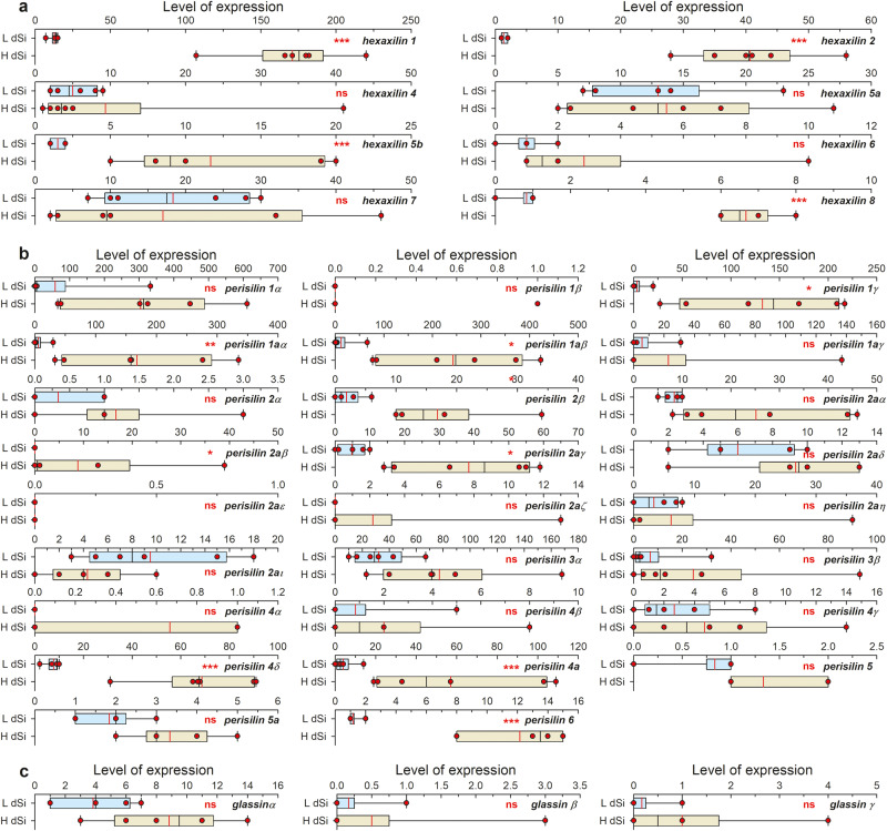
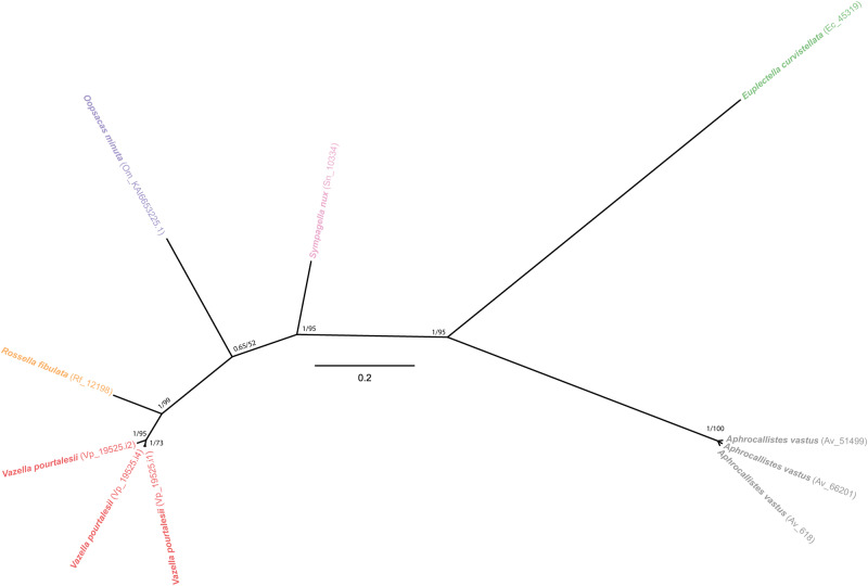
Metazoans use silicon traces but rarely develop extensive silica skeletons, except for the early-diverging lineage of sponges. The mechanisms underlying metazoan silicification remain incompletely understood, despite significant biotechnological and evolutionary implications. Here, the characterization of two proteins identified from hexactinellid sponge silica, hexaxilin and perisilin, supports that the three classes of siliceous sponges (Hexactinellida, Demospongiae, and Homoscleromorpha) use independent protein machineries to build their skeletons, which become non-homologous structures. Hexaxilin forms the axial filament to intracellularly pattern the main symmetry of the skeletal parts, while perisilin appears to operate in their thickening, guiding extracellular deposition of peripheral silica, as does glassin, a previously characterized hexactinellid silicifying protein. Distant hexaxilin homologs occur in some bilaterians with siliceous parts, suggesting putative conserved silicifying activity along metazoan evolution. The findings also support that ancestral Porifera were non-skeletonized, acquiring silica skeletons only after diverging into major classes, what reconciles molecular-clock dating and the fossil record.


Conflict of interest statement

The authors declare no competing interests.

Figures

Fig. 1
Fig. 1. Body shape, spicule complement, and relationships of the hexactinellid sponges Euplectella curvistellata and Vazella pourtalesii.
a Euplectella curvistellata voucher¸ republished from Shimizu et al. with PNAS permission. b, c Live (b) and longitudinal-sectioned (c) voucher of Vazella pourtalesii. Scale bars (bd): 2 cm. d Phylogenetic scheme of Hexactinellida illustrating the relationships of the genera hosting species with available genomic and transcriptomic data used in this study. Relationships are sensu Dohrmann, but updated according to the World Porifera Database https://www.marinespecies.org/porifera/, last visited on 13/01/23. e Schematic representation of spicules in E. curvistellata, as compiled from various sources,–, using Adobe Illustrator 2023 (version 27.1.1). 1: Parenchymalia-principalia hexactines; 2: Dermalia-choanosomalia stauractines; 3: Dermalia-choanosomalia pentactine; 4: Gastralia-canalaria pentactine; 5: Dermalia-choanosomalia paratetractine; 6: Large choanosomal stauractine; 7: Large choanosomal diactines of the sieve plate; 8: Anchorate basalia; 9: Dermalia-choanosomalia triactine; 10: Oscularia triactines; 11: Principalia diactine; 12: Dermal hexactine; 13: Choanosomal tauactine; 14: Oxyhexaster; 15: Floricome; 16: Graphicome. f Schematic representation of spicules in V. pourtalesii, as compiled from various sources, and personal skeletal observations, using Adobe Illustrator 2023 (version 27.1.1). 1: Hypodermal pentactine; 2: Dermal pentactine; 3: Dermal stauractine; 4: Atrial hexactine; 5: Atrial spicule with branching near central cross (tangential plane); 6: Choanosomal diactine; 7: Hexactines; 8: A fragment of tangential ray of hypodermal pentactine; 9: Hexaster; 10: Discohexaster; 11: Hexactines; 12: Prostalia monaxone.
Fig. 2
Fig. 2. Characterization and location of proteins occluded in the silica of the hexactinellids Euplectella curvistellata and Vazella pourtalesii.
a SDS-PAGE analysis of water-soluble and water-insoluble extracts from the silica of E. curvistellata¸ showing molecular-weight bands (kDa) for hexaxilin and glassin proteins. b Amino acid sequence of hexaxilin-1 obtained by translating the sequence of a PCR product amplified from E. curvistellata genomic DNA. c SDS-PAGE analysis of water-soluble and water-insoluble extracts from the silica of V. pourtalesii, a¸ showing bands of hexaxilin and perisilin proteins. d, e Amino acid sequence of hexaxilin-1 (d) and perisilin-1aα (e) from V. pourtalesii obtained by blast against V. pourtalesii’s transcriptome. Red letters in aa sequences represent peptides initially determined by the aa sequencer; blue letters indicate peptides used as immunogens for antibody production. fq Fractured spicules incubated with rabbit antisera against hexaxilin, perisilin and glassin of E. curvistellata and V. pourtalesii for spatial localization of these proteins after labeling primary antibodies with Alexafluor 488-conjugated goat anti-rabbit IgG. Figures show incubations with: (f, g) anti-E. curvistellata hexaxilin antiserum; (hj) anti-E. curvistellata glassin antiserum; (k, l) normal serum; (m, n) anti-V. pourtalesii hexaxilin affinity purified antibody; and (oq) anti-V. pourtalesii perisilin affinity purified antibody. (f, h, k, m, o) fluorescent images; (g, i, l, n and p) phase contrast images. Images (j) and (q) are Z-stack micrographs of spicules in cross-section captured under the laser-scanning confocal microscope. Note in images (f) and (m) a very faint extra-axial staining (yellow arrows), which corresponds to spicule zones in images g and n (purple arrows) where the silica is irregularly fractured, creating microcavities from which the fluorochrome is rinsed with greater difficulty after the incubations, leaving traces of it that account for the very faint extra-axial color signal (see Supplementary Fig. 2). Scale bar in image (f) represents 50 μm and it applies to all images from (f) to (q). SDS-PAGE (2a, 2c) and immunostainings (2fi, 2kp) were repeated at least three times with similar results. r Schematic drawing made with Adobe Illustrator 2023 (version 27.1.1) representing a three-dimensional side view (left) and a cross section (right) of a spicule to summarize the spatial distribution of hexaxilin, glassin and perisilin.
Fig. 3
Fig. 3. Diversity summary for hexaxilins, perisilins and glassins in Hexactinellida.
BUSCO completeness (metazoans) of the hexactinellid genomes/transcriptomes used in the present study is given in row 1, selecting for the most complete genome/ transcriptome if more than one were available to a species. Green and white cells indicate respectively the presence and absence of proteins. Numbers within green cells refer to number of different amino acid sequences found per protein in each species.
Fig. 4
Fig. 4. Evolutionary relationships of hexaxilin.
Bayesian Inference (BI) phylogenetic tree of hexaxilins obtained with MrBayes 3.2. (Maximum Likelihood tree is shown in Supplementary Fig. 5). Included sequences correspond to all blast hits for hexaxilin-1 of E. curvistellata (sequence Ec_20423) with a bit score larger than 50 (Supplementary Data 1). Hexactinellid species are represented by names in different color. Scale bar represents 0.4 amino acid substitutions per site. Posterior probability (pp) and bootstrap (b) values are given at each node as pp/b. Nodes with no bootstrap value were not recovered in the Maximum Likelihood phylogeny. Alignment data are available in fasta format in Source Data Fig. 4. Source Data Fig. 4.
Fig. 5
Fig. 5. Expression patterns of hexactinellid silica proteins.
Box plot of TMM normalized expression values (red dots) of hexaxilin (a), perisilin (b), and glassin (c) genes in the transcriptome of six individuals of V. pourtalesii exposed to naturally low dSi concentrations (light blue) versus that of six others exposed to high dSi concentrations (light brown). End of boxes define the 25th and 75th percentiles, with a black line at the median, a red line at the mean, and errors bars defining the 10th and 90th percentiles. Differentially expressed (DE) genes were determined through the quasi-likelihood F test implemented in the function GLMTreat of the Bioconductor package edgeR. Asterisks indicate the statistical significance of tests, following the criterion of at least a fourfold difference in expression and with the P-values corrected by false discovery rate (FDR): ***P < 0.001; **P < 0.01; *P < 0.05; ns= not significant. TMM expression values per individual, statistical tests, and exact probabilities of tests are detailed in Supplementary Data 3–4.
Fig. 6
Fig. 6. 3D models for the secondary structure of proteins extracted from the silica of the hexactinellid species Euplectella curvistellata and Vazella pourtalesii.
Models were inferred with both SWISS-MODEL and Alphafold2 software, which came into general agreement (Supplementary Fig. 7, 8, 12, 15). Protein alignment and templates for SWISS-MODEL are shown in Supplementary Fig. 6. SWISS-MODEL results are shown because they are more conservative, not considering those regions that are not available in the templates. Alphapfold2 generated full-length polypeptides, hypothesizing about the regions not covered by the templates and suggesting that those regions not inferred by SWISS-MODEL were basically random structures. a Hexaxilin-1 of E. curvistellata (ranging from aa 41 to aa 308). b Hexaxilin-1 of V. pourtalesii (ranging from aa 41 to aa 315). c Perisilin-1α of V. pourtalesii (ranging from aa 21 to aa197). d Glassin of E. curvistellata (ranging from aa 21 to aa 197). Sequences expected to form alpha-helices and beta-sheets are colored in blue and green, respectively.
Fig. 7
Fig. 7. Evolutionary relationships of perisilin.
Bayesian Inference (BI) phylogenetic tree of perisilins obtained with MrBayes 3.2. (Maximum Likelihood tree is shown in Supplementary Fig. 10). Included sequences correspond to all blast hits for perisilin-1α of V. pourtalesii (sequence Vp_1621.i8) with a bit score larger than 50, but very similar sequences for a given species in the ougroup have been collapsed in tree for the sake of clarity. All sequences are given in Supplementary Fig. 10 and Supplementary Data 5. Different hexactinellid species are represented by names in different color. Scale bar represents 0.3 amino acid substitutions per site. Posterior probability (pp) and bootstrap (b) values are given at each node as pp/b. Alignment data is available in fasta format in Source Data Fig. 7. Source Data Fig. 7.
Fig. 8
Fig. 8. Evolutionary relationships of glassin.
Unrooted Bayesian Inference (BI) phylogenetic tree of glassin obtained with MrBayes 3.2. (Maximum Likelihood tree is shown in Supplementary Fig. 13). Included sequences correspond to all blast hits for glassin of E. curvistellata (sequence Ec_45319) with a bit score larger than 50 (Supplementary Data 7). Each hexactinellid species is represented by a different color. Scale bar represents 0.2 amino acid substitutions per site. Posterior probability (pp) and bootstrap (b) values are given at each node as pp/b. Alignment data is available in fasta format in Source Data Fig. 8. Source Data Fig. 8.
Fig. 9
Fig. 9. TEM micrographs of silicification in Vazella pourtalesii.
a, b Cross-sections of early-developing stages of spicules (isp) still within the silica deposition vesicle (sd) in the cytoplasm (cy) of the sclerocytes (sc). The vesicle is limited by the silicalemma (lm), a membrane distinct from the plasmalemma (pm) of the sclerocyte. In these silicified ultra-thin sections, the silica (seen as an electron-dense material) of the intracellular (isp) and extracellular (esp) spicules has been crushed into small pieces by the diamond blade and dragged away. The hexactinellid sclerocytes show nucleated nuclei and are rich in electron-clear vesicles (containing dSi?) and mitochondria, being these features similar to those in sclerocytes of Demospongiae. cf Desilicified samples showing large empty holes within the silica deposition vesicle(sd) of the sclerocytes (sc) in which spicules where hosted prior to desilicfication in HF. Image (d) is a magnification of image c detailing an electron-dense zone of the sclerocyte cytoplasm(cy) adjacent to the silicalemma (sl), as also reported in previous literature. Image f details a longitudinal section of the space left by a large desilicified spicule that was enclosed within a sclerocyte (sc). Two nuclei (n) are seen in this sclerocyte section (i.e., sclerosyncytium). The sclerosyncytium is rich in mitochondria (mi), electron-clear vesicles (ev), and is connected to other cells through perforated plugs (pp). Findings derive from inspection of tissue from three different sponge individuals that were randomly collected but showed high silicate consumption rates during the high dSi treatment.

Similar articles

Cited by

References

    1. Sarikaya M, et al. Biomimetic model of a sponge-spicular optical fiber—mechanical properties and structure. J. Mater. Res. 2001;16:1420–1428. doi: 10.1557/JMR.2001.0198. - DOI
    1. Ehrlich H, et al. Supercontinuum generation in naturally occurring glass sponges spicules. Adv. Opt. Mater. 2016;4:1608–1613. doi: 10.1002/adom.201600454. - DOI
    1. Zhang M, et al. Robust and scalable in vitro surface mineralization of inert polymers with a rationally designed molecular bridge. ACS Appl. Mater. Interfaces. 2022;22:25. - PubMed
    1. Aizenberg J, et al. Skeleton of Euplectella sp.: Structural hierarchy from nanoscale to the macroscale. Science. 2005;309:275–278. doi: 10.1126/science.1112255. - DOI - PubMed
    1. Walter SL, Flinn BD, Mayer G. Mechanisms of toughening of a natural rigid composite. Mater. Sci. Eng. C. 2007;27:570–574. doi: 10.1016/j.msec.2006.05.020. - DOI

Publication types