Review
. 2024 Jan 8;9(1):13.
doi: 10.1038/s41392-023-01690-3.

Targeting proprotein convertase subtilisin/kexin type 9 (PCSK9): from bench to bedside

Affiliations
Review

Targeting proprotein convertase subtilisin/kexin type 9 (PCSK9): from bench to bedside

Xuhui Bao et al. Signal Transduct Target Ther. .

Abstract

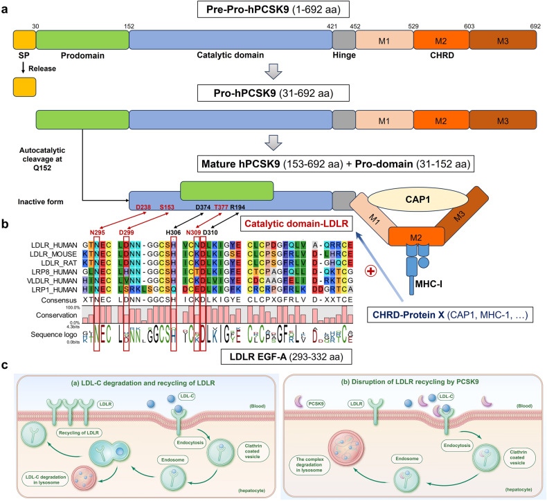
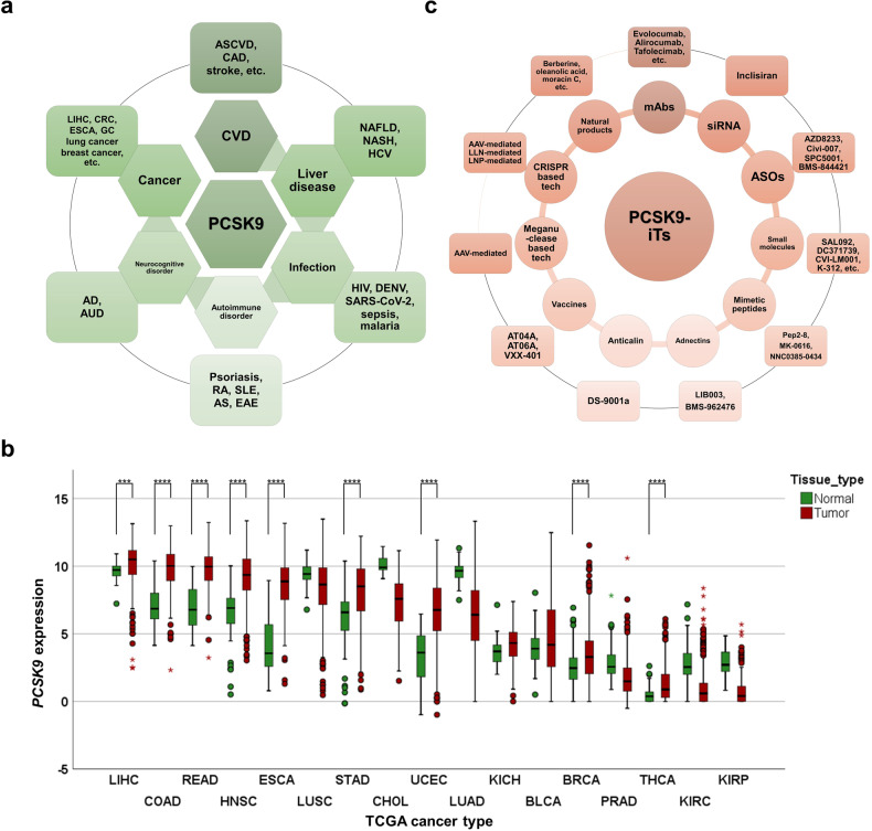
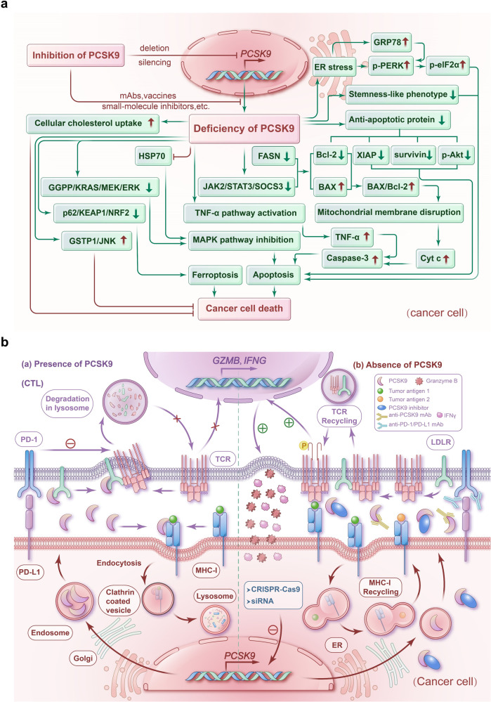
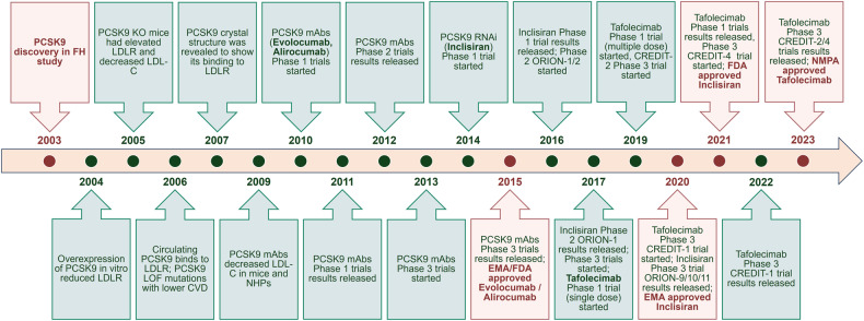
Proprotein convertase subtilisin/kexin type 9 (PCSK9) has evolved as a pivotal enzyme in lipid metabolism and a revolutionary therapeutic target for hypercholesterolemia and its related cardiovascular diseases (CVD). This comprehensive review delineates the intricate roles and wide-ranging implications of PCSK9, extending beyond CVD to emphasize its significance in diverse physiological and pathological states, including liver diseases, infectious diseases, autoimmune disorders, and notably, cancer. Our exploration offers insights into the interaction between PCSK9 and low-density lipoprotein receptors (LDLRs), elucidating its substantial impact on cholesterol homeostasis and cardiovascular health. It also details the evolution of PCSK9-targeted therapies, translating foundational bench discoveries into bedside applications for optimized patient care. The advent and clinical approval of innovative PCSK9 inhibitory therapies (PCSK9-iTs), including three monoclonal antibodies (Evolocumab, Alirocumab, and Tafolecimab) and one small interfering RNA (siRNA, Inclisiran), have marked a significant breakthrough in cardiovascular medicine. These therapies have demonstrated unparalleled efficacy in mitigating hypercholesterolemia, reducing cardiovascular risks, and have showcased profound value in clinical applications, offering novel therapeutic avenues and a promising future in personalized medicine for cardiovascular disorders. Furthermore, emerging research, inclusive of our findings, unveils PCSK9's potential role as a pivotal indicator for cancer prognosis and its prospective application as a transformative target for cancer treatment. This review also highlights PCSK9's aberrant expression in various cancer forms, its association with cancer prognosis, and its crucial roles in carcinogenesis and cancer immunity. In conclusion, this synthesized review integrates existing knowledge and novel insights on PCSK9, providing a holistic perspective on its transformative impact in reshaping therapeutic paradigms across various disorders. It emphasizes the clinical value and effect of PCSK9-iT, underscoring its potential in advancing the landscape of biomedical research and its capabilities in heralding new eras in personalized medicine.


Conflict of interest statement

The authors declare no competing interests.

Figures

Fig. 1
Fig. 1
The main structure and function of PCSK9. a PCSK9 comprises a signal peptide (SP, aa 1–30), a prodomain (aa 31–152), a catalytic domain (aa 153–421) with a hinge (aa 422–452), and a Cysteine-Histidine rich C-terminal domain (CHRD, aa 453–692) that can be further divided into three modules, M1 (aa 453–529), M2 (aa 530–603), and M3 (aa 604–692). In ER, proPCSK9 undergoes autocatalytic cleavage at Q152. The prodomain is then separated from the mature PCSK9, but remains associated with the catalytic domain, inhibiting the protease activity of the mature PCSK9. b There are five residues directly involved in the avidity of PCSK9:LDLR interface including S153:D299, R194:D310, D238:N295, D374:H306, and T377:N309, in which the hydrogen bonds between D238 and N295, T377 and N309, and a salt bridge between S153 and D299 contribute to the specificity of PCSK9 binding to the epidermal growth factor (EGF)-A domain of low-density lipoprotein receptor (LDLR) instead of other EGF-like domains. Primary sequence alignments of EGF-A domain from selected species (human, mouse, and rat) and human LDLR family members (LDLR related protein 8 [LRP8]/apolipoproteinE receptor 2 [ApoER2], very low-density lipoprotein receptor [VLDLR], and LRP1) were performed using the CLC workbench. Furthermore, cyclase associated actin cytoskeleton regulatory protein 1 (CAP1) and major histocompatibility complex class 1 (MHC-1) (e.g., human leukocyte antigen [HLA]-C) may be two strong candidates for “protein X” that can promote the degradation of PCSK9-LDLR complex in acidic cytosolic compartments. c (a) LDLRs are crucial in controlling levels of LDL cholesterol (LDL-C) in the blood by managing their removal from circulation. LDLRs bind to LDL-C and the resulting complexes are internalized into hepatocytes through endocytosis into clathrin-coated vesicles that can be subsequently fused with endosomes, whose acidic environment leads to the dissociation of the LDL-C particles to be transported to lysosomes to degrade into lipids and amino acids, while LDLRs can recycle back to the surface of the hepatocytes to transport and clear additional LDL-C from the circulation. (b) When PCSK9 is secreted from hepatocytes and binds to LDLRs on the cell surface, LDLR recycling to the cell surface is impeded. Due to a conformational change in LDLR caused by PCSK9, LDLR cannot get out of the endosome to recycle back to the cell surface. Instead, the PCSK9-LDLR-LDL-C complex traffics to the lysosome for degradation. By promoting LDLR degradation, PCSK9 decreases LDLR levels at the cell surface, increasing serum LDL-C. Panels were illustrated by Adobe Illustrator and Microsoft PowerPoint
Fig. 2
Fig. 2
The role of PCSK9 in various disorders, its aberrant expression in cancers, and the current PCSK9-iTs. a PCSK9 plays an important role in various disorders including cardiovascular diseases (CVDs), liver diseases, infection, autoimmune disorders, neurocognitive disorders, and cancer. CRC colorectal cancer, GC gastric cancer. b PCSK9 mRNA expression across different types of cancer in TCGA datasets. LIHC liver hepatocellular carcinoma, COAD colon adenocarcinoma, READ rectum adenocarcinoma, HNSC head and neck squamous cell carcinoma, ESCA esophageal carcinoma, LUSC lung squamous cell carcinoma, STAD stomach adenocarcinoma, CHOL cholangiocarcinoma, UCEC uterine corpus endometrial carcinoma, LUAD lung adenocarcinoma, KICH kidney chromophobe, BLCA bladder urothelial carcinoma, BRCA breast invasive carcinoma, PRAD prostate adenocarcinoma, THCA thyroid carcinoma, KIRC kidney renal clear cell carcinoma, and KIRP kidney renal papillary cell carcinoma ***P < 0.001, ****P < 0.0001. c Current PCSK9-iTs include monoclonal antibodies (mAbs), small interfering RNA (siRNA), antisense oligonucleotide (ASO), small-molecule inhibitors, mimetic peptides, adnectin, anticalin, vaccines, meganuclease based gene editing technology, clustered regularly interspaced short palindromic repeats (CRISPR) based gene editing technology, and natural products. Panels were illustrated by IBM SPSS Statistics and Microsoft PowerPoint
Fig. 3
Fig. 3
The potential mechanisms of PCSK9 on the regulation of cancer cell death and cancer immunity. a PCSK9 deficiency induces ER stress, leading to the dissociation of ER chaperone GRP78 from PERK, which causes downstream phosphorylation of eIF2α to trigger ER stress-induced apoptosis. PCSK9 deficiency can also suppress the development of stemness-like phenotype of cancer. In addition, deficiency of PCSK9 inhibits FA synthase (FASN) or Janus kinase 2/signal transducer and activator of transcription 3/suppressor of cytokine signaling 3 (JAK2/STAT3/SOCS3) pathway to downregulate Bcl-2 levels and upregulate Bax levels to increase Bax/Bcl-2 ratio leading to mitochondrial membrane disruption and subsequent release of cytochrome c (Cyt c) to activate caspase-3 in the cytosol, which can initiate caspase-dependent apoptosis. The caspase-3-initiated apoptotic signaling can also be initiated by the activation of the TNF-α pathway resulting from PCSK9 deficiency. In addition, a deficiency of PCSK9 can downregulate anti-apoptotic proteins X-linked inhibitor of apoptosis protein (XIAP), survivin, and phospho-protein kinase B (p-Akt) to cause cancer cell apoptosis. A deficiency of PCSK9 can also promote cancer cell apoptosis through the inhibition of the MAPK pathway via downregulating heat shock protein 70 (HSP70) levels or the geranylgeranyl diphosphate (GGPP)/KRAS/mitogen-activated extracellular signal-regulated kinase (MEK)/ERK signaling. Moreover, PCSK9 deficiency can downregulate the sequestome 1/Kelch-like ECH-associated protein 1/nuclear factor erythroid 2-related factor 2 (p62/KEAP1/NRF2) signaling pathway to cause cancer cell ferroptosis. However, PCSK9 deficiency may increase LDLR expression and cause cellular cholesterol uptake to increase, or upregulate the Jun N-terminal kinase (JNK) signaling via glutathione S-transferase Pi 1 (GSTP1), which may suppress cancer cell death. b (a) PCSK9’s presence can help cancer cells escape from T-cell recognition and elimination. In cancer cells, PCSK9 binds to MHC-I and facilitates its degradation through the endosomal/lysosomal pathway, thereby impeding its recycling to the cell surface. In cytotoxic T lymphocytes (CTLs), PCSK9 binds to LDLR in CTLs, which subsequently binds to the CD3 subunits of the T-cell receptor (TCR) complex and inhibits the recycling of the LDLR-TCR complex to the plasma membrane. Meanwhile, the interaction of programmed cell death 1 (PD-1) and programmed cell death ligand 1 (PD-L1) can drive CTLs to apoptosis or into a regulatory phenotype to lose killing function. (b) Instead, the absence of PCSK9 induced by antibodies, small-molecule inhibitors, or genetic depletion can restore the immune surveillance from CTLs against cancer cells. The recycling of MHC-I of cancer cells and the TCR complex of CTLs can proceed unimpededly, thereby maintaining their elevated levels on the cell surface, respectively. Therefore, cancer cells can more effectively present tumor-specific antigens, which in turn are more readily recognized by CTLs, to perform their antitumor activity. Moreover, if combined with the immune checkpoint inhibitor (ICI) to block the PD-1/PD-L1 axis, PCSK9-iTs can boost an enhanced synergistic antitumor immunity for significant cancer elimination. Panels were illustrated by Adobe Illustrator and Microsoft PowerPoint
Fig. 4
Fig. 4
The timeline of the development of PCSK9-iTs. The timeline of the discovery of PCSK9 and the development of its inhibitors. From the initial discovery in 2003, the function of PCSK9 and its inhibitors have been investigated in numerous preclinical and clinical studies over the past two decades, leading to the approval of three specific inhibitors of PCSK9, including three mAbs (Evolocumab and Alirocumab, EMA and US FDA in 2015; Tafolecimab, China’s NMPA in 2023) and one RNA interference (RNAi) (Inclisiran, EMA in 2020, and US FDA in 2021) to treat refractory hyperlipidemia in the clinic. NHP nonhuman primate. Panels were illustrated by Microsoft PowerPoint
Fig. 5
Fig. 5
The strategies to develop emerging PCSK9 inhibitors. a Strategies for the development of four representative PCSK9 inhibitors: PF-06815345 (a), CVI-LM001 (b), DC371739 (c), and carboxylic acid 9 (d). b The crystal structures of PCSK9 with small-molecule inhibitors. Crystal structures were adapted from https://www.rcsb.org/ (PDB ID: 6U3X, 6U2N, 6U2P, 6U38). c The co-crystal structures of PCSK9 with EGF-A (pink), LDLR peptide (green) (a) or PCSK9 with MK-0616 analog (b), and the strategy of development of macrocyclic PCSK9 inhibitor from mRNA display (c). Crystal structures were adapted from https://www.rcsb.org/ (PDB ID: 3BPS, 4NE9, 6XIE). d The crystal structures of the complex of Pep2–8 (a) and groove-binding Pep1 (b) with PCSK9 and strategies to develop the indicated PCSK9 inhibitors (a-c). Crystal structures were adapted from https://www.rcsb.org/ (PDB ID: 4NMX, 5VLP). AS/MS affinity selection/mass spectrometry, CETSA cellular thermal shift assay, SAR structure-activity relationships, ∆Tm melting temperature shift, Kd dissociation constant, Ki inhibition constant, Ab20 PCSK9-binding antibody 20, EGF-A epidermal growth-factor-like domain A, IC50 half-maximal inhibitory concentration. Panels were illustrated by ChemDraw and Microsoft PowerPoint

Similar articles

Cited by

References

    1. Steiner DF, Cunningham D, Spigelman L, Aten B. Insulin biosynthesis: evidence for a precursor. Science. 1967;157:697–700. doi: 10.1126/science.157.3789.697. - DOI - PubMed
    1. Steiner DF. On the discovery of precursor processing. Methods Mol. Biol. 2011;768:3–11. doi: 10.1007/978-1-61779-204-5_1. - DOI - PubMed
    1. Chretien M, Li CH. Isolation, purification, and characterization of gamma-lipotropic hormone from sheep pituitary glands. Can. J. Biochem. 1967;45:1163–1174. doi: 10.1139/o67-133. - DOI - PubMed
    1. Chretien M, et al. Isolation of peptides with opiate activity from sheep and human pituitaries: relationship to beta-lipotropin. Biochem. Biophys. Res. Commun. 1976;72:472–478. doi: 10.1016/S0006-291X(76)80066-6. - DOI - PubMed
    1. Chretien M. How the prohormone theory solved two important controversies in hormonal and neural Peptide biosynthesis. Front Endocrinol. (Lausanne) 2013;4:148. doi: 10.3389/fendo.2013.00148. - DOI - PMC - PubMed

MeSH terms