. 2024 Jan;602(2):333-354.
doi: 10.1113/JP285727. Epub 2024 Jan 7.

Two rare variants that affect the same amino acid in CFTR have distinct responses to ivacaftor

Affiliations

Two rare variants that affect the same amino acid in CFTR have distinct responses to ivacaftor

Hongyu Li et al. J Physiol. 2024 Jan.

Abstract

Some residues in the cystic fibrosis transmembrane conductance regulator (CFTR) channel are the site of more than one CFTR variant that cause cystic fibrosis. Here, we investigated the function of S1159F and S1159P, two variants associated with different clinical phenotypes, which affect the same pore-lining residue in transmembrane segment 12 that are both strongly potentiated by ivacaftor when expressed in CFBE41o- bronchial epithelial cells. To study the single-channel behaviour of CFTR, we applied the patch-clamp technique to Chinese hamster ovary cells heterologously expressing CFTR variants incubated at 27°C to enhance channel residence at the plasma membrane. S1159F- and S1159P-CFTR formed Cl- channels activated by cAMP-dependent phosphorylation and gated by ATP that exhibited thermostability at 37°C. Both variants modestly reduced the single-channel conductance of CFTR. By severely attenuating channel gating, S1159F- and S1159P-CFTR reduced the open probability (Po ) of wild-type CFTR by ≥75% at ATP (1 mM); S1159F-CFTR caused the greater decrease in Po consistent with its more severe clinical phenotype. Ivacaftor (10-100 nM) doubled the Po of both CFTR variants without restoring Po values to wild-type levels, but concomitantly, ivacaftor decreased current flow through open channels. For S1159F-CFTR, the reduction of current flow was marked at high (supersaturated) ivacaftor concentrations (0.5-1 μM) and voltage-independent, identifying an additional detrimental action of elevated ivacaftor concentrations. In conclusion, S1159F and S1159P are gating variants, which also affect CFTR processing and conduction, but not stability, necessitating the use of combinations of CFTR modulators to optimally restore their channel activity. KEY POINTS: Dysfunction of the ion channel cystic fibrosis transmembrane conductance regulator (CFTR) causes the genetic disease cystic fibrosis (CF). This study investigated two rare pathogenic CFTR variants, S1159F and S1159P, which affect the same amino acid in CFTR, to understand the molecular basis of disease and response to the CFTR-targeted therapy ivacaftor. Both rare variants diminished CFTR function by modestly reducing current flow through the channel and severely inhibiting ATP-dependent channel gating with S1159F exerting the stronger adverse effect, which correlates with its association with more severe disease. Ivacaftor potentiated channel gating by both rare variants without restoring their activity to wild-type levels, but concurrently reduced current flow through open channels, particularly those of S1159F-CFTR. Our data demonstrate that S1159F and S1159P cause CFTR dysfunction by multiple mechanisms that require combinations of CFTR-targeted therapies to fully restore channel function.

Keywords: CFTR chloride ion channel; CFTR inhibition; CFTR potentiation; cystic fibrosis; ivacaftor (VX-770); rare variant.


Conflict of interest statement

Competing interests

The authors have no conflicts of interest to declare.

Figures

Figure 1.
Figure 1.. Structure of human CFTR showing the rare variants at codon S1159 in the twelfth transmembrane segment
A, orthogonal views of PyMOL representations of a 3D model of phosphorylated, ATP-bound wild-type human CFTR complexed with ivacaftor (based on PDB id: 6O2P). The lefthand image shows the ivacaftor-binding site, while the righthand image has been rotated 180° to reveal the position of S1159 (see dashed box). The twelfth transmembrane segment (M12) is coloured wheat and M9 light blue. The R domain, unresolved in cryo-EM structures of CFTR (e.g. Liu et al., 2019), has been omitted. The position of the plasma membrane is shown with the intracellular (In) and extracellular (Out) sides indicated. The chemical structures of ATP and ivacaftor are displayed in green and magenta, respectively. BD, magnified views of the regions of M9 and M12 enclosed by the dashed box in A to show the interactions of D979 (pink) (M9) with S1159 (white), S1159F (red) and S1159P (orange). The hydrogen bond between S1159 and D979 is indicated by a yellow dashed line, with a bond length of 1.7 Å.
Figure 2.
Figure 2.. The multi-channel behaviour of S1159-CFTR variants
Representative multi-channel recordings and corresponding current amplitude histograms of wild-type (A), S1159F- (B) and S1159P-CFTR (C) in excised inside-out membrane patches from transiently transfected CHO-K1 cells. ATP (1 mM) and PKA (75 nM) were continuously present in the intracellular solution. Dotted lines indicate the closed channel state, arrowheads identify different open channel current levels and downward deflections correspond to channel openings. The labels C and O1 – O4 denote the closed and open channel amplitudes, respectively. Because openings of the S1159-CFTR variants to O3 and O4 were rare, they are not apparent in the current amplitude histograms. Unless otherwise indicated in this and subsequent figures, membrane voltage was clamped at −50 mV, a large Cl concentration gradient was imposed across the membrane patch ([Cl]int, 147 mM; [Cl]ext, 10 mM) and temperature was 37 °C.
Figure 3.
Figure 3.. The single-channel behaviour of S1159-CFTR variants
A, representative single-channel recordings and corresponding current amplitude histograms of wild-type, S1159F- and S1159P-CFTR in excised inside-out membrane patches from transiently transfected CHO-K1 cells. ATP (1 mM) and PKA (75 nM) were continuously present in the intracellular solution. Dotted lines indicate the closed channel state and downward deflections correspond to channel openings. The labels C and O denote the closed and open channel amplitudes, respectively. BE, summary single-channel current amplitude (i), interburst interval (IBI), mean burst duration (MBD) and open probability (Po) data determined from prolonged recordings (≥ 4 min) of wild-type, S1159F- and S1159P-CFTR for the experimental conditions described in A. Symbols represent individual values and columns represent means ± SD (wild-type: i and Po, n = 15; MBD and IBI, n = 8; S1159F: i and Po, n = 22; MBD and IBI, n = 15; S1159P: i and Po, n = 24; MBD and IBI, n = 13). [Panel B: ***P < 0.001 vs. wild-type CFTR; one-way ANOVA with Tukey post-hoc test; normality test (Shapiro–Wilk), P < 0.050 (failed); equal variance test (Brown-Forsythe), P = 0.350 (passed). Panel C: *P = 0.013 and **P = 0.001 vs. wild-type CFTR; Kruskal-Wallis one-way ANOVA on Ranks with Dunn’s post-hoc test; normality test (Shapiro–Wilk), P < 0.050 (failed). Panel D: ***P < 0.001 vs. wild-type CFTR; one-way ANOVA with Tukey post-hoc test; normality test (Shapiro–Wilk), P = 0.620 (passed); equal variance test (Brown-Forsythe), P = 0.050 (passed). Panel E: ***P < 0.001 vs. wild-type CFTR; one-way ANOVA with Tukey post-hoc test; normality test (Shapiro–Wilk), P = 0.490 (passed); equal variance test (Brown-Forsythe), P < 0.050 (failed)].
Figure 4.
Figure 4.. Impact of S1159-CFTR variants on the single-channel conductance of CFTR
A, representative single-channel recordings of wild-type, S1159F- and S1159P-CFTR in excised inside-out membrane patches from transiently transfected CHO-K1 cells acquired at the indicated voltages. ATP (1 mM) and PKA (75 nM) were continuously present in the intracellular solution. Dotted lines indicate the closed channel state and downward deflections correspond to channel openings. B and C, single-channel current-voltage (iV) relationships and summary slope conductance data of wild-type, S1159F- and S1159P-CFTR. Data are means ± SD (wild-type, n = 11–14; S1159F-CFTR, n = 3–5; S1159P-CFTR, n = 7–9). In B, the continuous lines are the fit of first order linear regression functions to mean data (r2 > 0.99), while in C, symbols represent individual values.
Figure 5.
Figure 5.. Dwell time histograms of S1159-CFTR variants
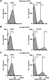
Representative dwell time histograms of wild-type (A), S1159F- (B) and S1159P-CFTR (C). Data are from experiments in which the excised inside-out membrane patch from transiently transfected CHO-K1 cells (B and C) [or stably transfected C127 cells (A)] contained only one active channel, studied in the presence of ATP (1 mM) and PKA (75 nM) in the intracellular solution; membrane voltage was −50 mV, a large Cl concentration gradient was imposed across the membrane patch ([Cl]int, 147 mM; [Cl]ext, 10 mM) and temperature was 37 °C. The continuous lines are the fit of one- or two-component exponential functions to the data and the dotted lines show the individual components of these functions. The vertical dashed lines indicate the mean values of the open (τO2) and closed (τC1, τC3) time constants. Logarithmic x-axes with 10 bins decade−1 were used for dwell time histograms.
Figure 6.
Figure 6.. Impact of S1159-CFTR variants on the ATP-dependent channel gating of CFTR
A, representative single-channel recordings of wild-type-, S1159F- and S1159P-CFTR in excised inside-out membrane patches from transiently transfected CHO-K1 cells acquired using the indicated intracellular ATP concentrations. PKA (75 nM) was continuously present in the intracellular solution. Dotted lines indicate the closed channel state and downward deflections correspond to channel openings. BD, relationship between ATP concentration and open probability (Po), mean burst duration (MBD) and interburst interval (IBI) for wild-type and S1159-CFTR variants; note that in D, the y-axis is plotted using a logarithmic scale. Data are means ± SD (wild-type: Po, n = 11–23; MBD and IBI, n = 6–8; S1159F: Po, n = 2–22; MBD and IBI, n = 2–15; S1159P: Po, S1159P, n = 11–24; MBD and IBI, n = 7–13) from experiments where ≥ 3 ATP concentrations were tested in each membrane patch. In B, the continuous lines are the fit of Michaelis–Menten functions to mean data (r2 ≥ 0.93).
Figure 7.
Figure 7.. Ivacaftor potentiates the channel gating of S1159-CFTR variants, but inhibits current flow through S1159F-CFTR
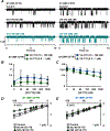
A, representative single-channel recordings of S1159F- and S1159P-CFTR in excised inside-out membrane patches from transiently transfected CHO-K1 cells in the absence and presence of ivacaftor. Ivacaftor (VX-770, 50 and 500 nM) was acutely added to the intracellular solution in the continuous presence of ATP (1 mM) and PKA (75 nM). Dotted lines indicate the closed channel state, downward deflections correspond to channel openings and teal is used to identify single-channel recordings acquired at a supersaturated concentration of ivacaftor. B and C, relationships between ivacaftor concentration and single-channel open probability (Po) and current amplitude (i) for S1159-CFTR variants at −50 mV. Data are means ± SD (S1159F, n = 3–5; S1159P, n = 8–9). D and E, single-channel current-voltage (iV) relationships of S1159F- and S1159P-CFTR in the absence and presence of ivacaftor (50 and 500 nM) in the intracellular solution. Data are means ± SD (S1159F: n = 2–3; S1159P: n = 2–4). In BE, triangles indicate supersaturated concentrations of ivacaftor. In CE, the continuous lines are the fit of first order linear regression functions to mean data (C, r2 > 0.90; D and E, r2 > 0.98), whereas in B, they are the fit of peak log normal functions to mean data.
Figure 8.
Figure 8.. Effects of ivacaftor on the single-channel behaviour of wild-type and S1159F-CFTR at different membrane voltages
A and B, representative single-channel recordings and corresponding current amplitude histograms of wild-type and S1159F-CFTR in excised inside-out membrane patches from CFTR-expressing C127 and CHO cells, respectively. The recordings were acquired at ±50 mV in the absence and presence of ivacaftor (VX-770; 100 and 1000 nM). Membrane patches were bathed in symmetrical 147 mM Cl solutions, ATP (1 mM) and PKA (75 nM) were continuously present in the intracellular solution and temperature was 37 °C. Dotted lines indicate the closed channel state and downward deflections at −50 mV and upward deflections at +50 mV correspond to channel openings. The labels C and O denote the closed and open channel amplitudes, respectively. In A and B, teal is used to identify single-channel recordings and current amplitude histograms acquired at a supersaturated concentration of ivacaftor. For ivacaftor (100 and 1000 nM), small leak currents at +50 mV shifted the current amplitude histograms of S1159F-CFTR by ~ 0.2 pA and 1.8 pA relative to that of the control. For summary single-channel current-voltage (iV) and open probability-voltage (PoV) relationships of wild-type and S1159F-CFTR, see Figure 10.
Figure 9.
Figure 9.. Actions of ivacaftor (50 nM) on the dwell time histograms of S1159F-CFTR
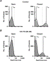
Representative dwell time histograms of S1159F-CFTR in the absence (A) and presence (B) of ivacaftor (VX-770, 50 nM). Data are from experiments in which the excised inside-out membrane patch from transiently transfected CHO-K1 cells contained only one active channel, studied in the presence of ATP (1 mM) and PKA (75 nM) in the intracellular solution; membrane voltage was −50 mV, a large Cl concentration gradient was imposed across the membrane patch ([Cl]int, 147 mM; [Cl]ext, 10 mM) and temperature was 37 °C. The continuous lines are the fit of one- or two-component exponential functions to the data and the dotted lines show the individual components of these functions. The black vertical dashed lines indicate the mean values of the open (τO2) and closed (τC1, τC3) time constants of S1159F-CFTR, while the grey vertical dotted lines indicate the same values for wild-type CFTR in the absence of ivacaftor. Logarithmic x-axes with 10 bins decade−1 were used for dwell time histograms.
Figure 10.
Figure 10.. Ivacaftor inhibition of current flow through S1159F-CFTR is voltage-independent
Single-channel open probability-voltage (PoV) relationships (A, D), current-voltage (iV) relationships (B, E) and summary slope conductance data (C, F) for wild-type (AC) and S1159F-CFTR (DF) in the absence and presence of ivacaftor (VX-770; 100 and 1000 nM). The data were acquired using excised inside-out membrane patches from C127 and CHO cells heterologously expressing wild-type and S1159F-CFTR, respectively, using the experimental conditions described in Figure 8. Data are means ± SD (wild-type, n = 5; S1159F, n = 4–6). In A, B, D and E, triangles indicate supersaturated concentrations of ivacaftor, while in C and F, these concentrations are identified by hatching. [Panel A, wild-type CFTR: ***P < 0.001 vs. control at ±50 mV; one-way ANOVA with Tukey post-hoc test; normality test (Shapiro–Wilk), P = 0.491 (passed); equal variance test (Brown-Forsythe), P < 0.050 (failed); †P = 0.0316 (two-tailed) vs. 100 nM VX-770 at −50 mV; Student’s paired t-test; normality test (Shapiro–Wilk), P = 0.524 (passed); †P = 0.0131 (two-tailed) vs. 100 nM VX-770 at +50 mV; Student’s paired t-test; normality test (Shapiro–Wilk), P = 0.557 (passed). Panel D, S1159F: **P = 0.003 vs. control at ±50 mV; one-way ANOVA with Tukey post-hoc test; normality test (Shapiro–Wilk), P = 0.271 (passed); equal variance test (Brown-Forsythe), P = 0.065 (passed). Panels B and E: the continuous lines are the fit of second order regression functions to mean data (r2 ≥ 0.98). Panel F, S1159F: ***P < 0.001 vs. control; †††P < 0.001 vs 100 nM VX-770; one-way ANOVA with Tukey post-hoc test; normality test (Shapiro–Wilk), P = 0.939 (passed); equal variance test (Brown-Forsythe), P = 0.739 (passed)].

Similar articles

Cited by

References

    1. Aleksandrov L, Aleksandrov AA, Chang X-B, & Riordan JR (2002). The first nucleotide binding domain of cystic fibrosis transmembrane conductance regulator is a site of stable nucleotide interaction, whereas the second is a site of rapid turnover. The Journal of Biological Chemistry 277, 15419–15425. - PubMed
    1. Anderson MP, & Welsh MJ (1992). Regulation by ATP and ADP of CFTR chloride channels that contain mutant nucleotide-binding domains. Science 257, 1701–1704. - PubMed
    1. Anderson MP, Berger HA, Rich DP, Gregory RJ, Smith AE, & Welsh MJ (1991). Nucleoside triphosphates are required to open the CFTR chloride channel. Cell 67, 775–784. - PubMed
    1. Avramescu RG, Kai Y, Xu H, Bidaud-Meynard A, Schnúr A, Frenkiel S, Matouk E, Veit G, & Lukacs GL (2017). Mutation-specific downregulation of CFTR2 variants by gating potentiators. Human Molecular Genetics 26, 4873–4885. - PMC - PubMed
    1. Bihler H, Sivachenko A, Millen L, Bhatt P, Thakerar Patel A, Chin J, Bailey V, Musisi I, LaPan A, Allaire NE, Conte J, Simon NR, Magaret AS, Raraigh KS, Cutting GR, Skach WR, Bridges RJ, Thomas PJ, & Mense M (2023). In vitro modulator responsiveness of 655 CFTR variants found in people with CF. bioRxiv DOI: 10.1101/2023.07.07.548159. - DOI - PubMed

Publication types