. 2024 Mar;115(3):937-953.
doi: 10.1111/cas.16062. Epub 2024 Jan 7.

L-type amino acid transporter 1 inhibitor JPH203 prevents the growth of cabazitaxel-resistant prostate cancer by inhibiting cyclin-dependent kinase activity

Affiliations

L-type amino acid transporter 1 inhibitor JPH203 prevents the growth of cabazitaxel-resistant prostate cancer by inhibiting cyclin-dependent kinase activity

Junryo Rii et al. Cancer Sci. 2024 Mar.

Abstract

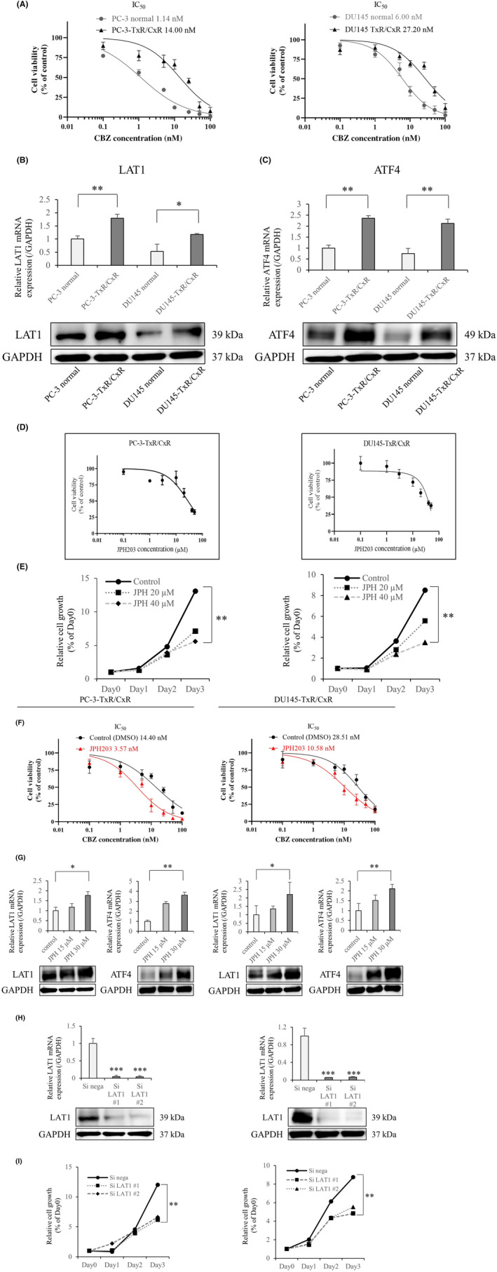
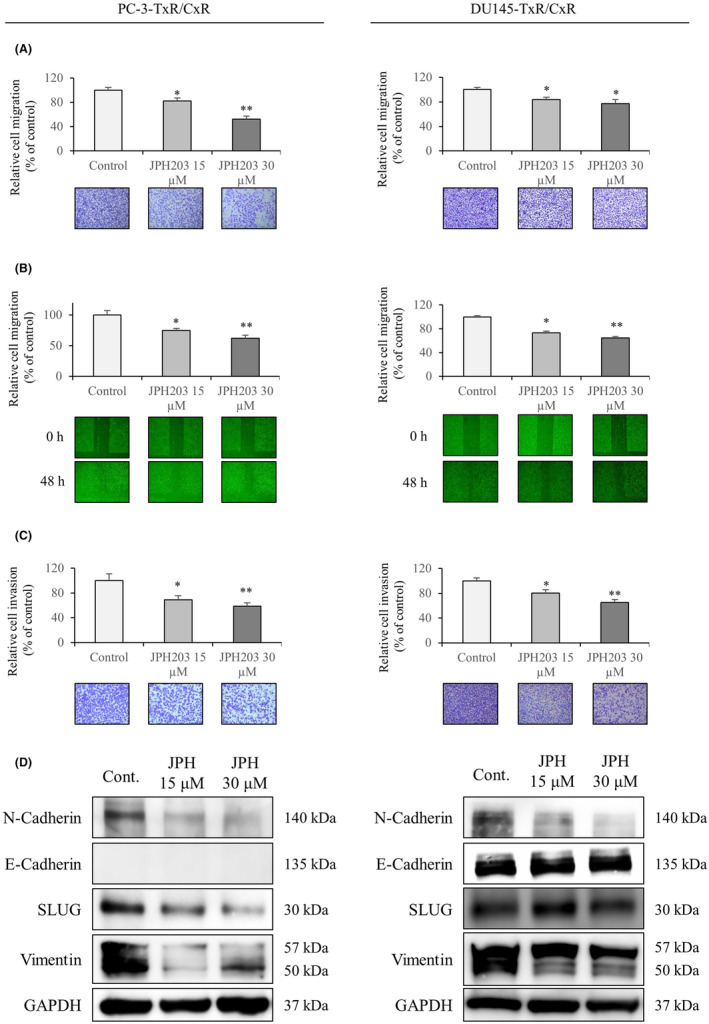
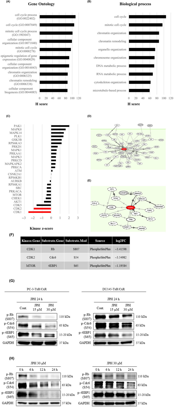
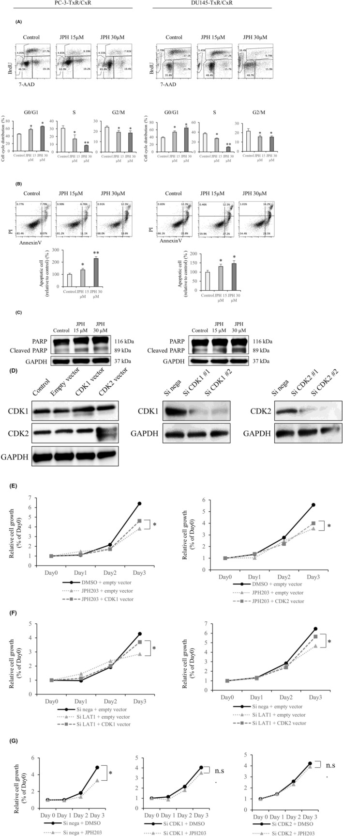
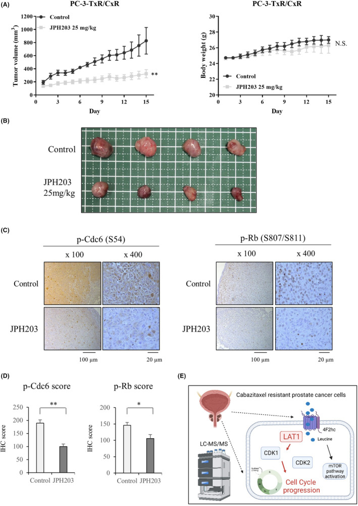
L-type amino acid transporter 1 (LAT1, SLC7A5) is an amino acid transporter expressed in various carcinomas, and it is postulated to play an important role in the proliferation of cancer cells through the uptake of essential amino acids. Cabazitaxel is a widely used anticancer drug for treating castration-resistant prostate cancer (CRPC); however, its effectiveness is lost when cancer cells acquire drug resistance. In this study, we investigated the expression of LAT1 and the effects of a LAT1-specific inhibitor, JPH203, in cabazitaxel-resistant prostate cancer cells. LAT1 was more highly expressed in the cabazitaxel-resistant strains than in the normal strains. Administration of JPH203 inhibited the growth, migration, and invasive ability of cabazitaxel-resistant strains in vitro. Phosphoproteomics using liquid chromatography-mass spectrometry to comprehensively investigate changes in phosphorylation due to JPH203 administration revealed that cell cycle-related pathways were affected by JPH203, and that JPH203 significantly reduced the kinase activity of cyclin-dependent kinases 1 and 2. Moreover, JPH203 inhibited the proliferation of cabazitaxel-resistant cells in vivo. Taken together, the present study results suggest that LAT1 might be a valuable therapeutic target in cabazitaxel-resistant prostate cancer.

Keywords: CDK; LAT1; amino acid transporter; phosphoproteomics; prostate cancer.


Conflict of interest statement

The authors declare no conflicts of interest.

Figures

FIGURE 1
FIGURE 1
Quantification of L‐type amino acid transporter 1 (LAT1) expression and the effects of LAT1 inhibition on cell proliferation. (A) Comparison of the ability of cabazitaxel‐resistant and normal strains to grow in response to cabazitaxel. (B) LAT1 expression in normal and TxR/CxR cells. (C) ATF4 expression in TxR/CxR cells. GAPDH was used as the loading control. (D) IC50 of JPH203 in TxR/CxR cells. (E) Cell proliferation over time with JPH203 administration. (F) Effect of JPH203 administration on IC50 for cabazitaxel (G) Changes in LAT1 and ATF4 expression upon JPH203 administration. (H) Confirmation of the decreased LAT1 expression by SiLAT1. (I) Effect of LAT1 knockdown on cell proliferation. Each bar represents the mean with the SEM. *p < 0.05; **p < 0.01, and ***p < 0.001 (unpaired Student's t‐test). “Si nega” is Si negative control.
FIGURE 2
FIGURE 2
Effects of JPH203 administration on cell migration, invasion, and the expression of epithelial–mesenchymal transition (EMT)‐related proteins. (A) Effect of JPH203 administration on the migration of TxR/CxR cells. (B) Effect of JPH203 administration in the wound healing assay. (C) Effect of JPH203 administration on the invasion of TxR/CxR cells. The data represent the results from three independent experiments. (D) JPH203‐induced changes in the expression of EMT‐related proteins in TxR/CxR cells. GAPDH was used as the loading control. Each bar represents the mean with the SEM. *p < 0.05 and **p < 0.01 (unpaired Student's t‐test).
FIGURE 3
FIGURE 3
Effects of JPH203 on intracellular phosphorylation in TxR/CxR cells. (A) The top 10 gene ontology (GO) terms and the H scores determined by KeyMolnet. (B) The top 10 biological process and the H scores determined by KeyMolnet. (C) Kinase scores determined by Kinase‐Substrate Enrichment Analysis (KSEA) in TxR/CxR cells with the administration of JPH203. Statistically significant changes are indicated in red. (D) Molecular network derived from CDK1 determined by KeyMolnet. (E) Molecular network derived from CDK2 determined by KeyMolnet. (F) Featured kinase‐substrate links by KSEA. (G) Confirmation by western blotting of the phosphorylated substrates that were decreased in the phosphoproteomics. (H) Time course of the phosphorylation changes by western blotting. GAPDH was used as the loading control. A p‐value <0.05 was considered statistically significant. CDK, cyclin‐dependent kinase.
FIGURE 4
FIGURE 4
Effects of JPH203 administration on the cell cycle and apoptosis in TxR/CxR cells. (A) Effect of JPH203 on the cell cycle. The bar charts show the percentages of JPH203‐treated cells in the G0/G1, S, and G2/M phases relative to the control cells. The data represent the results from three independent experiments. (B) Effect of JPH203 in apoptosis assays. (C) Western blotting of cleaved PARP, a marker of apoptosis. GAPDH was used as the loading control. (D) Verification of the effects of plasmid and SiRNA on CDK1 and CDK2. (E) Effect of JPH203 alone and JPH203 plus CDK plasmid administration on proliferation. (F) Influence of SiLAT1 alone and SiLAT1 plus CDK plasmid administration on proliferation. (G) Effects of JPH203 alone and JPH203 plus CDK knockdown on cell growth. Each bar represents the mean with the SEM. *p < 0.05; **p < 0.01. NS, not significant. CDK, cyclin‐dependent kinase.
FIGURE 5
FIGURE 5
Effects of JPH203 administration on the growth of PC‐3‐TxR/CxR cells in vivo. (A) Tumor diameter and body weight of mice in the control and JPH203‐treated groups. (B) Photograph of tumors removed from the control and JPH203‐treated groups. (C) Representative images of p‐cdc6 and p‐Rb immunostaining in tumors of the control and JPH203‐treated groups. (D) Immunohistochemistry (IHC) scores of the control and JPH203‐treated groups for p‐cdc6 and p‐Rb staining. Each bar represents the mean with the SEM. (E) Schematic illustration showing the function of LAT1 in cabazitaxel‐resistant prostate cancer cells. *p < 0.05; **p < 0.01. NS, not significant.

Similar articles

Cited by

References

    1. Gandaglia G, Leni R, Bray F, et al. Epidemiology and prevention of prostate cancer. Eur Urol Oncol. 2021;4:877‐892. - PubMed
    1. Pobel C, Auclin E, Procureur A, et al. Cabazitaxel schedules in metastatic castration‐resistant prostate cancer: a review. Future Oncol. 2021;17:91‐102. - PubMed
    1. Galletti G, Leach BI, Lam L, Tagawa ST. Mechanisms of resistance to systemic therapy in metastatic castration‐resistant prostate cancer. Cancer Treat Rev. 2017;57:16‐27. - PubMed
    1. Machioka K, Izumi K, Kadono Y, et al. Establishment and characterization of two cabazitaxel‐resistant prostate cancer cell lines. Oncotarget. 2018;9:16185‐16196. - PMC - PubMed
    1. Miyao T, Koike H, Sekine Y, Ohtsu A, Oka D, Suzuki K. YM155 reverses Cabazitaxel resistance in castration‐resistant prostate cancer by reducing Survivin expression. Anticancer Res. 2020;40:5091‐5095. - PubMed

Substances