Reconstruction of organ doses for patients undergoing computed tomography examinations in Canada 1992-2019
- PMID: 38186237
- PMCID: PMC10954068
- DOI: 10.1093/rpd/ncad315
Reconstruction of organ doses for patients undergoing computed tomography examinations in Canada 1992-2019
Abstract
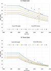
We derived the first comprehensive organ dose library for Canadian pediatric and adult patients who underwent computed tomography (CT) scans between 1992 and 2019 to support epidemiological analysis of radiation risk. We calculated organ absorbed doses for Canadian CT patients in two steps. First, we modeled Computed Tomography Dose Index (CTDI) values by patient age, scan body part, and scan year for the scan period between 1992 and 2019 using national survey data conducted in Canada and partially the United Kingdom survey data as surrogates. Second, we converted CTDI values to organ absorbed doses using a library of organ dose conversion coefficients built in an organ dose calculation program, the National Cancer Institute dosimetry system for CT. In result, we created a library of doses delivered to 33 organs and tissues by different patient ages and genders, scan body parts and scan years. In the scan period before 2000, the organs receiving the greatest dose in the head, chest and abdomen-pelvis scans were the active marrow (3.7-15.2 mGy), lungs (54.7-62.8 mGy) and colon (54.9-68.5 mGy), respectively. We observed organ doses reduced by 24% (pediatric head and torso scans, and adult head scans) and 55% (adult torso scans) after 2000. The organ dose library will be used to analyse the risk of radiation exposure from CT scans in the Canadian CT patient cohort.
© Published by Oxford University Press 2024.
Figures
References
-
- Hauptmann, M. et al. Brain cancer after radiation exposure from CT examinations of children and young adults: results from the EPI-CT cohort study. Lancet Oncol. 24, 45–53 (2023). - PubMed


