Case Reports
. 2023 Dec 6;15(12):e50079.
doi: 10.7759/cureus.50079. eCollection 2023 Dec.

Giant Villous Adenoma of the Rectum With Prolapse: Case Report

Affiliations
Case Reports

Giant Villous Adenoma of the Rectum With Prolapse: Case Report

Iurii Munteanu et al. Cureus. .

Abstract

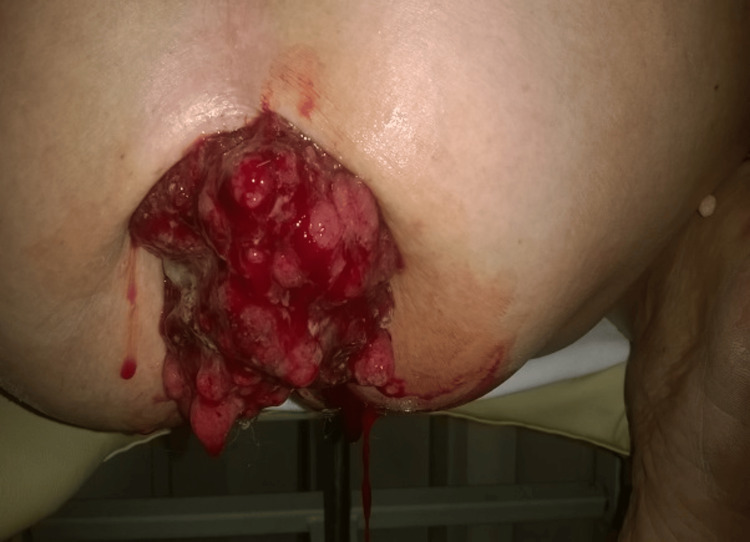
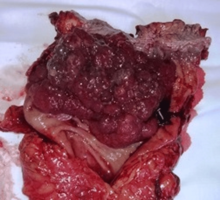
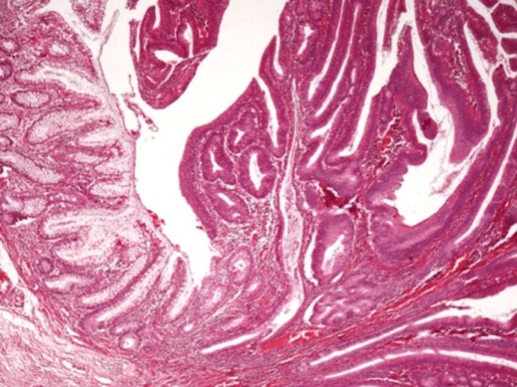
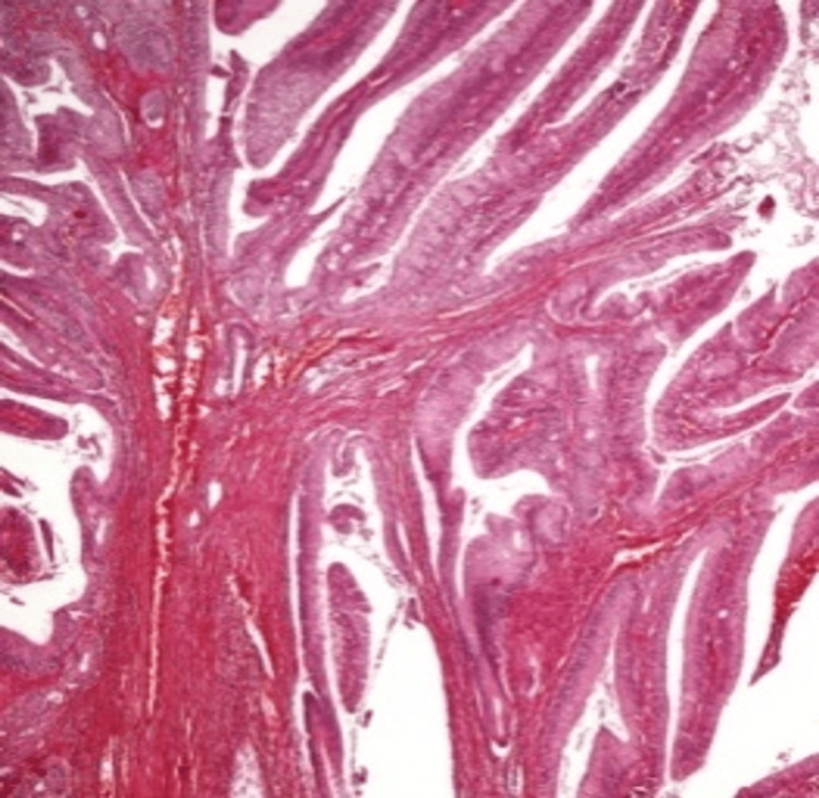
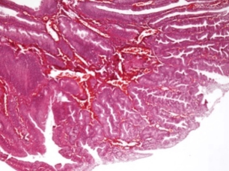
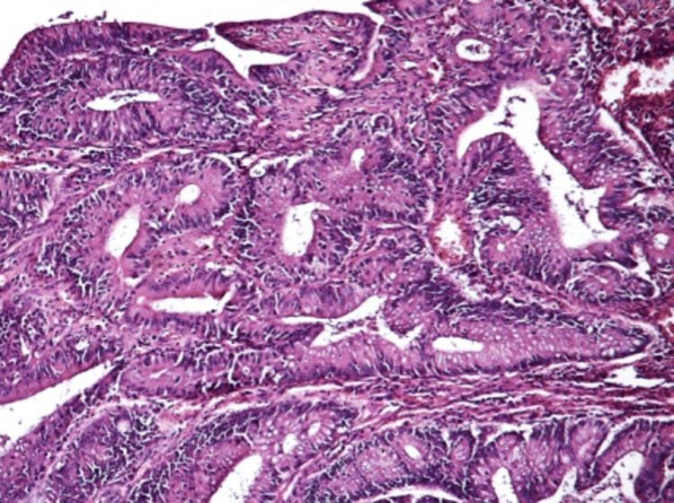
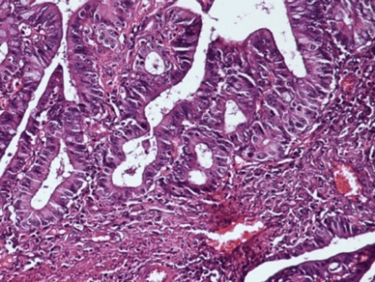
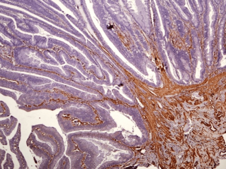
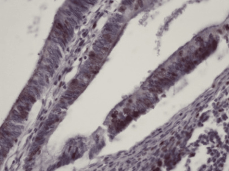
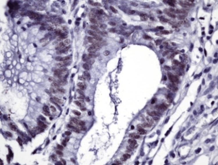
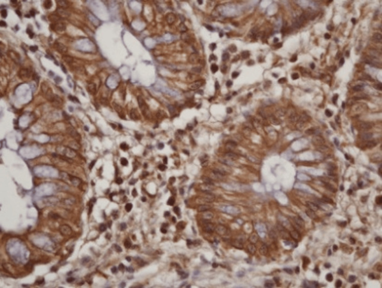
Colorectal polyps, frequently adenomas, are common in older adults, with villous adenomas being a notable subset due to their potential for significant malignancy risk. This case report highlights a rare instance of a giant villous adenoma in a 79-year-old female patient, challenging in both diagnosis and treatment. The patient, with a history of untreated essential arterial hypertension, was hospitalized for severe anemia following a massive rectal hemorrhage. An irreducible, prolapsed rectal mass was evident upon examination, and further investigations, including rectoscopy and abdominopelvic computed tomography scan, confirmed a villous adenoma with severe dysplasia. Given the tumor's substantial size, circumferential nature, and proximity to the dentate line, an abdominoperineal resection using the Miles technique was performed. The histopathological examination post-surgery confirmed the presence of a villous adenoma with high-grade epithelial neoplasia and localized areas of well-differentiated tubular adenocarcinoma. This case underscores the diagnostic and management complexities of large villous adenomas, emphasizing the need for meticulous surgical decision-making to ensure oncological safety and patient welfare, particularly when conservative resection may be inadequate.

Keywords: emergency gastroenterology and endoscopy; giant villous adenoma; inferior digestive hemorrhage; proctology; villous tumor.


Conflict of interest statement

The authors have declared that no competing interests exist.

Figures

Figure 1
Figure 1. Prolapsed rectal tumor: clinical presentation
A photograph showing the prolapsed rectal tumor as observed during physical examination.
Figure 2
Figure 2. Computed tomography scan of the anal-rectosigmoid mass
Coronal view showing the anal-rectosigmoid mass measuring 6.8 cm × 5.3 cm, with invasion into perirectal structures and multiple local-regional adenopathies. Degenerative disc and vertebral changes are highlighted with arrows indicating the tumor location.
Figure 3
Figure 3. Resected specimen of the rectum with intraluminal vegetative tumor
A photograph of the longitudinally cut specimen of the rectum, displaying the intraluminal vegetative tumor.
Figure 4
Figure 4. Microscopic evaluation of the villous adenoma
A microscopic view of the villous adenoma, detailing the general histopathological features.
Figure 5
Figure 5. Histopathology: fibrous septa with tumor epithelium
A microscopic view showing delicate fibrous septa covered by tumor epithelium without significant nuclear pleomorphism (hematoxylin and eosin stain, 100× magnification).
Figure 6
Figure 6. Histopathology: complex tumor architecture
Microscopic evaluation of the tumor showing more complex architecture with irregular glands, without significant pleomorphism and no tumor invasion (hematoxylin and eosin stain, 100× magnification).
Figure 7
Figure 7. Histopathology: epithelial proliferation with moderate nuclear atypia
Microscopic image showing epithelial proliferation of columnar cells with moderate nuclear atypia, displaying a glandular and alveolar pattern (hematoxylin and eosin stain, 20× magnification).
Figure 8
Figure 8. Histopathology: glandular architecture and stromal infiltration
Microscopic view revealing complex glandular architecture and stromal infiltration by small clusters of atypical cells in an area with a desmoplastic stromal response, with no invasion into the muscularis mucosa or submucosa (hematoxylin and eosin stain, 20× magnification).
Figure 9
Figure 9. Histopathology: no muscularis mucosa invasion
Microscopic evaluation showing no invasion of the muscularis mucosa (smooth muscle actin coloration, 100× magnification).
Figure 10
Figure 10. Immunohistochemistry: Ki67 cancer-marker evaluation
Microscopic view with immunohistochemistry showing relatively rare tumor nuclei positive for the Ki67 cancer marker (200× magnification).
Figure 11
Figure 11. Immunohistochemistry: microsatellite instability markers evaluation
Microscopic evaluation and immunohistochemistry showing tumor cells with various levels of positivity for microsatellite instability markers, indicating no microsatellite instability within the tumor.
Figure 12
Figure 12. Immunohistochemistry: comprehensive microsatellite instability analysis
Microscopic view with immunohistochemistry showing no microsatellite instability within the tumor. Tumor markers used include MutL homolog 1 (MLH1), postmeiotic segregation increased 2 (PMS2), MutS homolog 2 (MSH2), MutS homolog 6 (MSH6), and adenomatous polyposis coli (APC; 200× magnification).

Similar articles

Cited by

References

    1. Clinical significance of small polyps found during screening with flexible sigmoidoscopy. Farraye FA, Wallace M. Gastrointest Endosc Clin N Am. 2002;12:41–51. - PubMed
    1. [A new one in the classification of benign colon epithelial tumors (WHO, 2019, 5th edition)] Oleynikova NA, Malkov PG, Danilova NV. Arkh Patol. 2020;82:35–42. - PubMed
    1. McKittrick-Wheelock syndrome: a case series. Villanueva ME, Onglao MA, Tampo MM, Lopez MP. Ann Coloproctol. 2022;38:266–270. - PMC - PubMed
    1. McKittrick-Wheelock syndrome: a rare cause of chronic diarrhea. Jyala A, Mehershahi S, Shah N, Shaikh DH, Patel H. Cureus. 2021;13:0. - PMC - PubMed
    1. Giant villous adenoma of rectum- what is the malignant potential and what is the optimal treatment? A case and review of literature. Bains L, Lal P, Vindal A, Singh M. World J Surg Oncol. 2019;25:109–110. - PMC - PubMed

Publication types

LinkOut - more resources