

Ghrelin/GHSR Axis Induced M2 Macrophage and Alleviated Intestinal Barrier Dysfunction in a Sepsis Rat Model by Inactivating E2F1/NF- κ B Signaling
- PMID: 38187112
- PMCID: PMC10769719
- DOI: 10.1155/2023/1629777
Ghrelin/GHSR Axis Induced M2 Macrophage and Alleviated Intestinal Barrier Dysfunction in a Sepsis Rat Model by Inactivating E2F1/NF- κ B Signaling
Abstract
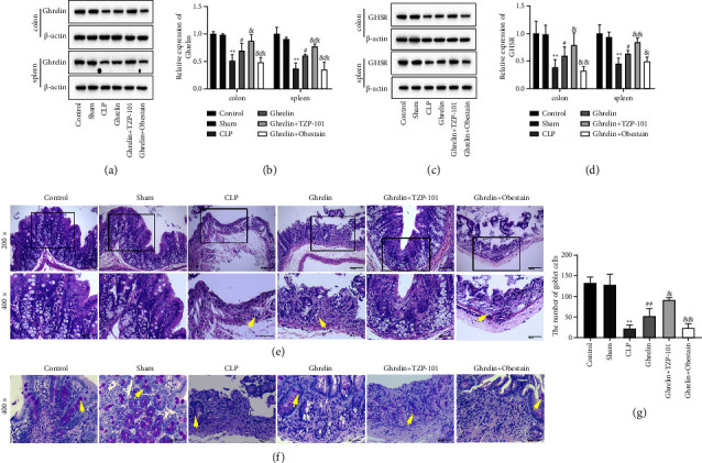
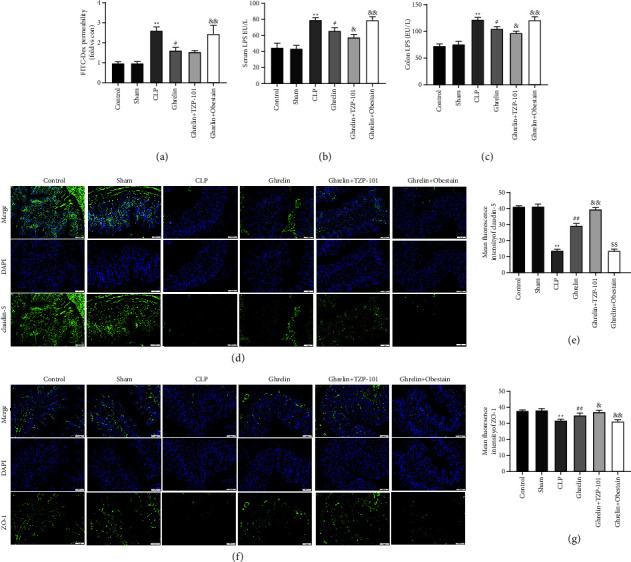
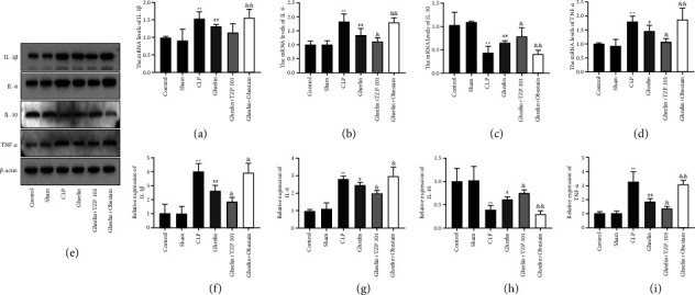
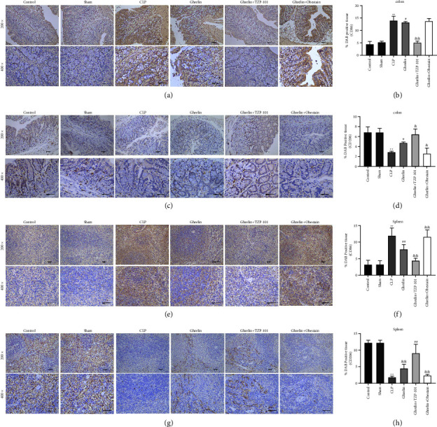
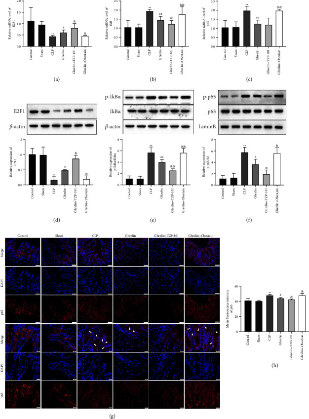
Sepsis is an inflammatory reaction disorder state that is induced by infection. The activation and regulation of the immune system play an essential role in the development of sepsis. Our previous studies have shown that ghrelin ameliorates intestinal dysfunction in sepsis. Very little is known about the mechanism of ghrelin and its receptor (GHSR) on the intestinal barrier and the immune function of macrophage regulation. Our research is to investigate the regulatory effect and molecular mechanism of the ghrelin/GHSR axis on intestinal dysfunction and macrophage polarization in septic rats. A rat model of sepsis was established by cecal ligation and puncture (CLP) operation. Then, the sepsis rats were treated with a ghrelin receptor agonist (TZP-101) or ghrelin inhibitor (obestatin). The results suggested that TZP-101 further enhanced ghrelin and GHSR expressions in the colon and spleen of septic rats and obestatin showed the opposite results. Ghrelin/GHSR axis ameliorated colonic structural destruction and intestinal epithelial tight junction injury in septic rats. In addition, the ghrelin/GHSR axis promoted M2-type polarization of macrophages, which was characterized by the decreases of IL-1β, IL-6, and TNF-α, as well as the increase of IL-10. Mechanistically, the ghrelin/GHSR axis promoted E2F2 expression and suppressed the activation of the NF-κB signaling pathway in septic rats. Collectively, targeting ghrelin/GHSR during sepsis may represent a novel therapeutic approach for the treatment of intestinal barrier injury.
Copyright © 2023 Lei Zhu et al.
Conflict of interest statement
The authors declare that they have no conflicts of interest.
Figures
Similar articles
-
HDAC5 promotes intestinal sepsis via the Ghrelin/E2F1/NF-κB axis.FASEB J. 2021 Jul;35(7):e21368. doi: 10.1096/fj.202001584R. FASEB J. 2021. PMID: 34125448
-
[Role of cholinergic anti-inflammatory pathway in Ghrelin regulation of peptide transporter 1 expression in small intestinal epithelium of septic rats].Zhonghua Wei Zhong Bing Ji Jiu Yi Xue. 2022 Nov;34(11):1132-1137. doi: 10.3760/cma.j.cn121430-20210817-01197. Zhonghua Wei Zhong Bing Ji Jiu Yi Xue. 2022. PMID: 36567554 Chinese.
-
Ghrelin alleviates neuropathic pain through GHSR-1a-mediated suppression of the p38 MAPK/NF-κB pathway in a rat chronic constriction injury model.Reg Anesth Pain Med. 2014 Mar-Apr;39(2):137-48. doi: 10.1097/AAP.0000000000000050. Reg Anesth Pain Med. 2014. PMID: 24513955
-
Mechanism of the inhibitory effect of ghrelin in sepsis.Hepat Med. 2010 Feb 23;2:33-8. doi: 10.2147/hmer.s7187. Hepat Med. 2010. PMID: 24367207 Free PMC article. Review.
-
Ghrelin/GHSR System in Depressive Disorder: Pathologic Roles and Therapeutic Implications.Curr Issues Mol Biol. 2024 Jul 10;46(7):7324-7338. doi: 10.3390/cimb46070434. Curr Issues Mol Biol. 2024. PMID: 39057075 Free PMC article. Review.
Cited by
-
Dexmedetomidine ameliorates diabetic intestinal injury by promoting the polarization of M2 macrophages through the MMP23B pathway.World J Diabetes. 2024 Sep 15;15(9):1962-1978. doi: 10.4239/wjd.v15.i9.1962. World J Diabetes. 2024. PMID: 39280187 Free PMC article.
-
Ghrelin improves small intestinal barrier damage in sepsis by promoting miR-143/ATG2B-mediated autophagy.PLoS One. 2025 Aug 7;20(8):e0329488. doi: 10.1371/journal.pone.0329488. eCollection 2025. PLoS One. 2025. PMID: 40773487 Free PMC article.
-
Intestinal injury signaling pathway in sepsis.Front Immunol. 2025 Jun 27;16:1620965. doi: 10.3389/fimmu.2025.1620965. eCollection 2025. Front Immunol. 2025. PMID: 40655155 Free PMC article. Review.
-
Affective-cognitive circuits in postoperative appetite reduction: an adaptive neuroimmune response to surgical stress.Front Neurosci. 2025 Aug 12;19:1654559. doi: 10.3389/fnins.2025.1654559. eCollection 2025. Front Neurosci. 2025. PMID: 40874114 Free PMC article. Review.
References
-
- Kell D. B., Pretorius E. To what extent are the terminal stages of sepsis, septic shock, systemic inflammatory response syndrome, and multiple organ dysfunction syndrome actually driven by a prion/amyloid form of fibrin? Seminars in Thrombosis and Hemostasis . 2018;44(03):224–238. doi: 10.1055/s-0037-1604108. - DOI - PMC - PubMed
MeSH terms
Substances
LinkOut - more resources
Full Text Sources
Medical
Molecular Biology Databases
Miscellaneous
