

Exosome-derived miR-23a-5p inhibits HCC proliferation and angiogenesis by regulating PRDX2 expression: MiR-23a-5p/PRDX2 axis in HCC progression
- PMID: 38187319
- PMCID: PMC10770527
- DOI: 10.1016/j.heliyon.2023.e23168
Exosome-derived miR-23a-5p inhibits HCC proliferation and angiogenesis by regulating PRDX2 expression: MiR-23a-5p/PRDX2 axis in HCC progression
Abstract
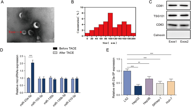
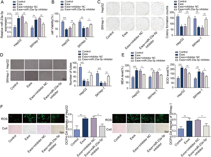
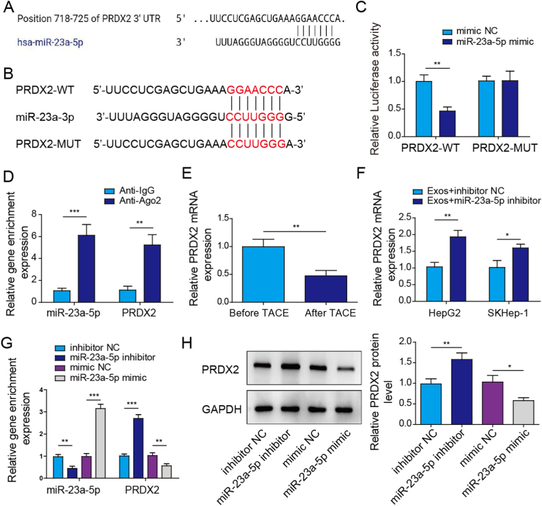
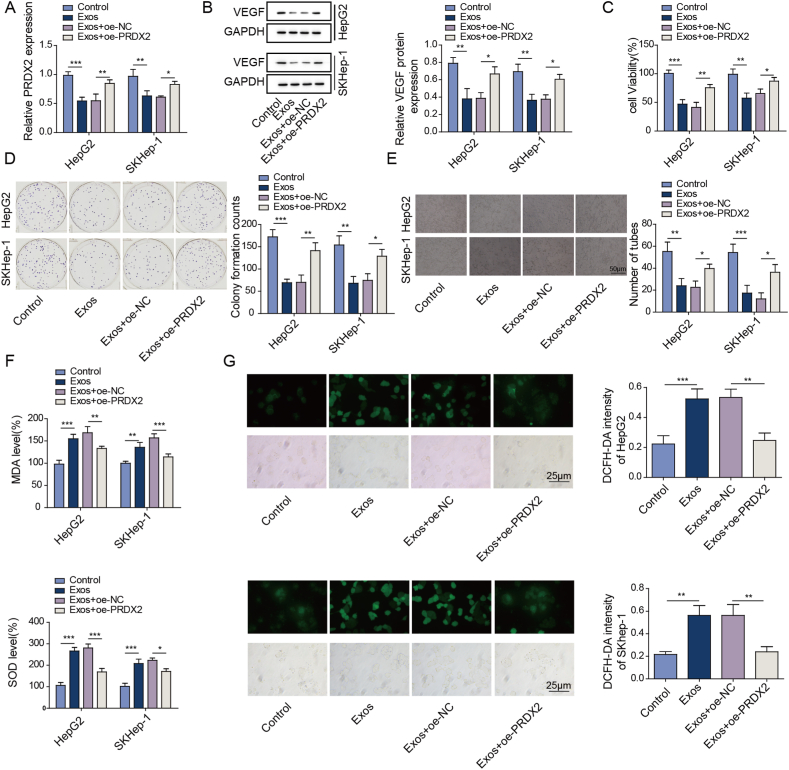
microRNAs (miRNAs) are closely related to the progression of hepatocellular carcinoma (HCC). Cancer-derived exosomes play an essential role in the establishment of the HCC microenvironment. However, the possible effects and underlying mechanisms of exosome (exo) microRNA-23a-5p (miR-23a-5p) in the progression of HCC remain unknown. In this study, we aimed to determine the role and specific molecular mechanism of exo miR-23a-5p in regulating HCC progression and to investigate whether exo miR-23a-5p levels can serve as an indicator of the prognosis of transarterial chemoembolization in patients with HCC. Our findings illustrated that miR-23a-5p was downregulated in exosomes separated from the serum of HCC patients and that miR-23a-5p carried by exosomes inhibited HCC cell proliferation and angiogenesis. Mechanistically, miR-23a-5p negatively targeted peroxiredoxin-2 (PRDX2). Functionally, PRDX2 overexpression relieved exosome-induced inhibition of HCC cell proliferation and angiogenesis by promoting vascular endothelial growth factor (VEGF) expression. In conclusion, Exo miR-23a-5p inhibited HCC proliferation and angiogenesis by regulating PRDX2 expression. Our results revealed the role and specific molecular mechanism of exo miR-23a-5p in regulating HCC progression.
Keywords: Angiogenesis; Exosomes; Hepatocellular carcinoma; PRDX2; miR-23a-5p.
© 2023 The Authors.
Conflict of interest statement
The authors declare that they have no known competing financial interests or personal relationships that could have appeared to influence the work reported in this paper.
Figures
Similar articles
-
Jianpi Huayu decoction inhibits the epithelial-mesenchymal transition of hepatocellular carcinoma cells by suppressing exosomal miR-23a-3p/Smad signaling.J Ethnopharmacol. 2022 Aug 10;294:115360. doi: 10.1016/j.jep.2022.115360. Epub 2022 May 11. J Ethnopharmacol. 2022. PMID: 35568116
-
Osteoclast-derived miR-23a-5p-containing exosomes inhibit osteogenic differentiation by regulating Runx2.Cell Signal. 2020 Jun;70:109504. doi: 10.1016/j.cellsig.2019.109504. Epub 2019 Dec 16. Cell Signal. 2020. PMID: 31857240
-
miR-660-5p-loaded M2 macrophages-derived exosomes augment hepatocellular carcinoma development through regulating KLF3.Int Immunopharmacol. 2021 Dec;101(Pt B):108157. doi: 10.1016/j.intimp.2021.108157. Epub 2021 Oct 18. Int Immunopharmacol. 2021. PMID: 34673296
-
Advances and challenges of exosome-derived noncoding RNAs for hepatocellular carcinoma diagnosis and treatment.Biochem Biophys Rep. 2024 Mar 24;38:101695. doi: 10.1016/j.bbrep.2024.101695. eCollection 2024 Jul. Biochem Biophys Rep. 2024. PMID: 38560049 Free PMC article. Review.
-
microRNA-23a in Human Cancer: Its Roles, Mechanisms and Therapeutic Relevance.Cancers (Basel). 2018 Dec 20;11(1):7. doi: 10.3390/cancers11010007. Cancers (Basel). 2018. PMID: 30577536 Free PMC article. Review.
Cited by
-
Nanoparticle/Engineered Bacteria Based Triple-Strategy Delivery System for Enhanced Hepatocellular Carcinoma Cancer Therapy.Int J Nanomedicine. 2024 Apr 29;19:3827-3846. doi: 10.2147/IJN.S453709. eCollection 2024. Int J Nanomedicine. 2024. PMID: 38708180 Free PMC article.
-
Exosomes, autophagy, and cancer: A complex triad.Int J Cancer. 2025 Aug 1;157(3):405-415. doi: 10.1002/ijc.35388. Epub 2025 May 2. Int J Cancer. 2025. PMID: 40318053 Free PMC article. Review.
-
Negative regulation of miRNA sorting into EVs is mediated by the capacity of RBP PCBP2 to impair the SYNCRIP-dependent miRNA loading.Elife. 2025 Jul 2;14:RP105017. doi: 10.7554/eLife.105017. Elife. 2025. PMID: 40601477 Free PMC article.
References
LinkOut - more resources
Full Text Sources
Research Materials
Miscellaneous
