[Preprint]. 2023 Dec 18:2023.12.18.571817.
doi: 10.1101/2023.12.18.571817.

Amyloid precursor protein induces reactive astrogliosis

Affiliations

Amyloid precursor protein induces reactive astrogliosis

Gretsen Velezmoro Jauregui et al. bioRxiv. .

Update in

  • Amyloid precursor protein induces reactive astrogliosis.
    Velezmoro Jauregui G, Vukić D, Onyango IG, Arias C, Novotný JS, Texlová K, Wang S, Kovačovicova KL, Polakova N, Zelinkova J, Čarna M, Lacovich V, Head BP, Havas D, Mistrik M, Zorec R, Verkhratsky A, Keegan L, O'Connell MA, Rissman R, Stokin GB. Velezmoro Jauregui G, et al. Acta Physiol (Oxf). 2024 Jun;240(6):e14142. doi: 10.1111/apha.14142. Epub 2024 Apr 8. Acta Physiol (Oxf). 2024. PMID: 38584589

Abstract

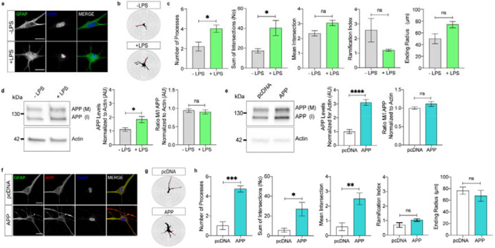
We present in vitro and in vivo evidence demonstrating that Amyloid Precursor Protein (APP) acts as an essential instigator of reactive astrogliosis. Cell-specific overexpression of APP in cultured astrocytes led to remodelling of the intermediate filament network, enhancement of cytokine production and activation of cellular programs centred around the interferon (IFN) pathway, all signs of reactive astrogliosis. Conversely, APP deletion in cultured astrocytes abrogated remodelling of the intermediate filament network and blunted expression of IFN stimulated gene (ISG) products in response to lipopolysaccharide (LPS). Following traumatic brain injury (TBI), mouse reactive astrocytes also exhibited an association between APP and IFN, while APP deletion curbed the increase in glial fibrillary acidic protein (GFAP) observed canonically in astrocytes in response to TBI. Thus, APP represents a molecular inducer and regulator of reactive astrogliosis.

Keywords: amyloid precursor protein; astrocytes; interferon pathway; lipopolysaccharide; reactive astrogliosis; traumatic brain injury.


Figures

Figure 1:
Figure 1:. APP promotes remodelling of GFAP-positive astrocytic profiles.
a. Representative images of untreated (-LPS) and LPS-treated (+LPS) primary human cortical astrocytes stained with GFAP and DAPI (Scale bar 20 μm). b. Representative image reconstructions of −LPS and +LPS GFAP-stained astrocytes based on the Sholl analysis using 5 μm spacing between the concentric circles. c. Comparison of key Sholl analysis parameters between −LPS and +LPS GFAP-stained astrocytes (5 to 8 cells per biological sample, N=3 biological samples per condition, Student t-test, * = p<0.05) d. Representative blot showing APP levels in −LPS and +LPS astrocytes normalised for actin levels as a loading control. Graphs show mean levels of APP normalised for actin (left) and the mean ratio between mature (M) versus immature (I) APP (right) (N=5 biological samples per condition, Student t-test, * = p<0.05). e. Representative blot showing APP levels in astrocytes transfected with either pcDNA3.1 (pcDNA) or APP770-pcDNA3.1 (APP) normalized for actin levels as a loading control. Graphs show average mean levels of APP normalised for actin (left) and the mean ratio of mature (M) versus immature (I) APP (right) (N=5 biological samples per condition, Student t-test, ns = not significant). f. Representative images of astrocytes transfected with either pcDNA3.1 or APP770-pcDNA3.1 and stained with GFAP, APP and DAPI (Scale bar 20 μm). g. Representative image reconstructions of GFAP-stained astrocytes transfected with either pcDNA3.1 or APP770-pcDNA3.1 based on the Sholl analysis using 5 μm spacing between the concentric circles. h. Comparison of key Sholl morphometric analysis of GFAP-stained astrocytes transfected either with pcDNA3.1 or with APP770-pcDNA3.1 (3–5 cells per biological sample, N = 4 biological samples per condition, Student t-test, * = p<0.05, ** = p < 0.01, *** = p < 0.001).
Figure 2:
Figure 2:. APP increases expression of cytokines in astrocytes.
a. Heatmap showing changes in abundances of cytokines in −LPS and +LPS astrocytes (N = 3 biological samples per condition, Student t-test, * = p < 0.05, ** = p < 0.01, *** = p < 0.001). Individual protein concentrations (pg/mg) were log2 transformed. b. Heatmap showing changes in abundances of cytokines in pcDNA3.1 (pcDNA), CMV, GFP-pdDNA3.1 (GFP) and APP770-pcDNA3.1 (APP) transfected astrocytes (N = 3 biological samples per condition, Student t-test, * = p < 0.05, ** = p < 0.01, *** = p < 0.001). Individual protein concentrations (pg/mg) were log2 transformed. c. Radar plot showing changes in cytokine profiles between LPS treated and APP770-pcDNA3.1 transfected astrocytes (N = 3 biological samples per condition, ANOVA with Tukey post-hoc test, * = p < 0.05, ** = p < 0.01). d. Bidirectionally clustered heatmap showing the expression of 1000 genes with the highest variability. Colour annotations differentiate individual astrocyte treatments and replicates. e. Principal Component Analysis (PCA) of transcriptomic profiles of APP770-pcDNA3.1 versus pcDNA3.1 empty vector transfected astrocytes. f. Volcano plot showing differentially expressed (DE) genes in astrocytes transfected with APP770-pcDNA3.1 and pcDNA3.1. The horizontal line indicates statistically significantly DE genes, vertical dotted lines indicate the log 2 FC of ±1.5. g. The most significantly changed REACTOME pathways by the DE genes with log 2 FC exceeding the cut off of ±1.5.
Figure 3:
Figure 3:. APP and IFNγ in astrocytes following TBI.
a. Cartoon depicting the controlled cortical impact (CCI) injury model of the traumatic brain injury (TBI). b. Representative confocal microscopy images of OPNC (omitted primary negative control) and GFAP-stained astrocytes in the corpus callosum of control and TBI mice (scale bar 20 μm). c. Representative reconstructed images of GFAP-immunoreactive astrocytes of the corpus callosum of control and TBI mice based on the Sholl analysis with a 5 μm interval of the concentric circles. d. Measurements of key Sholl parameters in GFAP-stained astrocytes in control and TBI mice (5 to 7 per animal, N = 4 mice, Student t-test, * = p < 0.05). e. Representative confocal microscopy images of APP- and IFNγ-immunoreactivities in GFAP-stained astrocytes in corpus callosum of control and TBI mice (Scale bar 20 μm). f. Graph showing mean APP- and IFNγ-immunoreactivities in GFAP-stained astrocytes in the corpus callosum of control and TBI mice (N = 4 mice, Student t-test, * = p < 0.05). g. Relationship between mean APP- and IFNγ-immunoreactivities in the GFAP-stained astrocytes in the corpus callosum of control and TBI mice (N = 4 mice, Pearson correlation coeficient ANOVA two-tailed **** = p < 0.0001 and ** = p < 0.01).
Figure 4:
Figure 4:. Deletion of APP hampers reactive astrogliosis in response to LPS.
a Representative western blot showing levels of APP normalised for actin as a loading control in untreated (-LPS) and LPS treated (+LPS) astrocytes transduced with multiMIR-Scramble (SCR) and multiMIR-APP (APP KD) lentiviral vectors. Graph showing mean APP levels normalized for actin (N = 5 biological replicates, ANOVA with Tukey’s multiple comparisons test, ns = not significant, * = p < 0.01, **** = p < 0.01). b. Representative confocal microscopy images of GFAP-stained −LPS and +LPS astrocytes transduced with multiMIR-Scramble and multiMIR-APP lentiviral vectors (Scale bar 20 μm). c. Representative reconstructed images of GFAP-stained −LPS and +LPS astrocytes transduced with multiMIR-Scramble and multiMIR-APP lentiviral vectors based on the Sholl analysis with a 5 μm interval of the concentric circles. d. Measurements of key Sholl parameters in GFAP-immunoreactive −LPS and +LPS astrocytes transduced with multiMIR-Scramble and multiMIR-APP lentiviral vectors (10 to 27 cells per treatment, N = 4 biological replicates/mice, ANOVA with Tukey’s multiple comparisons test, ** = p < 0.01). e. Representative western blot of ISG product levels and GAPDH levels as a loading control in −LPS and +LPS astrocytes transduced with multiMIR-Scramble and multiMIR-APP lentiviral vectors. f. Graph showing ratios of GAPDH normalised cellular ISG product levels (left, One Sample t-test, * = p < 0.05, ** = p < 0.01) and their geometrical differences presented as log2 fold changes (right, Student t-test with Benjamin-Hochberg correction, ** = p < 0.01, *** = p < 0.001) between multiMIR-Scramble and multiMIR-APP transduced astrocytes (N = 4 biological replicates). g. Graph showing ratios of GAPDH normalised cellular ISG product levels (left, One Sample t-test, * = p < 0.05, ** = p < 0.01) and their geometrical differences presented as log2 fold changes (right, Student t-test with Benjamin-Hochberg correction, *= p < 0.01, **= p < 0.01, *** = p < 0.001 **** = p < 0.0001 between −LPS and +LPS multiMIR-Scramble and multiMIR-APP transduced astrocytes (N = 4 biological replicates).

References

    1. Semyanov A, Verkhratsky A. Astrocytic processes: from tripartite synapses to the active milieu. Trends Neurosci. 2021;44 (10):781–792. doi: 10.1016/j.tins.2021.07.006. - DOI - PubMed
    1. Araque A, Parpura V, Sanzgiri RP, Haydon PG. Tripartite synapses: glia, the unacknowledged partner. Trends Neurosci. 1999;22 (5):208–215. doi: 10.1016/s0166-2236(98)01349-6. - DOI - PubMed
    1. Augusto-Oliveira M. Astroglia-specific contributions to the regulation of synapses, cognition and behaviour. Neurosci Biobehav Rev. 2020;118:331–357. doi: 10.1016/j.neubiorev.2020.07.039. - DOI - PubMed
    1. Durkee CA, Covelo A, Lines J, Kofuji P, Aguilar J, Araque A. G(i/o) protein-coupled receptors inhibit neurons but activate astrocytes and stimulate gliotransmission. Glia. 2019;67 (6):1076–1093. doi: 10.1002/glia.23589. - DOI - PMC - PubMed
    1. Südhof TC. Towards an Understanding of Synapse Formation. Neuron. 2018;100 (2):276–293. doi: 10.1016/j.neuron.2018.09.040. - DOI - PMC - PubMed

Publication types