[Preprint]. 2023 Dec 23:2023.12.21.572835.
doi: 10.1101/2023.12.21.572835.

Chromosome fusion and programmed DNA elimination shape karyotypes of parasitic nematodes

Affiliations

Chromosome fusion and programmed DNA elimination shape karyotypes of parasitic nematodes

James R Simmons et al. bioRxiv. .

Update in

Abstract

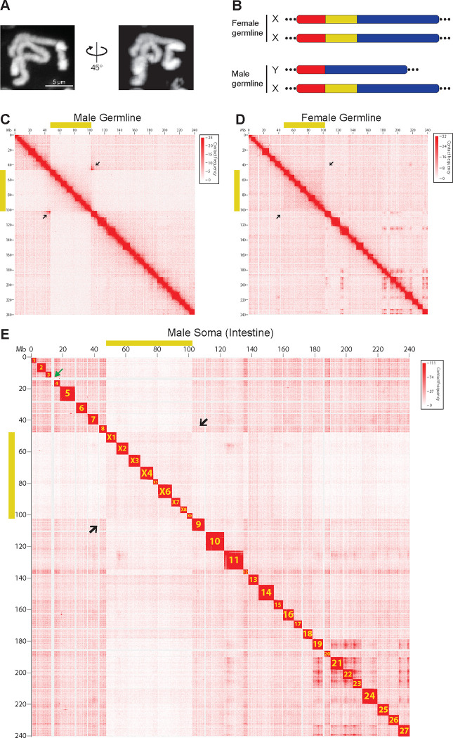
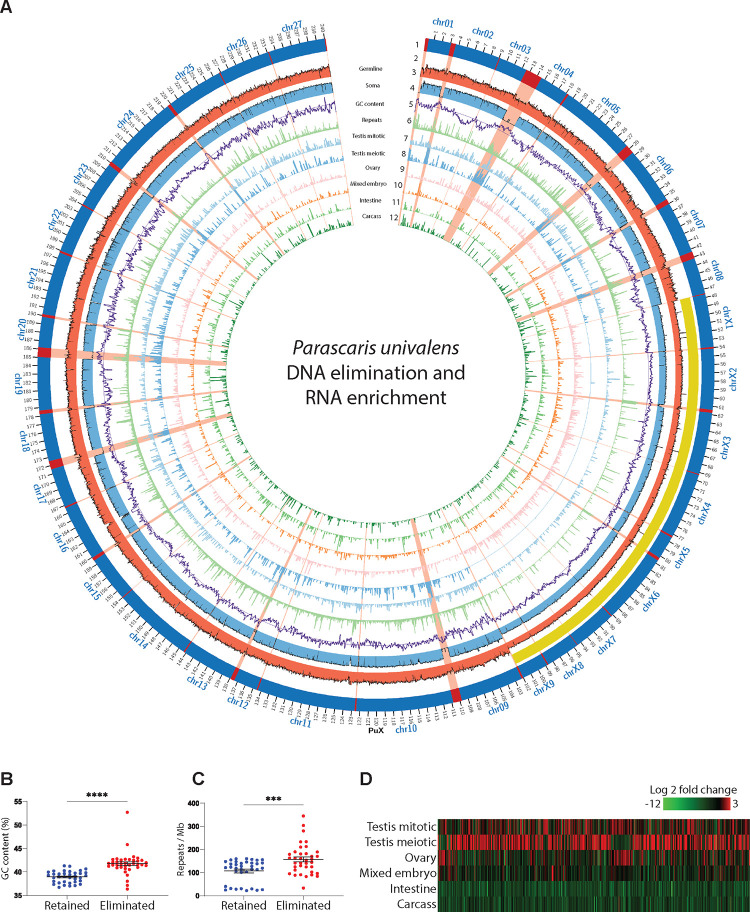
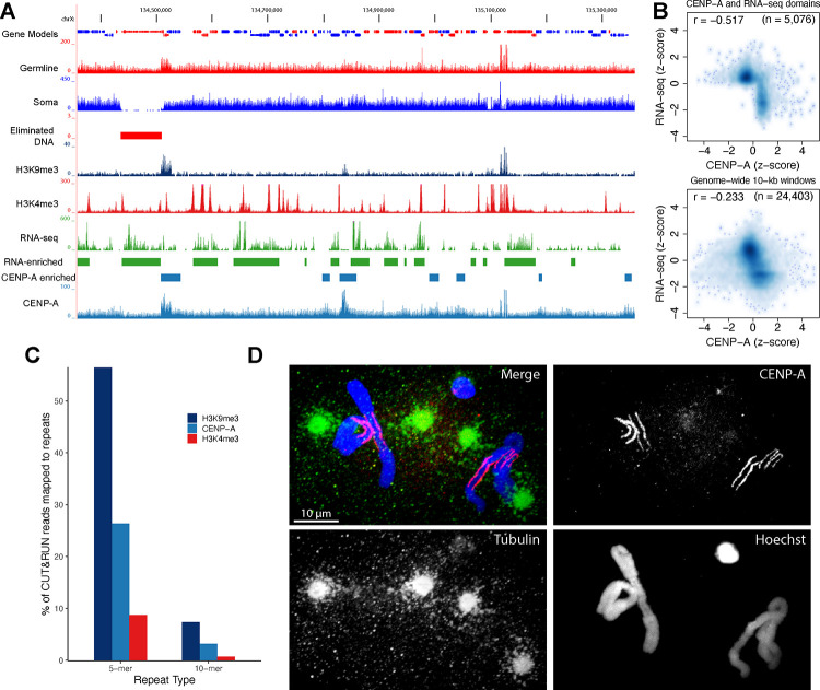
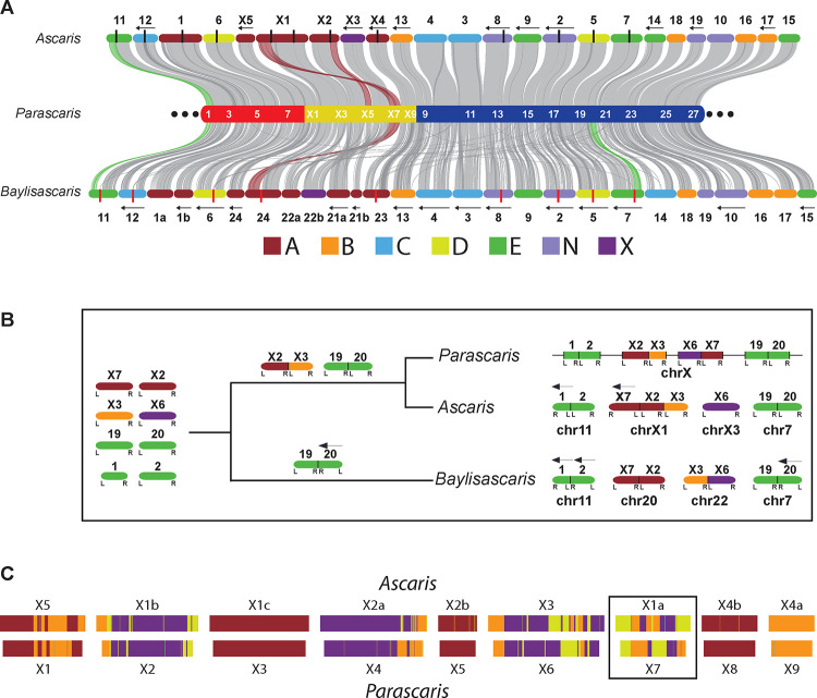
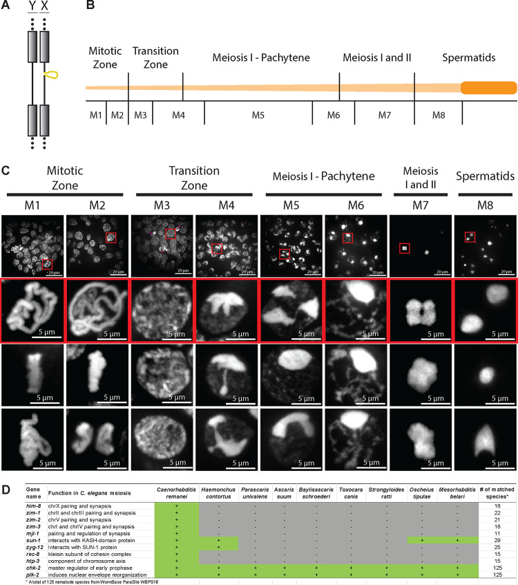
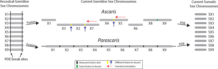
A growing list of metazoans undergo programmed DNA elimination (PDE), where a significant amount of DNA is selectively lost from the somatic genome during development. In some nematodes, PDE leads to the removal and remodeling of the ends of all germline chromosomes. In several species, PDE also generates internal breaks that lead to sequence loss and an increased number of somatic chromosomes. The biological significance of these karyotype changes associated with PDE and the origin and evolution of nematode PDE remain largely unknown. Here, we assembled the single germline chromosome of the horse parasite Parascaris univalens and compared the karyotypes, chromosomal gene organization, and PDE features among ascarid nematodes. We show that PDE in Parascaris converts an XX/XY sex-determination system in the germline into an XX/XO system in the somatic cells. Comparisons of Ascaris, Parascaris, and Baylisascaris ascarid chromosomes suggest that PDE existed in the ancestor of these parasites, and their current distinct germline karyotypes were derived from fusion events of smaller ancestral chromosomes. The DNA breaks involved in PDE resolve these fused germline chromosomes into their pre-fusion karyotypes, leading to alterations in genome architecture and gene expression in the somatic cells. Cytological and genomic analyses further suggest that satellite DNA and the heterochromatic chromosome arms play a dynamic role in the Parascaris germline chromosome during meiosis. Overall, our results show that chromosome fusion and PDE have been harnessed in these ascarids to sculpt their karyotypes, altering the genome organization and serving specific functions in the germline and somatic cells.

Keywords: DNA break; Parascaris; centromere; chromosome fusion; genome; karyotype; meiosis; nematode; programmed DNA elimination; satellite DNA.


Conflict of interest statement

Declaration of interests The authors declare no competing interests.

Figures

Figure 1.
Figure 1.. The single Parascaris germline X chromosome consists of both somatic autosomes and sex chromosomes.
A. Hoechst staining of the single, condensed pair of Parascaris univalens germline chromosomes from the mitotic region of the testis. B. Karyotypes for Parascaris germline chromosome derived from this study (see Figure 1C–E). Parascaris females have two X chromosomes while males have one X and a shorter Y chromosome that lacks the sex chromosome region (yellow). Autosomal regions are represented in red and blue. Coverage for PacBio long reads spanning the junctions of autosome and sex chromosome regions are shown. Dotted lines at the ends indicate the large heterochromatic arms (90% of the genome) that are mainly comprised of satellite repeats. C-E. Parascaris Hi-C interaction heatmaps reveal germline karyotypes and their changes after PDE. Yellow bars along the x and y axes indicate the sex chromosome region of the Parascaris X chromosome. Black arrows point to the boundaries between autosome and sex chromosome regions. C. Reduced Hi-C interactions (~50%) in the sex chromosome region and strong interactions at the junctions (arrows) suggest two distinct chromosomes (XY) in the male germline (see Figure 1B). D. Consistent Hi-C interactions across the regions with no elevated interactions at the junctions (arrows) indicate the same pair of chromosomes (XX) in the female germline. E. PDE breaks the germline genome into 36 distinct somatic chromosomes. Shown are Hi-C interactions from the male intestine (see Figure S1 for Hi-C data in the female intestine). The somatic chromosomes were named based on their order in the germline and by their autosome or sex chromosome identities. The green arrow between chromosomes 3 and 4 indicates the largest eliminated region (~2 Mb) of euchromatic DNA.
Figure 2.
Figure 2.. PDE breaks the Parascaris germline chromosome and silences germline-expressed genes.
A. Features of the Parascaris X chromosome illustrated in a circos plot. Descriptions of the features from outer to inner circles: 1). Retained (dark blue) and eliminated DNA (red); the eliminated regions are highlighted in red across all circles; 2). The sex chromosome region (yellow bar); 3–4). Genomic (Illumina) read coverage from testis (red) and male intestine (light blue); 5). GC content (purple) and the average GC line; 6). Repeat density (light green) and the average repeat line; and 7–12). Expression (normalized RNA-seq) data for male mitotic region (light blue), male meiotic region (blue), ovary (pink), mixed embryos (orange), intestine (green), and carcass (dark green). B. Eliminated regions have higher GC content. C. Eliminated DNA contains more repeats. D. Eliminated genes are only expressed in the germline - mainly in the testis. The heatmap shows 743 expressed eliminated genes (rpkm >= 5 in at least one of the tissues; see Table S1). P-values for B and C were determined using Welch’s t-tests (*** P < 0.01 and **** P < 0.001).
Figure 3.
Figure 3.. Centromere reorganization facilitates Parascaris meiosis and PDE.
A. CENP-A distribution in the Parascaris genome. A browser view of RNA-seq and CUT&RUN data from the ovary tissue for a genomic region of 1 Mb. B. Meta-analysis illustrating a genome-wide inverse relationship between RNA transcripts and CENP-A deposition. The top plot is derived from CENP-A and RNA-seq enriched genomic domains (see enriched regions in Figure 3A), showing a strong inverse relationship. The bottom plot is obtained from 10-kb windows for the entire genome showing a modest but consistent negative correlation. C. Differential CENP-A, H3K9me3, and H3K4me3 distribution on the pentanucleotide (5-mer) and decanucleotide (10-mer) repeats. D. CENP-A staining in early embryos shows CENP-A aligns at the outer side of the chromosomes towards centrosomes. Early embryos were stained with Hoechst (blue) and with antibodies against CENP-A (red) and tubulin (green). Note that CENP-A specifically labels the euchromatic regions of the chromosomes in both the germ cell and the pre-somatic cells. Tubulin staining shows centrosomes on both sides of the dividing chromosomes, with spindle microtubules contacting the CENP-A labeled regions.
Figure 4.
Figure 4.. Ascaris, Parascaris, and Baylisascaris have the same somatic chromosomes.
A. Synteny among three ascarid germline genomes. The Parascaris chromosome (middle) was colored as in Figure 1B, with the positions of every other somatic chromosome labeled in white. The Ascaris (top) and Baylisascaris (bottom) chromosomes were colored based on their predominant Nigon element (see legend for the seven Nigon groups). Their designated names for germline chromosomes are shown and their internal breaks for PDE are marked with black (Ascaris) or red (Baylisascaris) vertical lines. The left arrows on the top and bottom indicate chromosomes with reversed coordinates relative to Parascaris. See Figure S2 for synteny between Ascaris and Baylisascaris. B. An evolutionary model highlights various chromosome fusion events that lead to the different germline karyotypes observed in current ascarid genomes. All chromosomes were labeled on the top with designations from orthologous Parascaris somatic chromosomes; at the bottom are their current germline designations. The same Nigon color scheme was used. The L and R underneath each chromosome represent the left and right end of the ancestral chromosome, respectively. A chromosome with an R to L order indicates a reversed orientation relative to the Parascaris genome and is marked with a left arrow on the top of the chromosome. C. Ascaris and Parascaris sex chromosomes in the somatic cells were painted with the ancestral Nigon elements (see methods). The black box highlights the somatic chromosome X1a in Ascaris and X7 in Parascaris which have the most significant differences in their Nigon composition between the two species. See Fig S3 for highly conserved Nigon elements in the autosomes.
Figure 5.
Figure 5.. Common features of nematode chromosomes indicate fusions for ascarid germline chromosomes.
A. GC content (%) was plotted for 8 presumptive ancestral pre-fused chromosome pairs in Parascaris and Ascaris. The ancestral Nigon group for each chromosome (see Figure 4A) is indicated by the color of the border. B. SNP density, protein conservation, repeat density, and H3K9me3 (from the ovary) were plotted in presumptive ancestral pre-fused chromosome pairs from Parascaris and Ascaris. Representative autosomes (left) and sex chromosomes (right) are shown. See Figure S3 for all 36 chromosomes. Each data point represents a binned region of the genome (window sizes for the bins: GC and SNPs = 10 kb; repeats and H3K9me3 = 50 kb; and protein conservation = 10 genes). A few data points beyond the y-axis ranges were converted to the minimum or maximum values and were colored in magenta. The colored trend lines (red or blue) were based on the LOESS regression. The horizontal dashed lines represent the average values of all chromosomes (orange), autosomes (green), or sex chromosomes (purple). The length of each chromosome is indicated at the bottom. Some Ascaris chromosomes are reversed in orientation (chromosome length labeled on the left side) to match their orthologous chromosomes in Parascaris.
Figure 6.
Figure 6.. Dynamic organization of the Parascaris germline chromosome suggests a function for the heterochromatin arms during meiosis.
A. An illustration of the X and Y chromosomes in the male germline. The X chromosome has a 55-Mb unique sex chromosome region (bulge) not found in the Y chromosome. The grey boxes and dotted lines represent the heterochromatic arms that will be eliminated by PDE. B. Schematic of the Parascaris testis showing different developmental stages (top) and regions sampled (bottom). A fully untangled Parascaris testis can reach one meter in length. C. Hoechst staining illustrates the dynamic organization of the germline chromosomes during male gametogenesis. The top row shows representative fields of view. The insets (red boxes) beneath show enlarged examples of condensed chromosomes at each stage. Additional images from the same stages were shown in the bottom two rows. Images were produced through maximum projection of multiple z-planes. See Movie S1 for the 3D organization of the chromosomes. D. Many genes involved in C. elegans pairing and synapsis are not found in nematodes with PDE. Known genes involved in C. elegans meiosis were searched against 125 nematode genomes available in WormBase ParaSite. Selected genes and species were listed, with genes chk-2 and plk-2 and species C. remanei and H. contortus serving as controls for detection. See Table S4 for the full list of known C. elegans genes involved in meiosis in all available nematodes.
Figure 7.
Figure 7.. Fusion, PDE, and evolution of the sex chromosomes in parasitic nematodes.
A model showing the chromosome fusions and their split by PDE in sex chromosomes. In the ancestral state, all chromosomes likely undergo PDE at both ends of the chromosome (left). Chromosome fusion events lead to different germline chromosomes (and likely speciation) in nematodes. See the legend for the different types of fusion sites and chromosome orientations (middle). PDE splits different germline chromosomes into the same somatic chromosomes and restores the pre-fused germline karyotype (right). Autosomes, not shown in this model for simplicity, follow this same evolutionary trajectory and fate as the sex chromosomes.

Similar articles

References

    1. Wang J., and Davis R.E. (2014). Programmed DNA elimination in multicellular organisms. Curr Opin Genet Dev 27, 26–34. 10.1016/j.gde.2014.03.012. - DOI - PMC - PubMed
    1. Zagoskin M.V., and Wang J. (2021). Programmed DNA elimination: silencing genes and repetitive sequences in somatic cells. Biochem Soc Trans 49, 1891–1903. 10.1042/BST20190951. - DOI - PMC - PubMed
    1. Dedukh D., and Krasikova A. (2021). Delete and survive: strategies of programmed genetic material elimination in eukaryotes. Biol Rev Camb Philos Soc. 10.1111/brv.12796. - DOI - PMC - PubMed
    1. Drotos K.H.I., Zagoskin M.V., Kess T., Gregory T.R., and Wyngaard G.A. (2022). Throwing away DNA: programmed downsizing in somatic nuclei. Trends Genet 38, 483–500. 10.1016/j.tig.2022.02.003. - DOI - PubMed
    1. Boveri T. (1887). Ueber Differenzierung der Zellkerne wahrend der Furchung des Eies von Ascaris megalocephala. Anat. Anz. 2, 688–693.

Publication types