[Preprint]. 2023 Dec 22:2023.12.20.572615.
doi: 10.1101/2023.12.20.572615.

An AAV capsid reprogrammed to bind human Transferrin Receptor mediates brain-wide gene delivery

Affiliations

An AAV capsid reprogrammed to bind human Transferrin Receptor mediates brain-wide gene delivery

Qin Huang et al. bioRxiv. .

Update in

Abstract

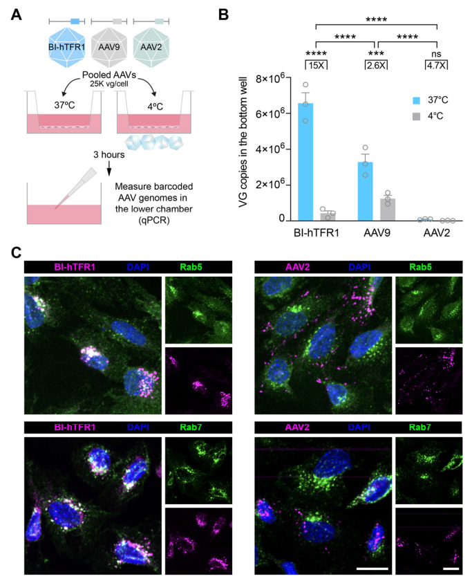
Developing vehicles that efficiently deliver genes throughout the human central nervous system (CNS) will broaden the range of treatable genetic diseases. We engineered an AAV capsid, BI-hTFR1, that binds human Transferrin Receptor (TfR1), a protein expressed on the blood-brain barrier (BBB). BI-hTFR1 was actively transported across a human brain endothelial cell layer and, relative to AAV9, provided 40-50 times greater reporter expression in the CNS of human TFRC knock-in mice. The enhanced tropism was CNS-specific and absent in wild type mice. When used to deliver GBA1, mutations of which cause Gaucher disease and are linked to Parkinson's disease, BI-hTFR1 substantially increased brain and cerebrospinal fluid glucocerebrosidase activity compared to AAV9. These findings establish BI-hTFR1 as a promising vector for human CNS gene therapy.


Conflict of interest statement

Competing interests B.E.D is a scientific founder and scientific advisory board member at Apertura Gene Therapy and a scientific advisory board member at Tevard Biosciences. B.E.D., A.J.B., K.Y.C., F.E.E., Q.H., J.W., and N.R.B.R. are named inventors on patent applications filed by the Broad Institute of MIT and Harvard related to this study. Remaining authors declare that they have no competing interests.

Figures

Fig. 1.
Fig. 1.. AAV9 can be programmed to bind human TfR1.
(A) An AAV9-based NNK capsid library of variants with random 7-mer insertions between VP1 residues 588–589 was screened for selective binding to human TfR1 in pull-down or cell binding assays. Individually produced human TfR1-binding variants carrying a CAG-NLS-mScarlet-P2A-Luciferase-WPRE-SV40pA construct exhibited enhanced species-specific (B) association with and (C) transduction (Luciferase activity) of CHO cells stably expressing TFRC (two-way ANOVA using AAV9 as the main comparison group for each cell line with Bonferroni multiple comparison correction: **** and *** indicate p ≤ 0.0001 and ≤ 0.001, respectively; n = 3 replicates, error bars indicate ± SEM). Values reported are normalized to AAV9 in each cell line. The TfR1-binding variants exhibited enhanced (D) association with and (E) transduction (Luciferase activity) of hBMVEC and hCMEC/D3 cells (one-way ANOVA using AAV9 as the comparison group for each cell line with Dunnett’s multiple comparison correction: ****, ***, and * indicate p ≤ 0.0001, ≤ 0.001, and ≤ 0.05, respectively; n = 4 replicates, error bars indicate ± SEM). Values are normalized to AAV9 in each cell line. (F) The binding kinetics between each capsid and full-length human TfR1 were assessed by BLI. AAVX probes were loaded with capsid and human TfR1 was used as an analyte. Sensorgram curve fits (black dash-dot lines) were generated by applying global 2:1 exponential association and decay models.
Fig. 2.
Fig. 2.. Human TfR1-targeted capsids transduce human brain endothelial cells via interactions with the apical domain of human TfR1.
(A) The plots show transduction (Luciferase, normalized RLU) of hCMEC/D3 cells incubated with 3 x 10⁸ vg/mL of the indicated AAV and the specified concentrations of the OKT9 or AF2474 antibody (two-way ANOVA using the no antibody control for each condition as the main comparison group with Bonferroni multiple comparison correction: **** indicates p ≤ 0.0001; n = 3 replicates, error bars indicate ± SEM). (B) The effect of Tf on BI-hTFR1 binding to full-length human TfR1 was assessed by BLI. AAVX probes were loaded with AAV9 or BI-hTFR1. Human TfR1 that either had or had not been pre-incubated with 300 nM holo-Tf was used as an analyte. 2:1 binding model curve fits (black dash-dot lines) are shown. (C) Biotinylated holo-Tf was immobilized on streptavidin-coated BLI probes (SA probe), introduced first into human TfR1, and then into buffer with or without the OKT9 antibody, and finally into BI-hTFR1 virus particles. Segments shaded in gray highlight the indicated association step. (D) BI-hTFR1 or AAV2 was incubated with hCMEC/D3 cells at 50,000 vg/cell for one hour at 4°C, with or without OKT9 (1 μg/mL) or Tf (1 μg/mL), and immunostained for AAV and TfR1. Scale bars = 15 μm. AAV9 binding to hCMEC/D3 cells was rarely detected therefore AAV2 was used as a control (fig. S3).
Fig. 3.
Fig. 3.. BI-hTFR1 is efficiently endocytosed and actively transported across human brain endothelial cells.
(A) Schematic shows the pooled transwell BBB model experimental design. (B) The vector genomes in the bottom chamber were quantified by qPCR (two-way ANOVA with Bonferroni multiple comparison correction: **** and *** indicate p ≤ 0.0001 and < 0.001 respectively; n = 3 transwell replicates, error bars indicate ± SEM). (C) BI-hTFR1 or AAV2 were incubated with hCMEC/D3 cells at 25,000 vg/cell for one hour at 37°C and stained for endosomal markers Rab5 and Rab7 as well as the AAVs. Scale bar = 15 μm.
Fig. 4.
Fig. 4.. BI-hTFR1 efficiently delivers genes to the CNS of TFRC KI mice.
(A) In TFRC KI mice, mouse Tfrc exons 4–19 encoding the extracellular region of TfR1 have been replaced by those of human TFRC. (B) BI-hTFR1 or AAV9 encoding CAG-NLS-mScarlet-P2A-Luciferase-WPRE-SV40pA were intravenously injected into adult female C57BL/6J or TFRC KI mice at 5 x 10¹¹ vg/mouse. AAV9 in C57BL/6J and BI-hTFR1 in TFRC KI mice had n = 4 mice per group. AAV9 in TFRC KI and BI-hTFR1 in C57BL/6J had n = 3 mice per group. The (C) biodistribution reported as vector genomes per mouse genome and (D) Luciferase activity within different organs are shown at three weeks post-injection (two-way ANOVA using BI-hTFR1 in TFRC KI mice as the main comparison group with Bonferroni multiple comparison correction: ****, ***, **, and * indicate p ≤ 0.0001, ≤ 0.001, ≤ 0.01, and ≤ 0.05, respectively; each data point represents an individual mouse, error bars indicate ± SEM).
Fig. 5.
Fig. 5.. BI-hTFR1 efficiently transduces neurons and astrocytes throughout the CNS.
Representative (A) whole brain and (B) spinal cord images from each group of mice at three weeks post-injection are shown. Representative images show cells transduced by BI-hTFR1 overlaid with (C) NeuN+ or (E) SOX9+ stained cells in the cortex, thalamus, and striatum of TFRC KI mice. The percentages of (D) NeuN+ neurons or (F) SOX9+ astrocytes that expressed mScarlet in the cortex, striatum, and thalamus are shown (two-way ANOVA using BI-hTFR1 in TFRC KI mice as the main comparison group with Bonferroni multiple comparison correction: **** indicates p ≤ 0.0001; each data point represents an individual mouse, error bars indicate ± SEM).
Fig. 6.
Fig. 6.. BI-hTFR1 efficiently delivered GBA1 and increased GCase activity in the brains of TFRC KI mice.
(A) Schematic of the experiment shows the ssDNA AAV genome expressing human glucocerebrosidase that was packaged into AAV9 or BI-hTFR1 and administered to TFRC KI transgenic mice at either 1 x 10¹⁴ vg/kg or 5 x 10¹² vg/kg. (B) The biodistribution of AAV genomes found in brain and liver tissue relative to AAV9 is shown (one-way ANOVA using AAV9 as the main comparison group with Sidak’s multiple comparison post-hoc test: **** and *** indicate p ≤ 0.0001 and ≤ 0.001, respectively; each data point represents an individual mouse, error bars indicate ± SEM). (C) Sagittal brain sections (top) from the mice in each group show GBA-HA (magenta) in whole brain sagittal sections. Scale bar = 1 mm. Images of immunostaining (bottom) show neurons (NeuN, green) and GBA-HA (magenta) in the substantia nigra pars compacta. Scale bar = 25 μm. (D) GCase enzyme activity levels in brain and liver tissue homogenate, CSF, and serum are shown (one-way ANOVA using AAV9 as the main comparison group with Sidak’s multiple comparison post-hoc test: **** indicates p ≤ 0.0001; each data point represents an individual mouse, error bars indicate ± SEM).

References

    1. Müller O. J., Kaul F., Weitzman M. D., Pasqualini R., Arap W., Kleinschmidt J. A., Trepel M., Random peptide libraries displayed on adeno-associated virus to select for targeted gene therapy vectors. Nat. Biotechnol. 21, 1040–1046 (2003). - PubMed
    1. Koerber J. T., Maheshri N., Kaspar B. K., Schaffer D. V., Construction of diverse adeno-associated viral libraries for directed evolution of enhanced gene delivery vehicles. Nat. Protoc. 1, 701–706 (2006). - PubMed
    1. Li W., Asokan A., Wu Z., Van Dyke T., DiPrimio N., Johnson J. S., Govindaswamy L., Agbandje-McKenna M., Leichtle S., Eugene Redmond D. Jr, McCown T. J., Petermann K. B., Sharpless N. E., Samulski R. J., Engineering and Selection of Shuffled AAV Genomes: A New Strategy for Producing Targeted Biological Nanoparticles. Mol. Ther. 16, 1252–1260 (2008). - PubMed
    1. Chen Y. H., Chang M., Davidson B. L., Molecular signatures of disease brain endothelia provide new sites for CNS-directed enzyme therapy. Nat. Med. 15, 1215–1218 (2009). - PMC - PubMed
    1. Deverman B. E., Pravdo P. L., Simpson B. P., Kumar S. R., Chan K. Y., Banerjee A., Wu W.-L., Yang B., Huber N., Pasca S. P., Gradinaru V., Cre-dependent selection yields AAV variants for widespread gene transfer to the adult brain. Nat. Biotechnol. 34, 204–209 (2016). - PMC - PubMed

Publication types