[Preprint]. 2023 Dec 23:2023.12.23.573178.
doi: 10.1101/2023.12.23.573178.

De novo design of drug-binding proteins with predictable binding energy and specificity

Affiliations

De novo design of drug-binding proteins with predictable binding energy and specificity

Lei Lu et al. bioRxiv. .

Update in

Abstract

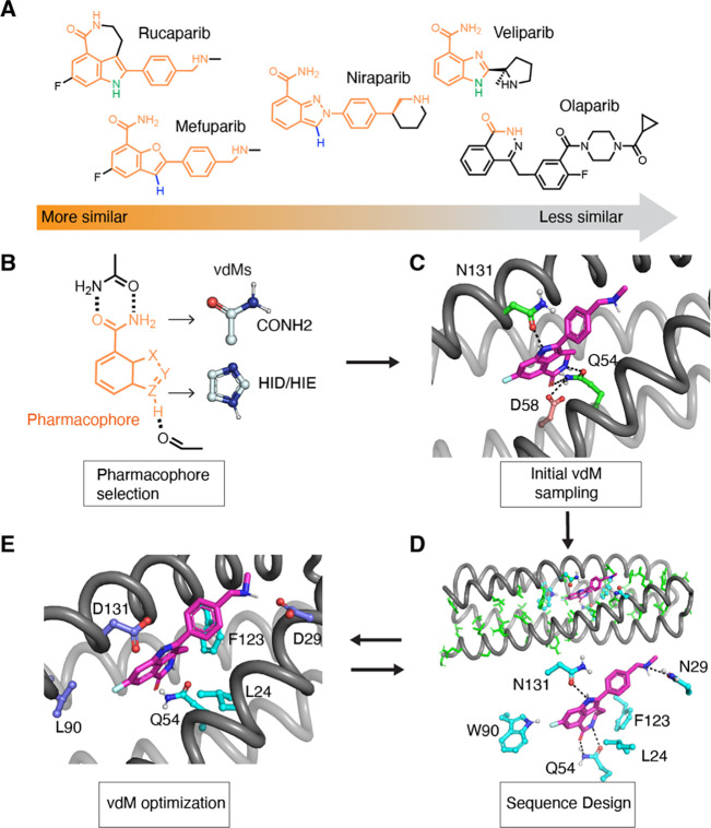
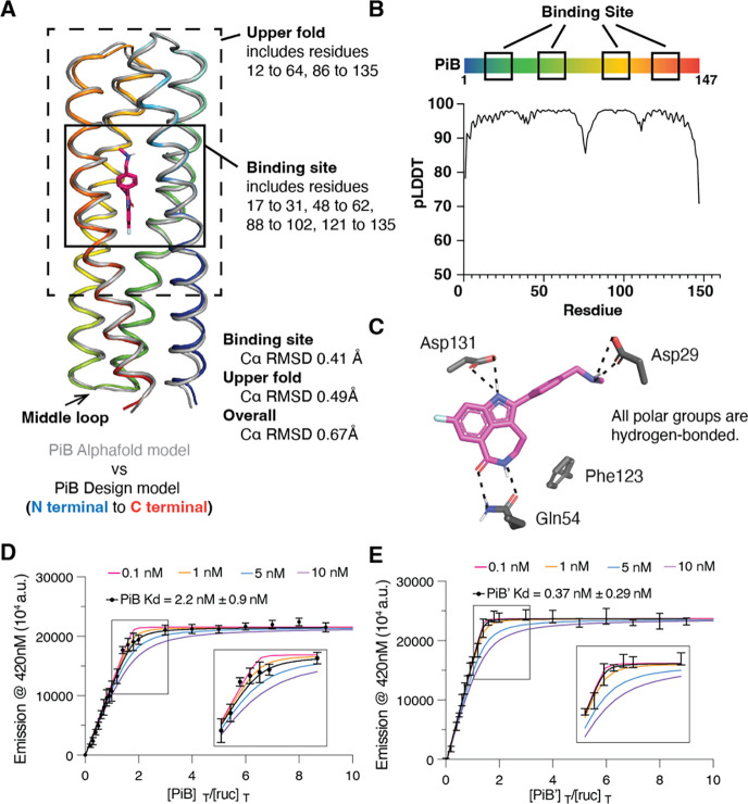
The de novo design of small-molecule-binding proteins has seen exciting recent progress; however, the ability to achieve exquisite affinity for binding small molecules while tuning specificity has not yet been demonstrated directly from computation. Here, we develop a computational procedure that results in the highest affinity binders to date with predetermined relative affinities, targeting a series of PARP1 inhibitors. Two of four designed proteins bound with affinities ranging from < 5 nM to low μM, in a predictable manner. X-ray crystal structures confirmed the accuracy of the designed protein-drug interactions. Molecular dynamics simulations informed the role of water in binding. Binding free-energy calculations performed directly on the designed models are in excellent agreement with the experimentally measured affinities, suggesting that the de novo design of small-molecule-binding proteins with tuned interaction energies is now feasible entirely from computation. We expect these methods to open many opportunities in biomedicine, including rapid sensor development, antidote design, and drug delivery vehicles.


Conflict of interest statement

Competing financial interests A.A. is a co-founder of Tango Therapeutics, Azkarra Therapeutics, Ovibio Corporation and Kytarro, a member of the board of Cytomx and Cambridge Science Corporation, a member of the scientific advisory board of Genentech, GLAdiator, Circle, Bluestar, Earli, Ambagon, Phoenix Molecular Designs, Yingli, ProRavel, Oric, Hap10 and Trial Library, a consultant for SPARC, ProLynx, Novartis and GSK, receives research support from SPARC, and holds patents on the use of PARP inhibitors held jointly with AstraZeneca from which he has benefited financially (and may do so in the future).

Figures

Figure 1.
Figure 1.. The computational design of poly(ADP-ribose) polymerase inhibitors (PARPi) protein binders.
(A) The PARPi analogues. The shared chemical features are marked in orange. Olaparib is used as a negative control in the design and binding assay. (B – E) The overall design strategy. (B, C) We first define the pharmacophore and use COMBS to sample vdMs on the selected protein backbones. We initially targeted the indole and carboxamide of the drug, and used COMBS to discover sidechains that would form first and second-shell hydrogen bonds to both of these chemical groups. We discovered a solution in which the carboxamide formed bidentate hydrogen bonds with sidechain of Gln54, and the drug’s indole NH interacted with the Asn131 (C, carbon atoms of protein green, those of rucaparib are purple). A second-shell interaction to Q54 that was discovered by COMBS was Asp58 (carbons brown). (D) We applied flexible backbone sequence design with a custom Rosetta script while fixing the interactions selected from COMBS. (E) Then we search vdM again based on the design output from the previous sequence step. The slightly different (~ 1 A Ca RMSD) backbone now preferred different vdMs at some locations (higher cluster scores) and these mutations were made. Three residues at 29, 90, 131 (deep blue) were changed based on COMBS results.
Figure 2.
Figure 2.. Assessing the computational model and experimental binding of PiB to rucaparib.
(A) The AlphaFold2 model agrees with the designed PiB very well, with the binding site Cα RMSD of 0.41 Å, the upper fold Cα RMSD of 0.49 Å and overall Cα RMSD of 0.67 Å. (B) The predicted local distance difference test scores (pLDDTs) concur with the trend of RMSD difference of the design model. For example, the N-terminal, C-terminal and the middle loop with low pLDDTs (<90) showed higher Cα RMSD. (C) The design model showing the polar groups of rucaparib are all hydrogen-bonded. (D) (E) A fluorescence titration shows that PiB and PiB’ bind rucaparib with KD < 5 nM. The fluorescence emission intensity at 420 nm of rucaparib (excitation wavelength 355 nm) was measured after titrating aliquots of PiB (D) or PiB’ (E) to a final concentration indicated in the abscissa. The data are well described by a single-site protein-ligand binding model, and a non-linear least squares fit to the data returned values of KD of 2.2 (± 0.9) nM for PIB, and 0.37 (± 0.29) nM for PIB’. Although the fitting error was relatively small, a sensitivity analysis, in which the value of KD was held constant at various values, showed that the data for both proteins were fit within experimental error so long as the KD is less than 5 nM. Therefore, while the most probable binding constants were 2 and 0.4 nM, respectively, we can confidently conclude that the values for PiB and PiB’ are less than 5 nM. The titration was carried out in in buffer containing 50 mM Tris, 100 mM NaCl (pH 7.4).
Figure 3.
Figure 3.. Spectral titrations and cell viability assay of PiB with PARPi.
(A) The values of KD of various drugs for PiB as obtained from global fit of a single-site binding model to the fluorescence changes (A, from Fig. 3) or absorbance changes as a function of the concentration of PiB. Indicated wavelengths for the titration were chosen to maximize the difference in absorption for the free versus bound drug. (B) Seven-day growth assays in DLD-1 BRCA2 mutated cells show that PiB alleviates the effects of rucaparib, mefuparib, niraparib and veliparib toxicity in a dose-dependent manner. The PARP inhibitors were pre-incubated with PiB in media at room temperature for 5 minutes at multiple concentration ratios (ligand : protein) of 1:0, 1:0.2, 1:1, 1:2.5, 1:5 and 1:10. (C) Cell viability assay as in Figure 4B showing that PiB had no effect on the olaparib dose response. (D) Table showing IC50 values for the inhibition of cell proliferation by PARPi drugs in the presence of increasing mole ratios of added PiB protein.
Figure 4.
Figure 4.. The structure of drug-bound PiB’ agrees with the design.
(A) The design model agrees well with the rucaparib-bound PiB’ crystal structure, with binding site (Fig. 3A) Cα RMSD range between 0.38–0.46 Å for the three monomers in the asymmetric unit. (B) The binding site of PiB’. A 2mFo-DFc composite omit map contoured at 1.6 σ. The map was generated from a model that omitted coordinates of rucaparib. Overlay of the design (gray) and the structure (protein in orange, rucaparib in pink). The sidechains of the binding pocket in rucaparib-bound PiB’ agrees with the design. Asp131 interacts with the indole NH via a bridging water as in MD simulations. (C) The structure of apo-PiB’ shows a preorganized open pocket filled with multiple waters, which are displaced in holo structure. (D) Reversal of the three designed substitutions from the vdM optimization procedure led to lower binding affinity (higher KD) for rucaparib by fluorescence emission titrations. (E) Alanine mutations of the direct binding residues decreased binding affinities confirmed by fluorescence emission titrations.
Figure 5.
Figure 5.. The MD simulations of PiB, PiB’, and mutants.
(A) Using unbiased molecular dynamics simulations in Amber, we calculated (in triplicate) the frequency with which the intermolecular hydrogen bonds formed between the protein scaffold and the bound drug molecule. PiB was found to form a hydrogen bond between Gln54 and the targeted drug carboxamide in 100% of all simulations for each drug complex. The charged ammonium groups of rucaparib and mefuparib interacted with Asp29 through a combination of direct and water-mediated hydrogen bonds, totaling to more than half of the full simulation time, which contrasts niraparib and veliparib’s inabilities to form equivalent hydrogen bonds (due to changes in chemical structure around the ammonium tail of the ligand). In a small fraction of each rucaparib and veliparib trajectory, Asp131 engaged in water-mediated hydrogen bonds to the drugs. (B) Using biased simulations in GROMACS, we calculated binding free energies for each ligand and found that ranked affinity for each drug is consistent with experimental results. (C) Comparison of ΔG binding from the GROMACS calculation with the experimental value from spectral titrations.

Similar articles

References

    1. Lassila J. K., Privett H. K., Allen B. D., Mayo S. L., Combinatorial methods for small-molecule placement in computational enzyme design. Proceedings of the National Academy of Sciences 103, 16710–16715 (2006). - PMC - PubMed
    1. Röthlisberger D., Khersonsky O., Wollacott A. M., Jiang L., DeChancie J., Betker J., Gallaher J. L., Althoff E. A., Zanghellini A., Dym O., Albeck S., Houk K. N., Tawfik D. S., Baker D., Kemp elimination catalysts by computational enzyme design. Nature 453, 190–195 (2008). - PubMed
    1. Privett H. K., Kiss G., Lee T. M., Blomberg R., Chica R. A., Thomas L. M., Hilvert D., Houk K. N., Mayo S. L., Iterative approach to computational enzyme design. Proceedings of the National Academy of Sciences 109, 3790–3795 (2012). - PMC - PubMed
    1. Yang W., Lai L., Computational design of ligand-binding proteins. Curr Opin Struct Biol 45, 67–73 (2017). - PubMed
    1. Schreier B., Stumpp C., Wiesner S., Höcker B., Computational design of ligand binding is not a solved problem. Proceedings of the National Academy of Sciences 106, 18491–18496 (2009). - PMC - PubMed

Publication types