[Preprint]. 2023 Dec 21:2023.12.20.572700.
doi: 10.1101/2023.12.20.572700.

Identification of new ciliary signaling pathways in the brain and insights into neurological disorders

Affiliations

Identification of new ciliary signaling pathways in the brain and insights into neurological disorders

Abdelhalim Loukil et al. bioRxiv. .

Update in

Abstract

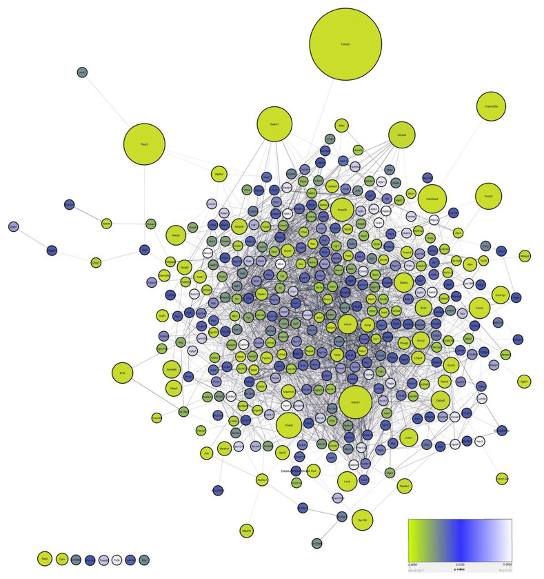
Primary cilia are conserved sensory hubs essential for signaling transduction and embryonic development. Ciliary dysfunction causes a variety of developmental syndromes with neurological features and cognitive impairment, whose basis mostly remains unknown. Despite connections to neural function, the primary cilium remains an overlooked organelle in the brain. Most neurons have a primary cilium; however, it is still unclear how this organelle modulates brain architecture and function, given the lack of any systemic dissection of neuronal ciliary signaling. Here, we present the first in vivo glance at the molecular composition of cilia in the mouse brain. We have adapted in vivo BioID (iBioID), targeting the biotin ligase BioID2 to primary cilia in neurons. We identified tissue-specific signaling networks enriched in neuronal cilia, including Eph/Ephrin and GABA receptor signaling pathways. Our iBioID ciliary network presents a wealth of neural ciliary hits that provides new insights into neurological disorders. Our findings are a promising first step in defining the fundamentals of ciliary signaling and their roles in shaping neural circuits and behavior. This work can be extended to pathological conditions of the brain, aiming to identify the molecular pathways disrupted in the brain cilium. Hence, finding novel therapeutic strategies will help uncover and leverage the therapeutic potential of the neuronal cilium.

Keywords: Biological sciences/Cell biology; iBioID; mature brain; neurodevelopmental disorders; neurons; primary cilia.


Figures

Figure 1:
Figure 1:. Targeting of the biotin ligase BirA2 to neuronal cilia in vivo
A. Experimental procedure to label the protein composition of the neuronal cilia in the brain. B. A simplified schematic of the targeting strategy for BirA2 using ciliary GPCRs. C. Immunohistochemical labeling with antibodies to HA (fused to BirA2, magenta), Biotin (green), and Arl13b (red) on mouse cerebral sections transduced with AAV-BirA2 or AAV-GPCRs-BirA2. DNA was stained with DAPI. Scale bars: 20 μm. D. Same as C with zoomed-in images. Scale bars: 5 μm. E. Streptavidin pulldowns were performed on total cell lysates from brains transduced with AAV-BirA2 or AAV-GPCRs-BirA2. Western blots were probed with Strepatavidin-680 and Arl13b antibodies.
Figure 2:
Figure 2:. Cilia iBioID identified neural-specific clusters in neuronal cilia in vivo
A. Volcano plot of SSTR3-proximate proteins labeled by BioID. The red dots show the proteins considered hits, delimited by the selected thresholds: log2 fold change ≥ 2 and significance ≥ 0.7. The top hit, Tnfrsf21, is highlighted in brown; GABA receptor signaling in green, axon guidance proteins in blue, and Eph/Ephrin signaling in black. The X-axis denotes the log2 fold change of SSTR3-BirA2: BirA2 control. In the Y-axis, significance displays the negative log10 transformed p-value for each protein. B. SIM acquisitions show that the top hit, Tnfrsf21, is enriched in the ciliary tip of primary mouse cortical neurons. Two representative examples are shown with cilia by Arl13b (red), centrosomes using Pericenrin (magenta), and Tnfrsf21 (green). Scale bars: 500 nm. C. Clustergram topologies of axon guidance hits D. GABAergic cluster enriched in Cilia iBioID.
Figure 3:
Figure 3:. The neuronal ciliary network.
This is a clustergram topology of all proteins enriched in SSTR3 iBioID. It is composed of protein networks, with proteins shown as spheres and their interactions as lines. Cytoscape software was deployed to generate this network, relying on its mining capabilities to extensively search the literature. The size of each sphere is correlated to the fold change value and the color shade to the p-value.
Figure 4:
Figure 4:. A distinct Eph/Ephrin signaling node is enriched in neuronal cilia
A. Clustergram topology of Eph/Ephrin proteins enriched in the cilia iBioID dataset The highlighted hits are validated in the following sections of this figure. B. Left panel: Representative immunofluorescence images acquired with the Structured illumination microscopy (SIM). Wild type primary cortical neurons are labeled with EphA4 (green) localizing to cilia co-stained with the ciliary marker Arl13bb (red) and centrosomal marker Pericentrin (magenta). Right panel also shows several examples of EphA4 ciliary enrichment (green) co-labeled with Arl13b and the neuronal marker, NeuN (magenta). DNA is stained with DAPI (blue). Arrows indicate areas of colocalization of EphA4 and Arl13b or Pericentrin. Scale bars: 1 μm. C. Representative immunofluorescence images of Efnb1, Efnb2, and EphB2 (green) enriched in neuronal primary cilia labeled with the ciliary marker Arl13b (red) and centrosomal Pericentrin (magenta). Arrows indicate the colocalization of Ephrin/Eph components with Arl13b. Scale bars: 1 μm. D. Representative immunofluorescence images of overexpressed EphA4-GFP and EphB3-V5 (green) in IMCD3 cells. Primary cilia are labeled with the ciliary marker AC3 or Arl13b (red) and centrosomal marker Pericentrin (magenta). Arrows show ciliary enrichment of EphA4-GFP and EphB3-V5. Scale bars: 1 μm.
Figure 5:
Figure 5:. GABAergic and CK1 proteins are enriched in neuronal cilia
A. Representative immunofluorescence images of GABRG2 or GABRA1 (green) localizing to neuronal primary cilia labeled with Arl13b (red). Wild type neurons are labeled with and identified by NeuN (magenta). Arrows indicate areas of colocalization of GABRG2 or GABRA1 and Arl13b. Scale bars: 1 μm. B. Representative immunofluorescence images of GABBR1 (green) enriched in neuronal primary cilia stained with Arl13b (red). Arrows denote enrichment of GABBR1 in neuronal cilia. Scale bars: 1 μm. C. Several examples of representative immunofluorescence of SLC6A1 (green) faintly enriched in neuronal primary cilia. White box denotes the ciliary region shown in the lower section. Arrows are ciliary regions with SLC6A1 enrichment within the neuronal cilium. Scale bars: 1 μm. D. CSNK1G1 and E. CSNK1G3 are labeled in primary cortical neurons and IMCD3 cells, respectively, with Arl13b (red) and Pericentrin (magenta). DNA was stained with DAPI (blue).
Figure 6:
Figure 6:. Insights into connections between neuronal cilia and brain disorders
A. Clustergram topology of hits associated with cognitive impairment (orange), autism (green), and neurodevelopmental disorders (NDD, red) B. Clustergram topology of proteins associated with seizure (orange), ataxia (green), and other movement disorders (red) C. Clustergram topology of hits associated with Parkinson’s disease (orange), Huntington’s disease (green), and Alzheimer’s disease (red). For all clustergrams, overlapping hits were shown in one of the categories.

References

    1. Goetz S. C. & Anderson K. V. The primary cilium: a signalling centre during vertebrate development. Nature Reviews Genetics 11, 331–344 (2010). - PMC - PubMed
    1. Huangfu D. et al. Hedgehog signalling in the mouse requires intraflagellar transport proteins. Nature 426, 83–87 (2003). - PubMed
    1. Haycraft C. J. et al. Gli2 and Gli3 localize to cilia and require the intraflagellar transport protein polaris for processing and function. PLoS Genet. 1, e53 (2005). - PMC - PubMed
    1. Taulman P. D., Haycraft C. J., Balkovetz D. F. & Yoder B. K. Polaris, a Protein Involved in Left-Right Axis Patterning, Localizes to Basal Bodies and Cilia. MBoC 12, 589–599 (2001). - PMC - PubMed
    1. Haycraft C. J., Swoboda P., Taulman P. D., Thomas J. H. & Yoder B. K. The C. elegans homolog of the murine cystic kidney disease gene Tg737 functions in a ciliogenic pathway and is disrupted in osm-5 mutant worms. Development 128, 1493–1505 (2001). - PubMed

Publication types