[Preprint]. 2023 Dec 21:2023.12.20.572679.
doi: 10.1101/2023.12.20.572679.

TipN's involvement with centromere segregation in Caulobacter crescentus

Affiliations

TipN's involvement with centromere segregation in Caulobacter crescentus

Morgan Letzkus et al. bioRxiv. .

Update in

Abstract

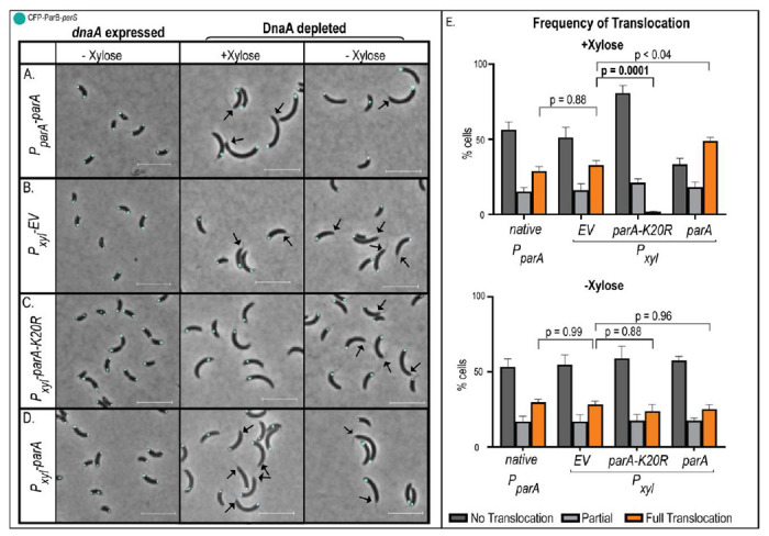
Bacteria's ability to maintain chromosomal integrity throughout their life cycle is crucial for their survival. In Caulobacter crescentus, the polar factor TipN has been proposed to be involved with the partitioning system ParABS. However, cells with tipN knocked out display subtle parS segregation defects. We hypothesized that TipN's role with parS segregation is obscured by other forces that are ParABS-independent. To test our hypothesis, we removed one of those forces - chromosome replication - and analyzed the role of TipN with ParA. We first demonstrate that ParA retains its ability to transport the centromeric region parS from the stalked pole to the opposite pole in the absence of chromosome replication. Our data revealed that in the absence of chromosome replication, TipN becomes essential for ParA's ability to transport parS. Furthermore, we identify a potential connection between the replication initiator DnaA and TipN. Although TipN is not essential for viability, tipN knockout cells lose viability when the regulation of DnaA levels is altered. Our data suggest that the DnaA-dependent susceptibility of tipN knockout cells is connected to parS segregation. Collectively, this work provides insights into the complex regulation involved in the coordination of chromosome replication and segregation in bacteria.


Conflict of interest statement

CONFLICT OF INTEREST The authors declare no conflict of interest.

Figures

Figure 1:
Figure 1:. Loss of TipN has no impact on growth in minimal media.
A) Schematic representation of C. crescentus highlighting ParB-parS (green), TipN (blue), and ParA dimers (brown) during chromosome replication and segregation of parS. (Bottom) Schematic of DNA-independent parS translocation. (B-C) Growth curves of wildtype (CB15N) cells expressing tipN (tipN+, black circles), ΔtipN (tipN-, orange squares), or with tipN complemented (tipN- Pxyl-tipN, gray triangles) in (B) rich (PYE) media and in (C) minimal media supplemented with 0.1% xylose. Small insert represents exponential growth phase. Each graph is representative of three independent trials. Error bars are ± SD for three replicates per trial.
Figure 2:
Figure 2:. Increase levels of ParA results in increased replication-independent translocation frequency of parS.
(A-D) Micrographs of mixed population of C. crescentus cells (parB::cfp-parB, ΔvanA, Pvan-dnaA, dnaA::Ω) expressing (A) parA from its native promoter (PparA-parA), (B) Pxyl-empty vector (EV), (C) Pxyl-parA-K20R, and (D) parA overexpression (Pxyl-parA) in minimal media. DnaA was either induced for expression or depleted for 3 hours prior to data analyses. Black arrows represent the location of the stalk in translocating cells. Scale bar – 5 μM. (E) Quantification of localization of the single CFP-ParB locus in A-D samples. Full translocation represents cells with CFP-ParB at the new cell pole. Data are mean averages from three independent trials. Error bars are ±SEM with two-way ANOVA analysis.
Figure 3:
Figure 3:. TipN is required for ParA to translocate parS in the absence of DNA replication.
A) Loss of tipN cause a subtle but significant increase in cells with partial parS segregation. Micrographs of exponentially growing cells (parB::cfp-parB, ΔvanA, Pvan-dnaA, dnaA::Ω ± tipN) grown in minimal media supplemented with 100 μM vanillate. Scale bars – 2.5 μM. Black arrows indicate partial segregation. Quantification using blinded samples of the mean average percentage of cells with CFP-ParB at both poles (bipolar), one pole (unipolar), or in between (partial). B) Quantification of parS translocation after 3-hour DnaA depletion. Cells (parB::cfp-parB, ΔvanA, Pvan-dnaA, dnaA::Ω) included tipN expression from native promoter (tipN+), ΔtipN (tipN-), tipN expressed from the xylose promoter in ΔtipN (tipN- Pxyl-tipN), or with overexpression of parA in ΔtipN (tipN- Pxyl-parA). 0.1% xylose was added to induce Pxyl expression. All data are from three independent trials. Error bars are ±SEM with 2-way ANOVA analyses.
Figure 4:
Figure 4:. DnaA-dependent loss of viability of ΔtipN cells.
Cell viability analyses using Colony forming unit (CFU) assays. Cells were serially diluted 1:10 and plated onto PYE plates supplemented with 100 μM vanillate, whenever noted. A) Comparison of cells expressing dnaA from native promoter (wildtype) and from inducible promoter vanillate (Pvan-dnaA) display no significant difference in CFUs. B) Comparison of wildtype cells CB15N cells with (tipN+) or without (tipN-) grown in the presence of vanillate reveals no impact on CFUs. C) The combination of dnaA expressed from inducible promoter (Pvan) and loss of tipN results in significant viability loss.
Figure 5:
Figure 5:. Characterization of DnaA-dependent susceptibility of ΔtipN cells.
A) The increased number of cells with >2 oris is observed in cells with or without tipN. Quantification of the number of CFP-ParB foci to represent number of ori per cell. Pxyl-parA was induced with 0.1% xylose for 3 hours in minimal media. Scale bars – 5 μM. Data are from three independent trials. Error bars are ±SEM with 2-way ANOVA analysis. B) Increased levels of ParA rescue DnaA-dependent viability defect of ΔtipN cells. Colony forming unit (CFU) viability assay in cells expressing dnaA from Pvan ± tipN and ± Pxyl-parA induction. Plates were supplemented with 100 μM vanillate and 0.1% xylose was added to induce Pxyl expression. CFUs are representatives of three independent trials.

Similar articles

References

    1. Austin S., & Abeles A. (1983). Partition of unit-copy miniplasmids to daughter cells. I. P1 and F miniplasmids contain discrete, interchangeable sequences sufficient to promote equipartition. J Mol Biol, 169(2), 353–372. 10.1016/s0022-2836(83)80055-2 - DOI - PubMed
    1. Ogura T., & Hiraga S. (1983). Partition mechanism of F plasmid: two plasmid gene-encoded products and a cis-acting region are involved in partition. Cell, 32(2), 351–360. 10.1016/0092-8674(83)90454-3 - DOI - PubMed
    1. Gerdes K., Larsen J. E., & Molin S. (1985). Stable inheritance of plasmid R1 requires two different loci. J Bacteriol, 161(1), 292–298. 10.1128/jb.161.1.292-298.1985 - DOI - PMC - PubMed
    1. Ely B. (1991). Genetics of Caulobacter crescentus. Methods Enzymol, 204, 372–384. 10.1016/0076-6879(91)04019-k - DOI - PubMed
    1. Niki H., Jaffe A., Imamura R., Ogura T., & Hiraga S. (1991). The new gene mukB codes for a 177 kd protein with coiled-coil domains involved in chromosome partitioning of E. coli. EMBO J, 10(1), 183–193. 10.1002/j.1460-2075.1991.tb07935.x - DOI - PMC - PubMed

Publication types