

Topical Application of Cell-Penetrating Peptide Modified Anti-VEGF Drug Alleviated Choroidal Neovascularization in Mice
- PMID: 38187905
- PMCID: PMC10771783
- DOI: 10.2147/IJN.S428684
Topical Application of Cell-Penetrating Peptide Modified Anti-VEGF Drug Alleviated Choroidal Neovascularization in Mice
Abstract
Background: Age-related macular degeneration (AMD) stands as the foremost cause of irreversible central vision impairment, marked by choroidal neovascularization (CNV). The prevailing clinical approach to AMD treatment relies on intravitreal injections of anti-vascular endothelial growth factor (VEGF) drugs. However, this method is encumbered by diverse complications, prompting exploration of non-invasive alternatives such as ocular administration via eye drops for anti-VEGF therapy.
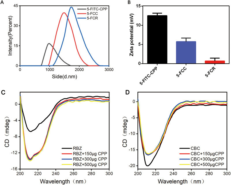
Methods: Two complexes, 5-FITC-CPP-Ranibizumab (5-FCR) and 5-FITC-CPP-Conbercept (5-FCC), were synthesized by incorporating the anti-VEGF drugs Ranibizumab (RBZ) or Conbercept (CBC) with cell-penetrating peptide (CPP). Circular dichroism spectrum (CD) facilitated complexes characterization. Eye drops was utilized to address laser-induced CNV in mice. Fluorescein fundus angiography (FFA) observe the CNV lesion, while FITC-dextran and IB4 dual fluorescent staining, along with hematoxylin-eosin (HE) staining, assessed in lesion size. Tissue immunofluorescence examined CD31 and VEGF expression in choroidal/retinal pigment epithelial (RPE) tissues. Biocompatibility and biosafety of 5-FCR and 5-FCC was evaluated through histological examination of various organs or cell experiments.
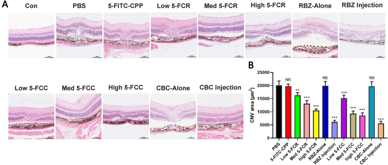
Results: Both 5-FCR and 5-FCC exhibited favorable biocompatibility and safety profiles. VEGF-induced migration of Human umbilical vein endothelial cells (HUVECs) significantly decreased post-5-FCR/5-FCC treatment. Additionally, both complexes suppressed VEGF-induced tube formation in HUVECs. FFA results revealed a significant improvement in retinal exudation in mice. Histological examination unveiled the lesion areas in the 5-FCR and 5-FCC groups showed a significant reduction compared to the control group. Similar outcomes were observed in histological sections of the RPE-choroid-sclera flat mounts.
Conclusion: In this study, utilizing the properties of CPP and two anti-VEGF drugs, we successfully synthesized two complexes, 5-FCR and 5-FCC, through a straightforward approach. Effectively delivering the anti-VEGF drugs to the target area in a non-invasive manner, suppressing the progression of laser-induced CNV. This offers a novel approach for the treatment of wet AMD.
Keywords: age-related macular degeneration; cell-penetrating peptide; choroidal neovascularization; conbercept; drug delivery; ranibizumab.
© 2024 Hu et al.
Conflict of interest statement
There are no conflicts of interest to declare.
Figures


Similar articles
-
Topical Delivery of Anti-VEGF Drugs to the Ocular Posterior Segment Using Cell-Penetrating Peptides.Invest Ophthalmol Vis Sci. 2017 May 1;58(5):2578-2590. doi: 10.1167/iovs.16-20072. Invest Ophthalmol Vis Sci. 2017. PMID: 28494491
-
Inhibition of YAP ameliorates choroidal neovascularization via inhibiting endothelial cell proliferation.Mol Vis. 2018 Jan 31;24:83-93. eCollection 2018. Mol Vis. 2018. PMID: 29422766 Free PMC article.
-
Topical Ophthalmic Liposomes Dual-Modified with Penetratin and Hyaluronic Acid for the Noninvasive Treatment of Neovascular Age-Related Macular Degeneration.Int J Nanomedicine. 2024 Feb 23;19:1887-1908. doi: 10.2147/IJN.S446425. eCollection 2024. Int J Nanomedicine. 2024. PMID: 38414529 Free PMC article.
-
Anti-vascular endothelial growth factor treatment for retinal conditions: a systematic review and meta-analysis.BMJ Open. 2019 May 28;9(5):e022031. doi: 10.1136/bmjopen-2018-022031. BMJ Open. 2019. PMID: 31142516 Free PMC article.
-
Aflibercept in wet AMD: specific role and optimal use.Drug Des Devel Ther. 2013 Aug 5;7:711-22. doi: 10.2147/DDDT.S40215. eCollection 2013. Drug Des Devel Ther. 2013. PMID: 23990705 Free PMC article. Review.
Cited by
-
Peptide-Bound Aflibercept Eye Drops for Treatment of Neovascular Age-Related Macular Degeneration in Nonhuman Primates.Adv Sci (Weinh). 2025 Mar;12(11):e2410744. doi: 10.1002/advs.202410744. Epub 2025 Jan 30. Adv Sci (Weinh). 2025. PMID: 39888276 Free PMC article.
-
Frontier applications of retinal nanomedicine: progress, challenges and perspectives.J Nanobiotechnology. 2025 Feb 25;23(1):143. doi: 10.1186/s12951-025-03095-6. J Nanobiotechnology. 2025. PMID: 40001147 Free PMC article. Review.
-
Mathematical Models of Topically and Intravitreally Applied Ranibizumab.Invest Ophthalmol Vis Sci. 2025 Aug 1;66(11):45. doi: 10.1167/iovs.66.11.45. Invest Ophthalmol Vis Sci. 2025. PMID: 40833326 Free PMC article.
-
Recent advancements in polymer science for retinal diseases: New frontiers in drug delivery systems.APL Bioeng. 2025 Jun 27;9(2):020902. doi: 10.1063/5.0264382. eCollection 2025 Jun. APL Bioeng. 2025. PMID: 40584817 Free PMC article.
References
MeSH terms
Substances
LinkOut - more resources
Full Text Sources
Medical
