

Cortical gray-white matter contrast abnormalities in male children with attention deficit hyperactivity disorder
- PMID: 38188507
- PMCID: PMC10768013
- DOI: 10.3389/fnhum.2023.1303230
Cortical gray-white matter contrast abnormalities in male children with attention deficit hyperactivity disorder
Abstract
Purpose: Presently, research concerning alterations in brain structure among individuals with attention deficit hyperactivity disorder (ADHD) predominantly focuses on entire brain volume and cortical thickness. In this study, we extend our examination to the cortical microstructure of male children with ADHD. To achieve this, we employ the gray-white matter tissue contrast (GWC) metric, allowing for an assessment of modifications in gray matter density and white matter microstructure. Furthermore, we explore the potential connection between GWC and the severity of disorder in male children by ADHD.
Methods: We acquired 3DT1 sequences from the public ADHD-200 database. In this study, we conducted a comparative analysis between 43 male children diagnosed with ADHD and 50 age-matched male controls exhibiting typical development trajectories. Our investigation entailed assessing differences in GWC and cortical thickness. Additionally, we explored the potential correlation between GWC and the severity of ADHD. To delineate the cerebral landscape, each hemisphere was subdivided into 34 cortical regions using freesurfer 7.2.0. For quantification, GWC was computed by evaluating the intensity contrast of non-normalized T1 images above and below the gray-white matter interface.
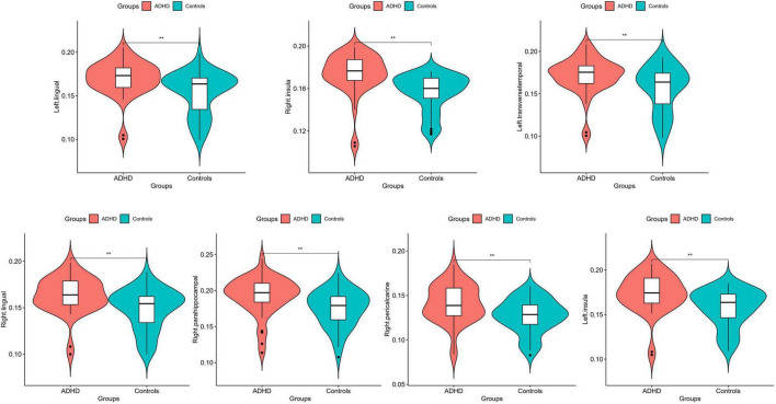
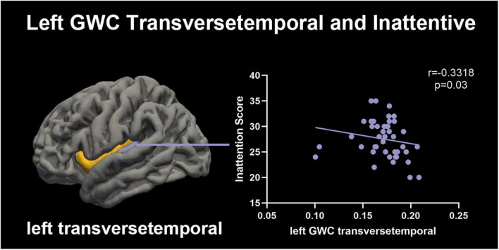
Results: Our findings unveiled elevated GWC within the bilateral lingual, bilateral insular, left transverse temporal, right parahippocampal and right pericalcarine regions in male children with ADHD when contrasted with their healthy counterparts. Moreover, the cortical thickness in the ADHD group no notable distinctions that of control group in all areas. Intriguingly, the GWC of left transverse temporal demonstrated a negative correlation with the extent of inattention experienced by male children with ADHD.
Conclusion: Utilizing GWC as a metric facilitates a more comprehensive assessment of microstructural brain changes in children with ADHD. The fluctuations in GWC observed in specific brain regions might serve as a neural biomarker, illuminating structural modifications in male children grappling with ADHD. This perspective enriches our comprehension of white matter microstructure and cortical density in these children. Notably, the inverse correlation between the GWC of the left transverse temporal and inattention severity underscores the potential role of structural and functional anomalies within this region in ADHD progression. Enhancing our insight into ADHD-related brain changes holds significant promise in deciphering potential neuropathological mechanisms.
Keywords: attention deficit hyperactivity disorder; cortical thickness; gray-white matter tissue contrast; inattention; male children.
Copyright © 2023 Wang, Shen, Cheng, Zhu, Lv, Zhang, Feng, Yang and Zhao.
Conflict of interest statement
The authors declare that the research was conducted in the absence of any commercial or financial relationships that could be construed as a potential conflict of interest.
Figures

Similar articles
-
Gray/White Matter Contrast in Parkinson's Disease.Front Aging Neurosci. 2018 Mar 27;10:89. doi: 10.3389/fnagi.2018.00089. eCollection 2018. Front Aging Neurosci. 2018. PMID: 29636679 Free PMC article.
-
Differential brain development with low and high IQ in attention-deficit/hyperactivity disorder.PLoS One. 2012;7(4):e35770. doi: 10.1371/journal.pone.0035770. Epub 2012 Apr 20. PLoS One. 2012. PMID: 22536435 Free PMC article.
-
Cortical gray matter in attention-deficit/hyperactivity disorder: a structural magnetic resonance imaging study.J Am Acad Child Adolesc Psychiatry. 2010 Mar;49(3):229-38. doi: 10.1016/j.jaac.2009.11.008. J Am Acad Child Adolesc Psychiatry. 2010. PMID: 20410712 Free PMC article.
-
Neural alterations in ADHD children as indicated by voxel-based cortical thickness and morphometry analysis.Brain Dev. 2017 May;39(5):403-410. doi: 10.1016/j.braindev.2016.12.002. Epub 2017 Jan 2. Brain Dev. 2017. PMID: 28057397
-
Association between gray/white matter contrast and white matter microstructural alterations in medication-naïve obsessive-compulsive disorder.Neuroimage Clin. 2022;35:103122. doi: 10.1016/j.nicl.2022.103122. Epub 2022 Jul 19. Neuroimage Clin. 2022. PMID: 35872436 Free PMC article.
Cited by
-
Prenatal Tobacco Exposure, Brain Subcortical Volumes, and Gray-White Matter Contrast.JAMA Netw Open. 2024 Dec 2;7(12):e2451786. doi: 10.1001/jamanetworkopen.2024.51786. JAMA Netw Open. 2024. PMID: 39699892 Free PMC article.
References
-
- Albajara Saenz A., Villemonteix T., Massat I. (2019). Structural and functional neuroimaging in attention-deficit/hyperactivity disorder. Dev. Med. Child Neurol. 61 399–405. - PubMed
-
- Almeida Montes L. G., Prado Alcantara H., Martinez Garcia R. B., De La Torre L. B., Avila Acosta D., Duarte M. G. (2013). Brain cortical thickness in ADHD: Age, sex, and clinical correlations. J. Atten. Disord. 17 641–654. - PubMed
LinkOut - more resources
Full Text Sources