

Anatomical variant of anteriorly extending mental foramen: A case report
- PMID: 38188843
- PMCID: PMC10766552
- DOI: 10.1002/ccr3.8341
Anatomical variant of anteriorly extending mental foramen: A case report
Erratum in
-
Correction to Anatomical Variant of Anteriorly Extending Mental Foramen: A Case Report.Clin Case Rep. 2024 Nov 26;12(12):e9562. doi: 10.1002/ccr3.9562. eCollection 2024 Dec. Clin Case Rep. 2024. PMID: 39605849 Free PMC article.
Abstract
Key clinical message: By sharing this case, we aim to enhance the understanding of the mental foramen's intricate morphology, ultimately promoting safer and more successful surgical practices in the field of oral and maxillofacial surgery.
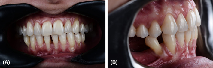
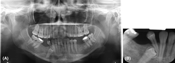
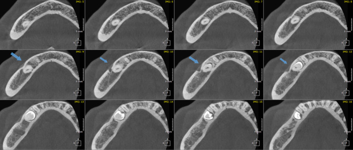
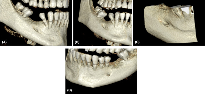
Abstract: In surgical procedures near the mental foramen, preserving this vital structure and its contents is crucial. Surgical treatments, including procedures like implants, orthognathic surgery, and tooth extractions, can potentially lead to injuries of the mental nerve, resulting in sensory disturbances such as numbness or tingling in the lower lip and chin. This case report highlights an uncommon anterior extension of the mental foramen, posing a risk to the patient if unnoticed. Variations in this structure are possible, emphasizing the need for comprehensive three-dimensional radiographic analysis before surgery to ensure patient safety. This report sheds light on the significance of identifying and understanding such variations to enhance the safety and precision of oral and maxillofacial interventions.
Keywords: anatomical variation; case report; mental foramen; patient safety; superior anterior.
© 2024 The Authors. Clinical Case Reports published by John Wiley & Sons Ltd.
Conflict of interest statement
The authors declare there are no competing interests.
Figures
Similar articles
-
The mental foramen and nerve: clinical and anatomical factors related to dental implant placement: a literature review.J Periodontol. 2006 Dec;77(12):1933-43. doi: 10.1902/jop.2006.060197. J Periodontol. 2006. PMID: 17209776 Review.
-
Use of Adult Dry Human Mandibles to Study of Mandibular Foramen from Various Bony Landmarks.J Pharm Bioallied Sci. 2024 Feb;16(Suppl 1):S666-S668. doi: 10.4103/jpbs.jpbs_927_23. Epub 2023 Dec 20. J Pharm Bioallied Sci. 2024. PMID: 38595526 Free PMC article.
-
A Rare Anatomical Finding: Bilateral Accessory Mental Foramen.Case Rep Dent. 2021 Aug 21;2021:6020515. doi: 10.1155/2021/6020515. eCollection 2021. Case Rep Dent. 2021. PMID: 34471550 Free PMC article.
-
Accessory Mental Foramen and Maxillofacial Surgery.J Craniofac Surg. 2018 May;29(3):e216-e217. doi: 10.1097/SCS.0000000000004094. J Craniofac Surg. 2018. PMID: 29077686
-
Accessory mandibular foramen during dental implant placement: case report and review of literature.Implant Dent. 2014 Apr;23(2):116-24. doi: 10.1097/ID.0000000000000056. Implant Dent. 2014. PMID: 24637530 Review.
Cited by
-
A rare case of an anatomical variant of nonexistent mental foramen.Radiol Case Rep. 2024 Nov 28;20(2):1082-1086. doi: 10.1016/j.radcr.2024.11.009. eCollection 2025 Feb. Radiol Case Rep. 2024. PMID: 39659692 Free PMC article.
References
-
- Reda R, Zanza A, Bhandi S, Biase A, Testarelli L, Miccoli G. Surgical‐anatomical evaluation of mandibular premolars by CBCT among the Italian population. Dent Med Probl. 2022;59(2):209‐216. - PubMed
-
- Cellina M, Martinenghi C, De Nardi S, et al. Anatomy of the mental foramen: relationship among different metrical parameters for accurate localization. Applied Sciences. 2023;13(16):9235.
-
- Goyushov S, Tözüm MD, Tözüm TF. Assessment of morphological and anatomical characteristics of mental foramen using cone beam computed tomography. Surg Radiol Anat. 2018;40:1133‐1139. - PubMed
-
- Phillips JL, Weller RN, Kulild JC. The mental forman: part I. Size, orientation, and positional relationship to the mandibular second premolar. J Endod. 1990;16(5):221‐223. - PubMed
Publication types
LinkOut - more resources
Full Text Sources
