A computed tomography radiomics-based model for predicting osteoporosis after breast cancer treatment
- PMID: 38190012
- PMCID: PMC10963549
- DOI: 10.1007/s13246-023-01360-2
A computed tomography radiomics-based model for predicting osteoporosis after breast cancer treatment
Abstract
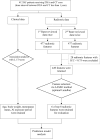
Many treatments against breast cancer decrease the level of estrogen in blood, resulting in bone loss, osteoporosis and fragility fractures in breast cancer patients. This retrospective study aimed to evaluate a novel opportunistic screening for cancer treatment-induced bone loss (CTIBL) in breast cancer patients using CT radiomics. Between 2011 and 2021, a total of 412 female breast cancer patients who received treatment and were followed up in our institution, had post-treatment dual-energy X-ray absorptiometry (DXA) examination of the lumbar vertebrae and had post-treatment chest CT scan that encompassed the L1 vertebra, were included in this study. Results indicated that the T-score of L1 vertebra had a strongly positive correlation with the average T-score of L1-L4 vertebrae derived from DXA (r = 0.91, p < 0.05). On multivariable analysis, four clinical variables (age, body weight, menopause status, aromatase inhibitor exposure duration) and three radiomic features extracted from the region of interest of L1 vertebra (original_firstorder_RootMeanSquared, wavelet.HH_glcm_InverseVariance, and wavelet.LL_glcm_MCC) were selected for building predictive models of L1 T-score and bone health. The predictive model combining clinical and radiomic features showed the greatest adjusted R2 value (0.557), sensitivity (83.6%), specificity (74.2%) and total accuracy (79.4%) compared to models that relied solely on clinical data, radiomic features, or Hounsfield units. In conclusion, the clinical-radiomic predictive model may be used as an opportunistic screening tool for early identification of breast cancer survivors at high risk of CTIBL based on non-contrast CT images of the L1 vertebra, thereby facilitating early intervention for osteoporosis.
Keywords: Breast cancer; Cancer treatment-induced bone loss; Computed tomography; Osteoporosis; Predictive modelling; Radiomics.
© 2024. The Author(s).
Conflict of interest statement
The authors declare no commercial affiliation, conflict of interest or any potential financial interests involved in this study.
Figures
References
MeSH terms
LinkOut - more resources
Full Text Sources
Medical


