. 2024 Jan 8;18(1):e0011872.
doi: 10.1371/journal.pntd.0011872. eCollection 2024 Jan.

A novel Trichinella spiralis serine proteinase disrupted gut epithelial barrier and mediated larval invasion through binding to RACK1 and activating MAPK/ERK1/2 pathway

Affiliations

A novel Trichinella spiralis serine proteinase disrupted gut epithelial barrier and mediated larval invasion through binding to RACK1 and activating MAPK/ERK1/2 pathway

Yan Yan Song et al. PLoS Negl Trop Dis. .

Abstract

Background: Gut epithelium is the first natural barrier against Trichinella spiralis larval invasion, but the mechanism by which larval penetration of gut epithelium is not completely elucidated. Previous studies showed that proteases secreted by T. spiralis intestinal infective larvae (IIL) degraded tight junctions (TJs) proteins of gut epithelium and mediated larval invasion. A new T. spiralis serine proteinase (TsSPc) was identified in the IIL surface proteins and ES proteins, rTsSPc bound to the intestinal epithelial cell (IECs) and promoted larval invasion of IECs. The aim of this study was to characterize the interacted proteins of TsSPc and IECs, and to investigate the molecular mechanisms of TsSPc mediating larval invasion of gut mucosa.

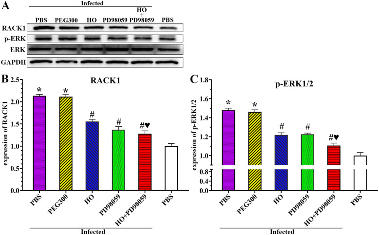
Methodology/principal finding: IIFT results showed natural TsSPc was detected in infected murine intestine at 6, 12 hours post infection (hpi) and 3 dpi. The results of GST pull-down, mass spectrometry (MS) and Co-IP indicated that rTsSPc bound and interacted specifically with receptor for activated protein C kinase 1 (RACK1) in Caco-2 cells. rTsSPc did not directly hydrolyze the TJs proteins. qPCR and Western blot showed that rTsSPc up-regulated RACK1 expression, activated MAPK/ERK1/2 pathway, reduced the expression levels of gut TJs (occludin and claudin-1) and adherent protein E-cad, increased the paracellular permeability and damaged the integrity of intestinal epithelial barrier. Moreover, the RACK1 inhibitor HO and ERK1/2 pathway inhibitor PD98059 abolished the rTsSPc activating ERK1/2 pathway, they also inhibited and abrogated the rTsSPc down-regulating expression of occludin, claudin-1 and E-cad in Caco-2 monolayer and infected murine intestine, impeded larval invasion and improved intestinal epithelial integrity and barrier function, reduced intestinal worm burdens and alleviated intestinal inflammation.

Conclusions: rTsSPc bound to RACK1 receptor in gut epithelium, activated MAPK/ERK1/2 pathway, decreased the expression of gut epithelial TJs proteins and disrupted the epithelial integrity, consequently mediated T. spiralis larval invasion of gut epithelium. The results are valuable to understand T. spiralis invasion mechanism, and TsSPc might be regarded as a vaccine target against T. spiralis invasion and infection.


Conflict of interest statement

The authors have declared that no competing interests exist.

Figures

Fig 1
Fig 1. Binding of native TsSPc with enteral epithelium of T. spiralis-infected mice at various times after infection.
Intestinal sections from infected mice at different times after infection were probed by anti-rTsSPc immune serum and infection serum, and green fluorescence immunostaining on enteral epithelium was detected at 6 and 12 hpi, and 3 dpi. Cell nuclei were stained blue with DAPI. Each experiment was performed in triplicate. Scale bars: 100 μm.
Fig 2
Fig 2. Binding proteins of rTsSPc and Caco-2 cells by GST-pull down.
rTsSPc was co-incubated with soluble Caco-2 cell proteins to capture their interacting proteins. M: Low molecular weight protein marker. Each experiment was performed in triplicate. The arrow points to two protein bands with 36 and 55 kDa from the Caco-2 cell proteins captured and interacted with rTsSPc.
Fig 3
Fig 3. IIFT and Co-IP detection of co-localization and interaction of rTsSPc and RACK1 in Caco-2 cells.
A: IIFT assay of co-localization of rTsSPc and RACK1 in Caco-2 cells. Caco-2 cells were pre-incubated with rTsSPc and IIL ES antigens at 37 °C for 2 h. After washing, the Caco-2 cells was probed by anti-RACK1 antibody, anti-rTsSPc serum, infected serum, or pre-immune serum, and then stained with goat anti-mouse IgG-Alexa Fluor 488 conjugate and goat anti-rabbit IgG-CY3 conjugate. The cellular nuclei were stained as blue with DAPI. Scale bars: 10 μm. B: Co-IP assay of interaction between rTsSPc and RACK1 in Caco-2 cells. Caco-2 cells were first incubated with rTsSPc. Then, anti-rTsSPc antibodies and protein A/G were added and incubated for 6 h. The bound proteins were separated on SDS-PAGE and transferred to the PVDF membrane. The membrane was probed by anti-rTsSPc antibody and anti-RACK1 antibody. Lane 1: Immune co-precipitation complex (rTsSPc, anti-rTsSPc antibody and RACK1 in Caco-2 cell protein); Lane 2: Normal mouse IgG; Lane 3: Caco-2 cell protein after incubation with rTsSPc. Each experiment was carried out in triplicate.
Fig 4
Fig 4. Expression levels of TJs protein in Caco-2 cell monolayers incubated with rTsSPc by IIFT and qPCR and Western blot.
A: Caco-2 cell monolayers were incubated at 37 °C for 2 h with rTsSPc (20 μg/ml). ΔrTsSPc represents the heating inactivated rTsSPc. PMSF+rTsSPc represents rTsSPc after treated with PMSF (serine protease inhibitor). Trypsin and IIL ES antigens were used as the positive control to evaluate the hydrolyzing or regulatory effect of rTsSPc on tight junctions (TJs) expression in Caco-2 cells. The cells were fixed with 4% paraformaldehyde, permeabilized for 10 min with 0.25% Triton X-100 and blocked with 1% BSA. After washing, the cells were probed with primary antibodies to occludin, claudin-1, claudin-2 or E-cad and then incubated with FITC- or Cy3-conjugated secondary antibodies. Cell nuclei were stained blue with DAPI. The cells were observed under a fluorescence microscope. Scale bars: 10 μm. B: Quantitative analysis of the fluorescence. C: qPCR analysis of transcription level of TJs protein in rTsSPc-treated Caco-2 cells. D: Western blot analysis of expression levels of TJs proteins in rTsSPc-treated Caco-2 cells. Density metrology analysis of protein expression of E-cad, occludin, claudin-1 and claudin-2 obtained from Western blotting compared to the GAPDH expression. Each experiment was carried out in triplicate. * in the Figures represents P < 0.0001 compared to the PBS group.
Fig 5
Fig 5. The rTsSPc up-regulated expression of RACK1 and p-ERK1/2 in Caco-2 cells by Western blotting.
A: rTsSPc up-regulated expression of RACK1. After Caco-2 cells were treated with rTsSPc, trypsin or IIL ES antigens, the RACK1 expression was obviously up-regulated compared to the PBS group. B: rTsSPc up-regulated expression of p-ERK1/2. ERK1/2 phosphorylation level in Caco-2 cells treated with rTsSPc was detected by Western blot. Compared to the PBS group, the ERK1/2 MAPK signaling pathway was activated and ERK1/2 phosphorylation level was evidently increased in Caco-2 cells treated with rTsSPc, trypsin and IIL ES antigens. ΔrTsSPc represents the heating inactivated rTsSPc at 100 °C for 5 min, and * in the figure represents P < 0.0001 compared to the PBS group. Each experiment was carried out in triplicate.
Fig 6
Fig 6. Assessment of direct hydrolysis of Caco-2 monolayer TJs proteins by rTsSPc.
A: Western blotting of direct hydrolysis of TJs in Caco-2 cell proteins incubated with rTsSPc, direct hydrolysis of E-cad, occludin, claudin-1 and claudin-2 was observed only in Caco-2 cell proteins incubated with IIL ES antigens and trypsin groups. B: Quantitative analysis of Western blot results in Figure 6A showed that rTsSPc had no direct degradation role on E-cad, occludin, claudin-1 and claudin-2, relative to the PBS group. ΔrTsSPc represents the heating inactivated rTsSPc, and MTsSPc shows the rTsSPc with enzymatic active site mutation which was prepared in our laboratory. * in the figure represents P < 0.0001 compared to the PBS group. Each experiment had three replicates.
Fig 7
Fig 7. rTsSPc disrupted the integrity of Caco-2 monolayer barrier.
When the Caco-2 cells grew to fully the confluence, the integrity of the cell monolayer was assessed by measuring TEER. The apical-to-basolateral flux of FD4TC-dextran through the cell monolayers cultured in Transwell system was measured for 2 h after the addition of dextran in the presence of rTsSPc. Data showed the mean ± SD of three independent experiments. A: TEER measure of Caco-2 monolayer before treatment with different doses of rTsSPc. B: TEER of Caco-2 monolayer before treatment with rTsSPc and IIL antigens. C: Effect of different concentrations of rTsSPc on the permeability of Caco-2 monolayer for diverse incubation times. D: Effect of rTsSPc and IIL ES antigens on the permeability of the cell monolayer for diverse incubation times. E: Permeability of cell monolayer treated with various doses of rTsSPc for 120 min. F: Permeability of cell monolayer treated with rTsSPc and IIL antigens for 120 min. Each experiment had triplicate. ΔrTsSPc represents the heating inactivated rTsSPc at 100 °C for 5 min. *P < 0.0001 relative to the PBS control group.
Fig 8
Fig 8. Inhibitors and antibody abrogated the rTsSPc down-regulating TJs expression in Caco-2 cells.
A: IIFT assay of TJs expression proteins in Caco-2 cells incubated with rTsSPc after pretreatment with inhibitors and anti-RACK1 antibody. Caco-2 monolayer was pre-treated with HO, PD98059 or anti-RACK1 at 37 °C for 24 h, and then incubated with rTsSPc for 2 h. PBS was used as a negative control, IIL ES antigens was used as a positive control. The regulatory effect of rTsSPc on expression of TJs proteins in Caco-2 cells was assessed by IIFT. The treated cells were fixed with 4% paraformaldehyde, permeated with 0.25% Triton X-100 for 10 min, and blocked with 1% BSA. After washing, the cells were probed with primary antibodies against occludin, claudin-1, claudin-2 or E-cad and then incubated with FITC- or Cy3-conjugated secondary antibodies. Cell nuclei were stained blue with DAPI. The cells were observed under a fluorescence microscope (1000×). Scale bars: 10 μm. B: Quantitative analysis of the fluorescence. C: qPCR analysis of transcription level of TJs protein E-cad, occludin, claudin-1, and claudin-2 genes relative to the PBS group. D: Western blotting of expression level of TJs proteins in Caco-2 cells incubated with rTsSPc after pretreatment with HO, PD98059, and anti-RACK1 antibody. Expression level of E-cad, occludin, claudin-1 and claudin-2 in Caco-2 cells was compared to the PBS group, only rTsSPc or IIL ES antigen alone group. Each experiment was performed in triplicate. ΔrTsSPc represents the heating inactivated rTsSPc, MTsSPc shows mutant rTsSPc. *P < 0.05 compared to the PBS group, #P < 0.05 compared to the rTsSPc group; P < 0.05 relative to the IIL ES antigen group.
Fig 9
Fig 9. RACK1 receptor and ERK1/2 pathway inhibitors abrogated rTsSPc activating ERK1/2 pathway.
Caco-2 monolayer was pretreated with two inhibitors HO and PD98059, and then incubated with rTsSPc, the ERK1/2 pathway was not activated in inhibitors-pretreated Caco-2 cells. A: Western blotting analysis of RACK1 expression level in Caco-2 cells. B: Western blotting analysis of ERK1/2 expression level in Caco-2 cells. Quantitative analysis of protein bands in Western blot was performed by using ImageJ software. ΔrTsSPc represents the heating inactivated rTsSPc. Each experiment was performed in triplicate. * in the figure represents P < 0.05 compared to the PBS group. #P < 0.05 compared to the rTsSPc group; P < 0.05 relative to the IIL ES antigen group.
Fig 10
Fig 10. Inhibitors and anti-RACK1 antibody suppressed larval invasion and reduced intestinal worm burden in infected mice.
A: Inhibitors and anti-RACK1 antibody inhibited larval invasion of Caco-2 cells. The muscle larvae were first activated into the IIL with 5% porcine bile at 37 °C for 2 h, and then were added onto the Caco-2 monolayer pretreated with inhibitors or anti-RACK1 antibody and incubated for 24 h. The larval invasion was observed under a microscope. B: The data were expressed as the inhibition (%) normalized to the PBS control group. HO and PD98059 also reduced intestinal IIL and adult burden in infected mice at 12 hours and 5 days after infection. C: 12 h IIL burden (n = 10); D: Intestinal adult worm burden (n = 10). Each experiment was carried out in triplicate, and the data of each group were represented as mean ± standard deviation. *P < 0.05 relative to the PBS group, #P < 0.05 relative to the only HO or PD98059 alone group.
Fig 11
Fig 11. HO and PD98059 inhibited up-regulation of RACK1 and p-ERK1/2 caused by T. spiralis infection.
A: Western blotting of expression of RACK1 and p-ERK in infected mouse intestine. B: HO and PD98059 inhibited the RACK1 expression in infected mice. C: HO and PD98059 decreased up-regulation of p-ERK1/2 in infected mice. Each test had three replicates. *P < 0.0001 compared to the uninfected PBS group; #P < 0.001 relative to the infected PBS group; P < 0.05 compared to the only HO or PD98059 alone group.
Fig 12
Fig 12. HO and PD98059 up-regulated the transcription and expression of gut TJs proteins in infected mice.
A: qPCR analysis of transcription level of TJs proteins in infected mice pretreated by HO and PD98059. B: Western blotting of TJs protein expression in infected mice pretreated by HO and PD98059. HO and PD98059 pretreatment increased the expression levels of TJs (E-cad, occludin and claudin-1) and decreased claudin-2 expression in T. spiralis-infected murine intestine. Each test had three replicates. * P < 0.05 compared with the uninfected PBS group; # P < 0.05 compared with the infected PBS group; P < 0.05 compared to the only HO or PD98059 alone group.
Fig 13
Fig 13. HO and PD98059 alleviated intestinal inflammation caused by T. spiralis infection.
A and B: HO and PD98059 inhibited and abrogated the elevated transcription level of pro-inflammatory cytokines (TNF-α and IL-1β) caused by T. spiralis infection. C and D: HO and PD98059 up-regulated further the transcription level of anti-inflammatory cytokines (IL-4 and IL-10) caused by T. spiralis infection. Each experiment was carried out in triplicate. * P < 0.05 compared to the uninfected PBS group; # P < 0.05 compared to the infected PBS group; P < 0.05 compared to the only HO or PD98059 alone group.
Fig 14
Fig 14. HO and PD98059 suppressed and abrogated the increased intestinal permeability caused by T. spiralis infection.
HO and PD98059 reduced intestinal FD-4 flow. Each assay had six replicates in each group, * P < 0.0001 relative to the uninfected PBS group; #P < 0.0001 in comparison with infected PBS group. P <0.0001 in comparison with the only HO or PD98059 alone group.

References

    1. Pozio E. The broad spectrum of Trichinella hosts: From cold- to warm-blooded animals. Vet Parasitol. 2005; 132: 3–11. doi: 10.1016/j.vetpar.2005.05.024 - DOI - PubMed
    1. van der Giessen J, Deksne G, Gómez-Morales MA, Troell K, Gomes J, Sotiraki S, et al.. Surveillance of foodborne parasitic diseases in Europe in a One Health approach. Parasite Epidemiol Control. 2021; 13. doi: 10.1016/j.parepi.2021.e00205 - DOI - PMC - PubMed
    1. Ribicich MM, Farina FA, Aronowicz T, Ercole ME, Bessi C, Winter M, et al.. A review on Trichinella infection in South America. Vet Parasitol. 2020; 285: 109234. doi: 10.1016/j.vetpar.2020.109234 - DOI - PubMed
    1. Zhang XZ, Wang ZQ and Cui J. Epidemiology of trichinellosis in the People’s Republic of China during 2009–2020. Acta Trop. 2022; 229: 106388. doi: 10.1016/j.actatropica.2022.106388 - DOI - PubMed
    1. Xu YXY, Zhang XZ, Weng MM, Cheng YK, Liu RD, Long SR, et al.. Oral immunization of mice with recombinant Lactobacillus plantarum expressing a Trichinella spiralis galectin induces an immune protection against larval challenge. Parasit Vectors. 2022; 15: 475. doi: 10.1186/s13071-022-05597-w - DOI - PMC - PubMed