. 2024 Mar 4;84(5):675-687.
doi: 10.1158/0008-5472.CAN-23-1459.

AXL/WRNIP1 Mediates Replication Stress Response and Promotes Therapy Resistance and Metachronous Metastasis in HER2+ Breast Cancer

Affiliations

AXL/WRNIP1 Mediates Replication Stress Response and Promotes Therapy Resistance and Metachronous Metastasis in HER2+ Breast Cancer

Mauricio Marquez-Palencia et al. Cancer Res. .

Abstract

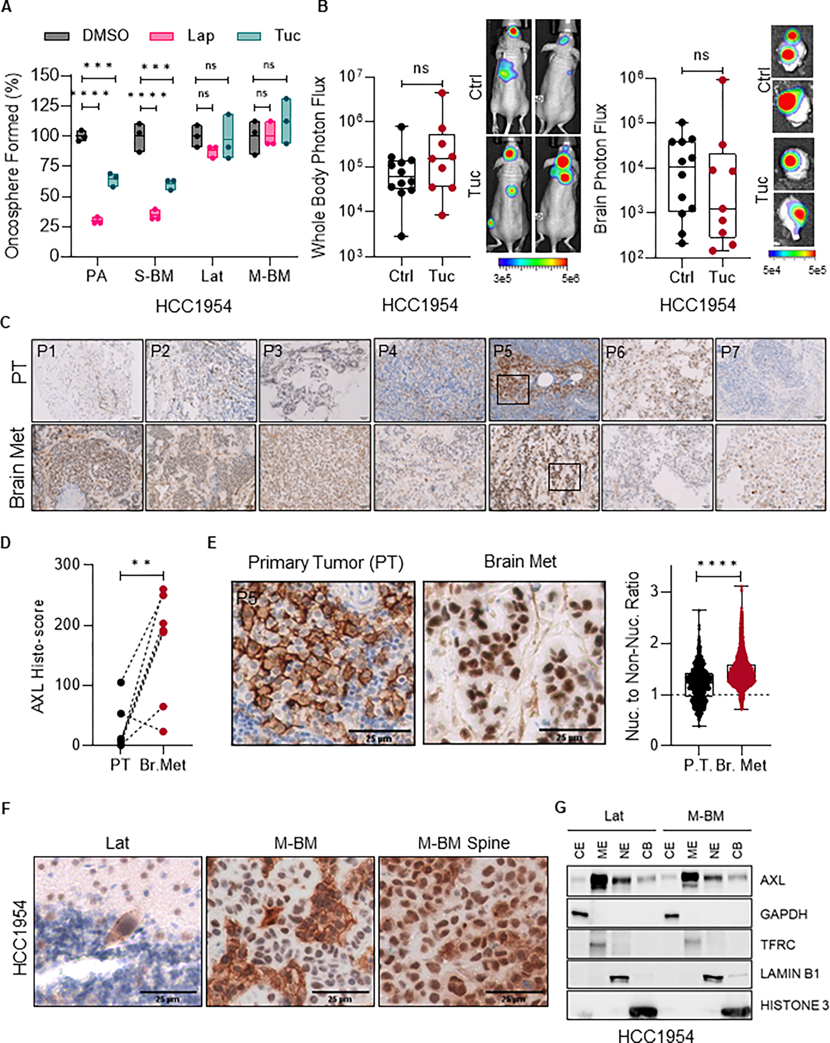
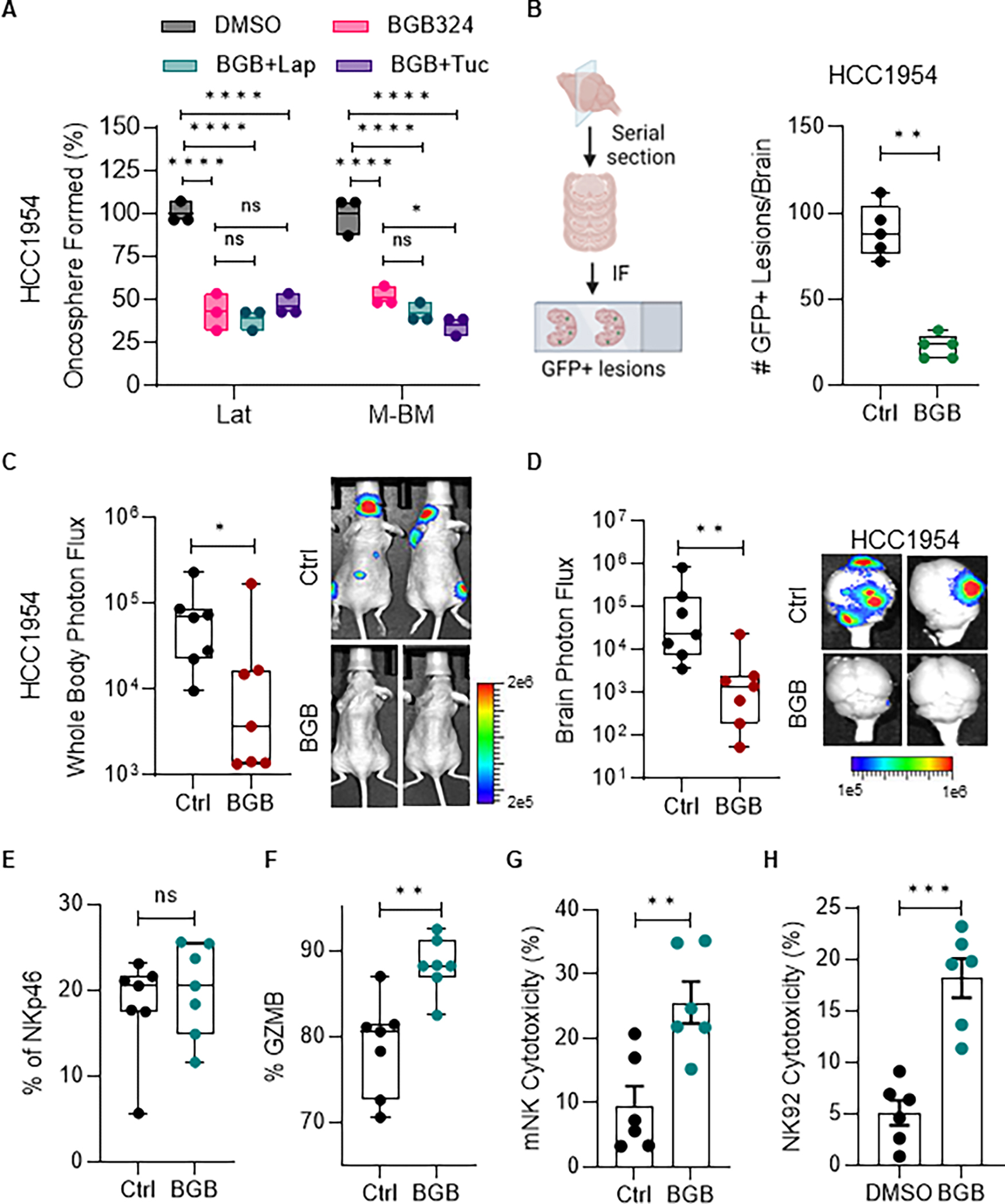
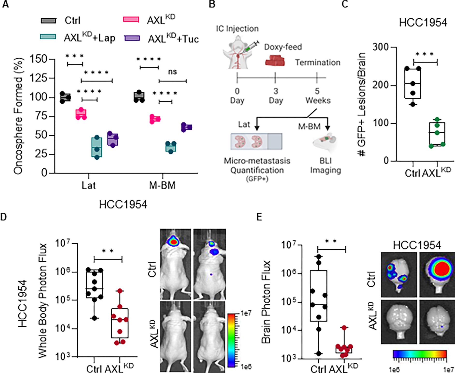
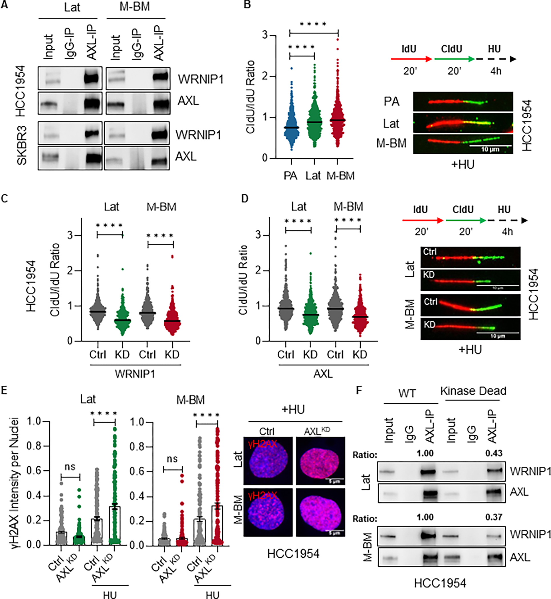
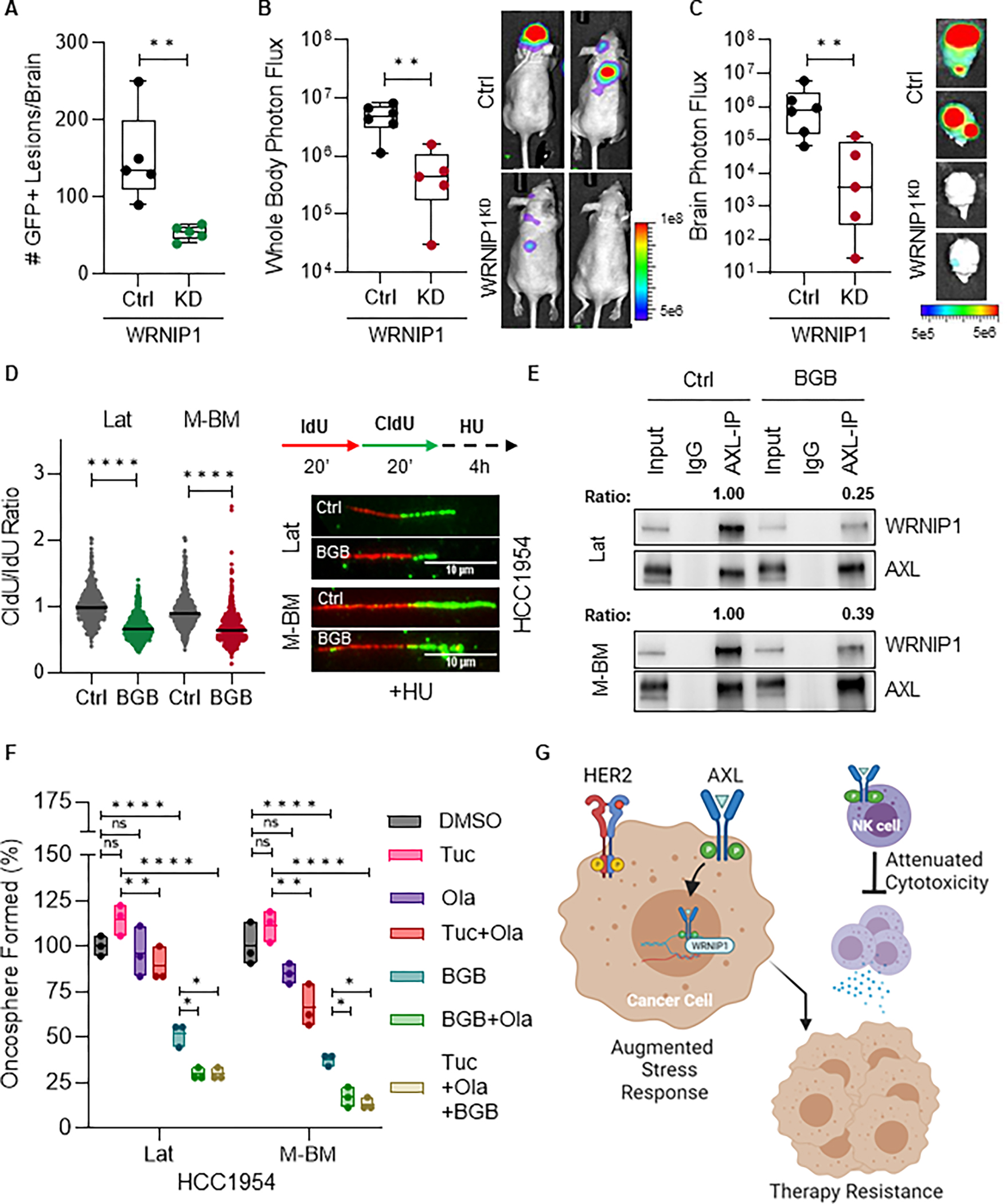
Therapy resistance and metastatic progression are primary causes of cancer-related mortality. Disseminated tumor cells possess adaptive traits that enable them to reprogram their metabolism, maintain stemness, and resist cell death, facilitating their persistence to drive recurrence. The survival of disseminated tumor cells also depends on their ability to modulate replication stress in response to therapy while colonizing inhospitable microenvironments. In this study, we discovered that the nuclear translocation of AXL, a TAM receptor tyrosine kinase, and its interaction with WRNIP1, a DNA replication stress response factor, promotes the survival of HER2+ breast cancer cells that are resistant to HER2-targeted therapy and metastasize to the brain. In preclinical models, knocking down or pharmacologically inhibiting AXL or WRNIP1 attenuated protection of stalled replication forks. Furthermore, deficiency or inhibition of AXL and WRNIP1 also prolonged metastatic latency and delayed relapse. Together, these findings suggest that targeting the replication stress response, which is a shared adaptive mechanism in therapy-resistant and metastasis-initiating cells, could reduce metachronous metastasis and enhance the response to standard-of-care therapies.

Significance: Nuclear AXL and WRNIP1 interact and mediate replication stress response, promote therapy resistance, and support metastatic progression, indicating that targeting the AXL/WRNIP1 axis is a potentially viable therapeutic strategy for breast cancer.


Conflict of interest statement

Authors declare no potential conflicts of interest.

Figures

Figure 1.
Figure 1.. Nuclear AXL is enriched in HER2+ therapy resistance latent and metachronous HER2+ breast cancer brain metastatic cells.
A, Relative quantification of oncospheres formed by HCC1954 PA, S-BM, Lat and M-BM cells seven days post-treatment with Tucatinib (3μM) and Lapatinib (1μM) compared to DMSO control. Data are presented as mean ± SEM. One-way ANOVA was used, followed by the Dunnett test. ****, P < 0.0001. ns: not significant. B, Whole body and ex-vivo brain metastasis incidence as measured by bioluminescence imaging (BLI) signal from athymic nude mice bearing HCC1954 M-BM cells that were intracardially injected. Vehicle (Ctrl) and Tucatinib (Tuc, 50mg/kg, once a day) was administrated orally 3 days post-injection daily up to 5 weeks. Control (n = 12) and Tucatinib (n = 9). Data are presented as mean ± SEM. Mann-Whitney U test, ns: not significant. Two representative BLI images of whole-body mice and ex-vivo brains of their respective conditions. C-E, AXL immunohistochemistry (IHC) in matched primary tumors and brain metastatic lesions (n = 7) from HER2+ breast cancer patients. (C). Representative AXL IHC images of matched primary and brain metastatic lesions from HER2+ breast cancer patients. (D). Histology-score analysis of AXL IHC. Data are presented as mean ± SEM. Paired Student t test. **, P < 0.01 (E). Representative images from patient-5 (P5) of Figure 1C highlighting nuclear AXL expression and quantification of AXL nuclear to non-nuclear ratio in matched primary and brain metastatic lesions. Data are presented as mean ± SEM. Unpaired Student t test. ****, P < 0.001. PT: primary tumor. Nuc.: nuclear. F, Representative images of AXL IHC on brain and spinal metastatic lesions in mice bearing HCC1954 Lat and M-BM cells. G, Immunoblot analysis of subcellular fractions from HCC1954 Lat and M-BM cells showing AXL localization and purity of subcellular fractions. GAPDH (CE, cytoplasmic extract), TFRC (ME, membrane extract), Lamin-B1 (NE, soluble nuclear extract) and Histone-3 (CB, chromatic bound extract).
Figure 2.
Figure 2.. AXL inhibition attenuates brain metastasis and enhances NK cytotoxicity.
A, Relative quantification of oncospheres formed by HCC1954 Lat and M-BM cells treated with BGB324 (BGB, 1μM) alone or in combination with Lapatinib (Lap, 1μM) and Tucatinib (Tuc, 3μM) for seven days. Data are presented as mean ± SEM. One-way ANOVA was used, followed by the Dunnett test. *, P < 0.05, ****, P < 0.0001, ns: not significant. B, Illustration showing brain tissue processing to quantify GFP+ lesions. Quantification of GFP+ cells/lesions from brains of mice bearing HCC1954 Lat cells (n = 5 in both groups). C-D, Whole body and ex-vivo brain metastasis incidence as measured by BLI signal from athymic nude mice bearing HCC1954 M-BM cells. BGB324 (BGB, 50mg/kg, BID) or vehicle (Ctrl) was administrated orally 3 days post-injection for 5 weeks (n = 7 in both groups). Data are presented as mean ± SEM. Mann–Whitney U test, *, P < 0.05, **, P < 0.01. Two representative BLI images of whole-body mice and ex-vivo brains of their respective conditions. E-F, Quantification of NK cells (NKp46) and cytotoxic NK cells (GZMB) in peripheral blood collected from mice experiment in Figure 2C–D. Data are presented as mean ± SEM. Unpaired Student t test. ns: not significant. G, Quantification of mouse NK cell cytotoxicity against HCC1954 PA cells. Mouse NK cells were isolated from spleens of mice treated with vehicle or BGB324 in figure 2C–D. Data are presented as mean ± SEM. Unpaired Student t test. **, P < 0.01. H, Quantification of human NK cell (NK92) cytotoxicity after BGB324 (1μM) treatment on HCC1954 PA cells. Data are presented compared to DMSO (Ctrl) as mean ± SEM. Unpaired Student t test. ***, P < 0.001. (B, Created with BioRender.com).
Figure 3.
Figure 3.. AXL depletion attenuates latent and metachronous brain metastasis.
A, Relative quantification of oncospheres formed by HCC1954 Lat and M-BM cells upon AXL knockdown (AXLKD) and Lapatinib (1μM) or Tucatinib (3μM) treatment for seven days. Data are presented as mean ± SEM. One-way ANOVA was used, followed by the Dunnett test. *, P < 0.05, ***, P < 0.001, ****, P < 0.0001, ns: not significant. B, Experimental scheme to assess the impact of AXL depletion on metastatic latency and relapse. C, Quantification of GFP+ brain metastatic lesions in mice bearing HCC1954 AXLKD Lat cells (n = 5 per group). Mice were on doxycycline feed 3 days post injection until study termination. Data are presented as mean ± SEM. Mann–Whitney U test, ***, P < 0.001. D-E, Whole body and ex-vivo brain metastasis incidence as measured by BLI signal from athymic nude mice bearing HCC1954 AXLKD M-BM cells. (Ctrl group n = 9 and AXLKD group n = 8). Mice were on doxycycline feed 3 days post injection until study termination. Data are presented as mean ± SEM. Mann–Whitney U test, **, P < 0.01. Two representative BLI images of whole-body mice and ex-vivo brains of their respective conditions. (B, Created with BioRender.com).
Figure 4.
Figure 4.. AXL-WRNIP1 interaction mediates replication stress response in Lat and M-BM cells.
A, Immunoblot showing AXL-WRNIP1 interaction in HCC1954 Lat and M-BM cells under normal (Ctrl) and replication stress induced condition (in presence of 4mM HU for 5hrs). B, Relative fork degradation measured by the CldU/IdU ratio in presence of 4mM HU. Representative images of DNA fibers from HCC1954 PA, Lat and M-BM cells are also shown. IdU (red color) and CldU (green color). N = 3 for all experiments. Data are presented as mean ± SEM. Mann–Whitney U test, ****, P < 0.0001. C-D, Relative fork degradation measured by the CldU/IdU ratio in presence of 4mM HU upon WRNIP1 knockdown (WRNIP1KD) and AXL knockdown (AXLKD) in HCC1954 Lat and M-BM cells. Representative images of DNA fibers are also shown. IdU (red color) and CldU (green color). N = 3 for all experiments. Data are presented as mean ± SEM. Mann–Whitney U test, ****, P < 0.0001. E, γH2AX intensity quantification of HCC1954 Lat and M-BM cells treated with 4mM HU for 5hrs. Representative image of γH2AX intensity under HU. Data are presented as mean ± SEM. One-way ANOVA was used, followed by the Dunnett test. ****, P < 0.001, ns: not significant. F, Immunoblot showing reduced interaction between kinase dead AXL and WRNIP1. This assay was performed by reintroducing AXL (WT) and kinase dead mutant in HCC1954 AXLKD Lat and M-BM cells. AXL immunoprecipitation was performed and ratio was determined by normalizing WRNIP1 expression to immune-precipitated AXL.
Figure 5.
Figure 5.. Augmenting replication stress attenuates metastatic latency and recurrence
A, Quantification of GFP+ metastatic lesions in mice bearing HCC1954 WRNIP1 knockdown (WRNIP1KD) Lat cells (n = 5 per group). Doxycycline feed was given 3 days post injection and continued till study termination. Data are presented as mean ± SEM. Mann–Whitney U test, ***, P < 0.001. B-C, Whole body and ex-vivo brain metastasis incidence as measured by BLI signal from athymic nude mice bearing HCC1954 WRNIP1KD M-BM cells. (Ctrl group n = 6 and WRNIP1KD group n = 5). Doxycycline feed was given 3 days post injection and continued till study termination. Data are presented as mean ± SEM. Mann–Whitney U test, **, P < 0.01. Two representative BLI images of whole-body mice and ex-vivo brains of their respective conditions. D, Relative fork degradation measured by the CldU/IdU ratio in presence of 4mM HU upon BGB324 treatment in HCC1954 Lat and M-BM cells. Representative images of DNA fibers are also shown. IdU (red color) and CldU (green color). N = 3 for all experiments. Data are presented as mean ± SEM. Mann–Whitney U test, ****, P < 0.0001. E, Immunoblot showing reduced AXL-WRNIP1 interaction in the presence of BGB324 (1μM for 5 hours). Ratio was determined by normalizing WRNIP1 expression to immunoprecipitated AXL. F, Relative quantification of oncospheres formed by HCC1954 Lat and M-BM cells treated with DMSO, BGB324 (BGB: 1μM), Olaparib (Ola: 5μM), Tucatinib (Tuc: 3μM) and combinations. Data are presented as mean ± SEM. One-way ANOVA was used, followed by the Dunnett test. *, P < 0.05, **, P < 0.01, ****, P < 0.0001, ns: not significant. G, Graphical abstract summarizing key findings of AXL-WRNIP1 mediated replication stress response promoting latent and metachronous metastasis. (G, Created with BioRender.com).

References

    1. Kim K, Marquez-Palencia M, Malladi S. Metastatic Latency, a Veiled Threat. Front Immunol 2019;10:1836. - PMC - PubMed
    1. Lambert AW, Pattabiraman DR, Weinberg RA. Emerging Biological Principles of Metastasis. Cell 2017;168:670–91 - PMC - PubMed
    1. Massague J, Ganesh K. Metastasis-Initiating Cells and Ecosystems. Cancer Discov 2021;11:971–94 - PMC - PubMed
    1. Pantel K, Hayes DF. Disseminated breast tumour cells: biological and clinical meaning. Nat Rev Clin Oncol 2018;15:129–31 - PubMed
    1. Peitzsch C, Tyutyunnykova A, Pantel K, Dubrovska A. Cancer stem cells: The root of tumor recurrence and metastases. Semin Cancer Biol 2017;44:10–24 - PubMed

Publication types

MeSH terms

Substances