. 2024 Jan 8;12(1):e008086.
doi: 10.1136/jitc-2023-008086.

Clinically relevant orthotopic pancreatic cancer models for adoptive T cell transfer therapy

Affiliations

Clinically relevant orthotopic pancreatic cancer models for adoptive T cell transfer therapy

Natalie K Horvat et al. J Immunother Cancer. .

Abstract

Background: Pancreatic ductal adenocarcinoma (PDAC) is an aggressive tumor. Prognosis is poor and survival is low in patients diagnosed with this disease, with a survival rate of ~12% at 5 years. Immunotherapy, including adoptive T cell transfer therapy, has not impacted the outcomes in patients with PDAC, due in part to the hostile tumor microenvironment (TME) which limits T cell trafficking and persistence. We posit that murine models serve as useful tools to study the fate of T cell therapy. Currently, genetically engineered mouse models (GEMMs) for PDAC are considered a "gold-standard" as they recapitulate many aspects of human disease. However, these models have limitations, including marked tumor variability across individual mice and the cost of colony maintenance.

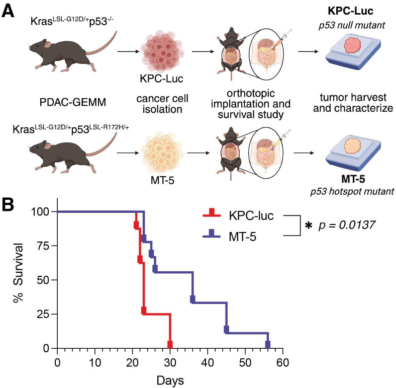
Methods: Using flow cytometry and immunohistochemistry, we characterized the immunological features and trafficking patterns of adoptively transferred T cells in orthotopic PDAC (C57BL/6) models using two mouse cell lines, KPC-Luc and MT-5, isolated from C57BL/6 KPC-GEMM (KrasLSL-G12D/+p53-/- and KrasLSL-G12D/+p53LSL-R172H/+, respectively).

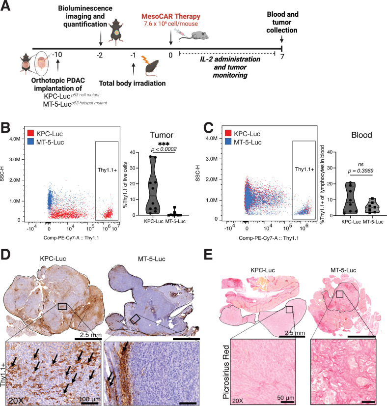
Results: The MT-5 orthotopic model best recapitulates the cellular and stromal features of the TME in the PDAC GEMM. In contrast, far more host immune cells infiltrate the KPC-Luc tumors, which have less stroma, although CD4+ and CD8+ T cells were similarly detected in the MT-5 tumors compared with KPC-GEMM in mice. Interestingly, we found that chimeric antigen receptor (CAR) T cells redirected to recognize mesothelin on these tumors that signal via CD3ζ and 41BB (Meso-41BBζ-CAR T cells) infiltrated the tumors of mice bearing stroma-devoid KPC-Luc orthotopic tumors, but not MT-5 tumors.

Conclusions: Our data establish for the first time a reproducible and realistic clinical system useful for modeling stroma-rich and stroma-devoid PDAC tumors. These models shall serve an indepth study of how to overcome barriers that limit antitumor activity of adoptively transferred T cells.

Keywords: CD4-CD8 ratio; T-lymphocytes; receptors, chimeric antigen; receptors, immunologic; tumor microenvironment.


Conflict of interest statement

Competing interests: GBL has consulted for ProDa Biotech and received compensation. GBL has also received research funding through a sponsored research agreement between Emory University and Merck and Co, Bristol-Myers Squibb, Boehringer Ingelheim, and Vaccinex. CP has received research funding through a sponsored research agreement between the Medical University of South Carolina and Obsidian, Lycera, and ThermoFisher, and is the cofounder of Ares Immunotherapy.

Figures

Figure 1
Figure 1
Orthotopic PDAC tumors recapitulate the TME of GEMM tumors. (A) Experimental schematic of cell isolation and orthotopic longitudinal survival study. Immortalized cell lines were isolated from KPC-GEMMs. (B) Survival kinetics of immunocompetent mice bearing KPC-Luc or MT-5 orthotopic PDAC tumors (n=10 per group). One-way ANOVA: *p<0.05. ANOVA, analysis of variance; GEMM, genetically engineered mouse model; PDAC, pancreatic ductal adenocarcinoma; TME, tumor microenvironment.
Figure 2
Figure 2
Orthotopic MT-5 PDAC tumors parallel the immune landscape of KPC-GEMM tumors. (A) Whole tissue (left) and 20× magnification (right) of H&E-stained FFPE KPC-GEMM (left), KPC-Luc (middle), and MT-5 (right) tumor sections harvested at endpoint. (B) Picrosirius red and (C) ɑ-SMA immunohistochemistry staining of KPC-GEMM (left), KPC-Luc (middle), and MT-5 (right) FFPE tumor sections. Quantification of stromal fibrosis by picrosirius red (D) and myofibroblastic positive stain by ɑ-SMA (E) as measured by per cent positive cells of the total tumor area. The activated stroma index (F) was calculated by dividing the values of the ɑ-SMA quantification by the picrosirius red values. (G) Representative images of CD4+ (red) and CD8+ (green) T cell immunofluorescence on whole tissue sections of KPC-GEMM, KPC-Luc, and MT-5 tumors. Quantification of CD4+ or CD8+ T cell infiltration (below) (% positive cells out of the total cell count in the tumor area). Percentage of CD45+ cells (H), percentage of CD3+, Ly6G+CD11b+, and Ly6C+ out of the total CD45+ cells (I), and quantification of dendritic cells (DCs), monocytes, tumor-associated macrophages (TAMs), and monocytic myeloid-derived suppressor cells (M-MDSCs) out of the total Ly6C+ population (J) from KPC-Luc and MT-5 tumors by flow cytometry. Kruskal-Wallis (D, E, F, G, H) or one-way ANOVA (J) test: *p<0.05, **p<0.01, ***p<0.001. α-SMA, alpha-smooth muscle actin; ANOVA, analysis of variance; FFPE, formalin-fixed paraffin-embedded; GEMM, genetically engineered mouse model; PDAC, pancreatic ductal adenocarcinoma.
Figure 3
Figure 3
Trp53 hotspot mutant tumor mice reduce CAR T cell infiltration in mice. (A) Study schematic. C57BL/6 mice were implanted with either KPC-Luc or MT-5-Luc PDAC tumors (n=10/cell line), and after 10 days CD3+ mesoCAR T cell treatment was administered for 7 days. Tumors were harvested for either histological or flow cytometry analysis. Quantification of donor CAR T cells infiltrating the tumor tissue (B) or circulating in blood (C) as measured by flow cytometry. Data represented as percentage of Thy1.1 cells out of the total live tumor cells (left) and as percentage of Thy1.1 cells out of the total blood lymphocytes (right). (D) IHC representative images of Thy1.1+ stain in whole tissue and 20× magnification of KPC-Luc and MT-5-Luc FFPE tumor sections. Black arrows indicate positive Thy1.1 cells found in the tumor parenchyma (left) or in the tumor periphery (right). (E) Representative images of picrosirius red staining. Mann-Whitney test: ***p<0.001. CAR, chimeric antigen receptor; FFPE, formalin-fixed paraffin-embedded; IHC, immunohistochemistry; IL-2, interleukin-2; PDAC, pancreatic ductal adenocarcinoma; SSC, side scatter.

References

    1. Cancer facts & figures. American Cancer Society, Available: https://www.cancer.org/research/cancer-facts-statistics/all-cancer-facts...
    1. Rahib L, Smith BD, Aizenberg R, et al. . Projecting cancer incidence and deaths to 2030: the unexpected burden of thyroid, liver, and Pancreas cancers in the United States. Cancer Res 2014;74:2913–21. 10.1158/0008-5472.CAN-14-0155 - DOI - PubMed
    1. Ansari D, Gustafsson A, Andersson R. Update on the management of Pancreatic cancer: surgery is not enough. World J Gastroenterol 2015;21:3157–65. 10.3748/wjg.v21.i11.3157 - DOI - PMC - PubMed
    1. Grossberg AJ, Chu LC, Deig CR, et al. . Multidisciplinary standards of care and recent progress in Pancreatic Ductal adenocarcinoma. CA Cancer J Clin 2020;70:375–403. 10.3322/caac.21626 - DOI - PMC - PubMed
    1. Foley K, Kim V, Jaffee E, et al. . Current progress in Immunotherapy for Pancreatic cancer. Cancer Lett 2016;381:244–51. 10.1016/j.canlet.2015.12.020 - DOI - PMC - PubMed

Publication types

Substances