. 2024 Jan 9;14(1):853.
doi: 10.1038/s41598-023-50932-5.

Understanding the molecular mechanism of pathogenic variants of BIR2 domain in XIAP-deficient inflammatory bowel disease

Affiliations

Understanding the molecular mechanism of pathogenic variants of BIR2 domain in XIAP-deficient inflammatory bowel disease

Juhwan Lee et al. Sci Rep. .

Abstract

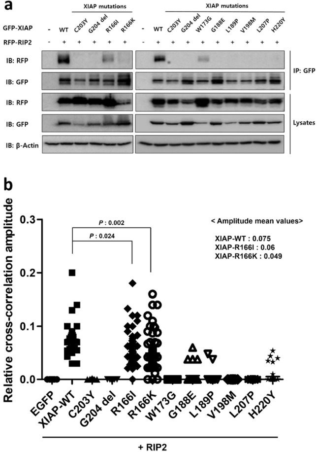
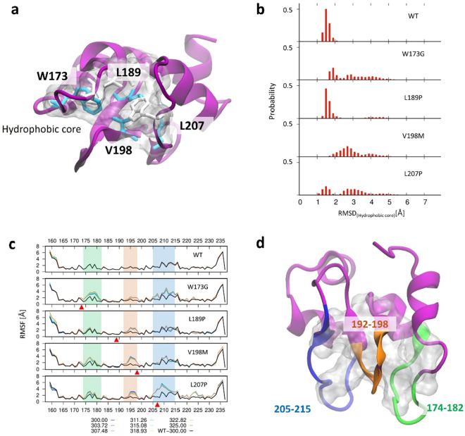
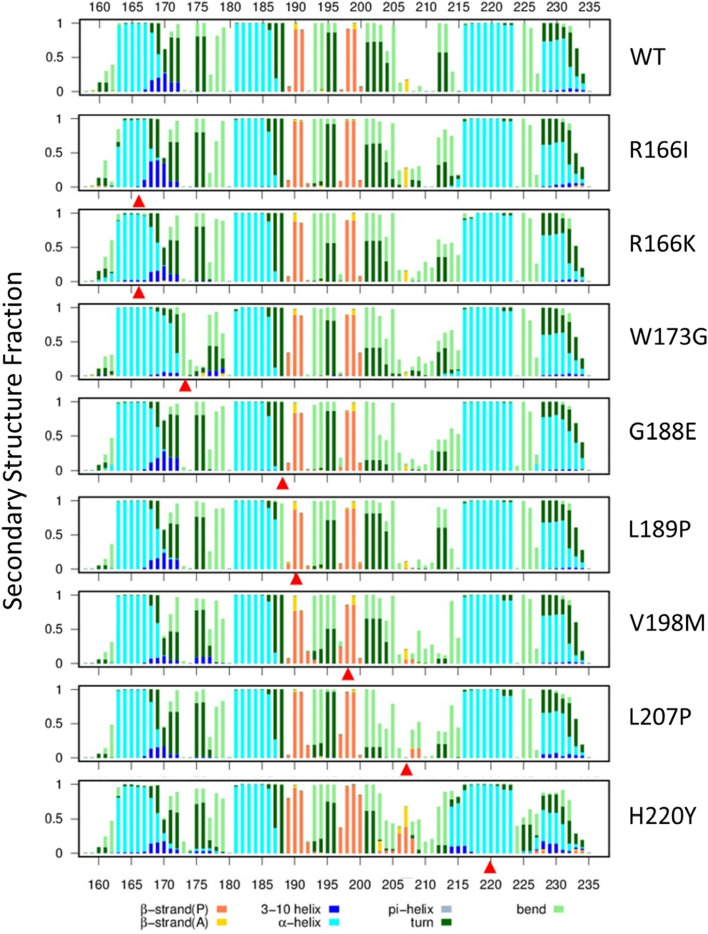
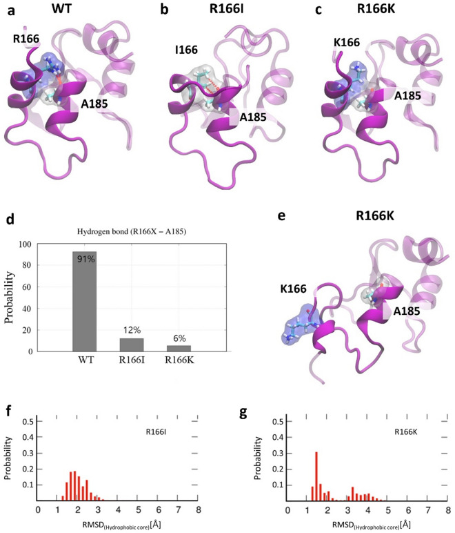
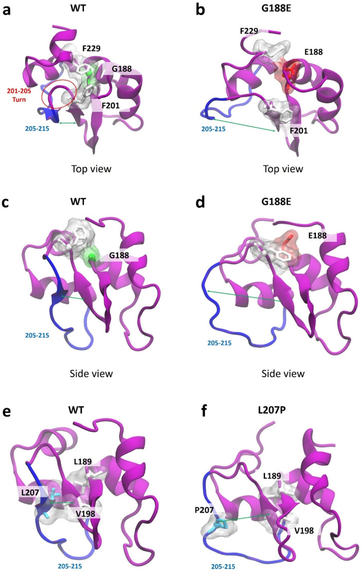
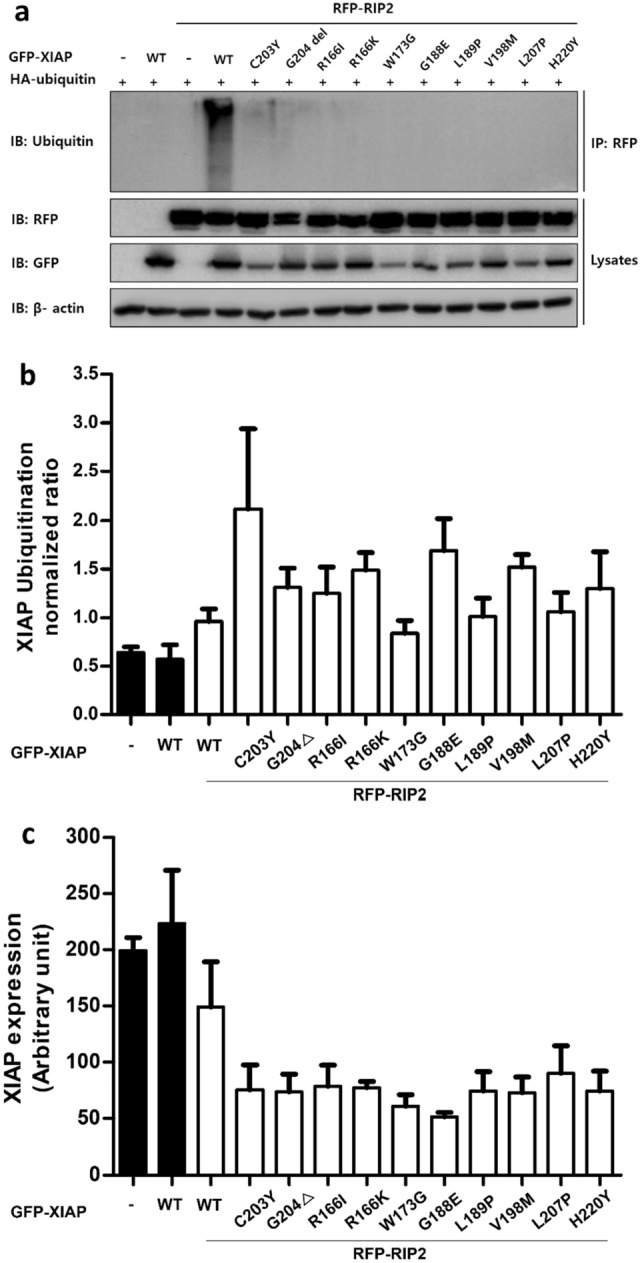
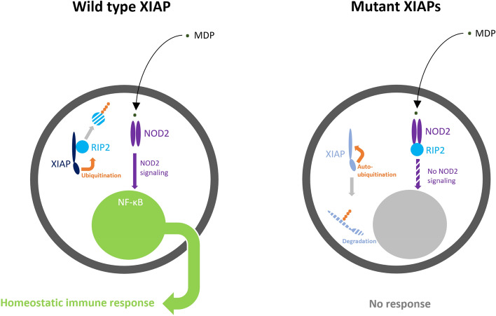
X-linked inhibitor of apoptosis protein (XIAP) deficiency causes refractory inflammatory bowel disease. The XIAP protein plays a pivotal role in the pro-inflammatory response through the nucleotide-binding oligomerization domain-containing signaling pathway that is important in mucosal homeostasis. We analyzed the molecular mechanism of non-synonymous pathogenic variants (PVs) of XIAP BIR2 domain. We generated N-terminally green fluorescent protein-tagged XIAP constructs of representative non-synonymous PVs. Co-immunoprecipitation and fluorescence cross-correlation spectroscopy showed that wild-type XIAP and RIP2 preferentially interacted in live cells, whereas all non-synonymous PV XIAPs failed to interact properly with RIP2. Structural analysis showed that various structural changes by mutations, such as hydrophobic core collapse, Zn-finger loss, and spatial rearrangement, destabilized the two loop structures (174-182 and 205-215) that critically interact with RIP2. Subsequently, it caused a failure of RIP2 ubiquitination and loss of protein deficiency by the auto-ubiquitination of all XIAP mutants. These findings could enhance our understanding of the role of XIAP mutations in XIAP-deficient inflammatory bowel disease and may benefit future therapeutic strategies.


Conflict of interest statement

The authors declare no competing interests.

Figures

Figure 1
Figure 1
Mutant XIAP BIR2 with non-synonymous PVs failed to interact with RIP2. (a) Co-IP of GFP-tagged XIAP WT or mutants, and RFP-tagged RIP2 confirmed their interaction. The tagged XIAP was immunoprecipitated from lysates of HEK 293 T cells transfected with plasmids encoding the indicated GFP-tagged XIAP WT or mutants, and western blot images were cropped for clarification. (b) Comparison of relative correlation amplitudes of live HEK 293 T cells expressing the indicated XIAP mutants. The relative correlation amplitudes between GFP–XIAP mutants and RFP-RIP2 were determined by confocal imaging during FCCS. The negative control indicates a mean value of relative correlation amplitude of 0, obtained from cells co-expressing EGFP and RIP2. The mean amplitudes of XIAP mutants were significantly lower than that of the positive control (XIAP-WT + RIP2).
Figure 2
Figure 2
Effect of W173G, L189P, V198M, and L207P mutation on the hydrophobic core. (a) Major hydrophobic core of BIR2 domain of XIAP and hydrophobic amino acids members (BIR2: purple cartoon, Hydrophobic core: white transparent surface, Mutated amino acids: cyan sticks, other amino acids: white sticks) (b) The root mean square deviation (RMSD) of BIR2- WT and mutants hydrophobic cores. (c) The root mean square fluctuation (RMSF) of BIR2- WT and mutants. More fluctuated regions than WT are highlighted in green, orange, and blue. (d) XIAP-BIR2 cartoon structure (purple) and fluctuated regions (green, orange, yellow) by mutations.
Figure 3
Figure 3
Secondary structure fraction of WT and mutant BIR2 of XIAP protein. Secondary structure fraction of WT and eight mutants over the amino acid count. Mutation points are pointed using red arrows.
Figure 4
Figure 4
Effect of R166I and R166K mutations on the two alpha-helices. (a) Representative structure of WT BIR2, R166, and A185 form a strong hydrogen bond. (b) Representative structure of BIR2-R166I mutant. I166 and A185 form a weak hydrogen bond. (c) Representative structure of BIR2-R166K mutant. K166 and A185 form a weak hydrogen bond. (d) Hydrogen bond formation probability between amino acid on 166 and A185. (e) Structural change of R166K between two helices. (f) Root mean square deviation (RMSD) of BIR2-R116I hydrophobic core. (g) The RMSD of BIR2-R166K hydrophobic core.
Figure 5
Figure 5
Effect of G188E and L207P mutations on beta structure. (a) BIR2-WT structure. Red circle indicates a turn structure between two beta structures. (b) BIR2-G188E structure. (c) BIR2-WT structure from a different angle. (d) BIR2-G188E structure from a different angle. (e) BIR2-WT structure from a different angle. (f) BIR2-L207P structure from a different angle. Two beta structures were also separated by L207P mutation.
Figure 6
Figure 6
Analysis of XIAP-mediated ubiquitination of XIAP and RIP2. (a) Ub conjugates were purified with Protein A or G magnetic beads from lysates of HEK 293 T cells transfected as indicated and examined by immunoblotting. RIP2 ubiquitination was defective in HEK 293 T cells for all XIAP mutants. (b) XIAP expression was analyzed in HEK 293 T cells transfected GFP-tagged XIAP WT or mutant constructs, along with RFP-tagged RIP2 by LC–MS. XIAP mutants expression levels were significantly lower compared to XIAP WT expression level. (c) XIAP ubiquitination was analyzed in HEK 293 T cells transfected with GFP-tagged XIAP WT or mutant constructs, along with RFP-tagged RIP2 by MS. Auto-ubiquitination of XIAP mutants were significantly higher compared to that of XIAP WT.
Figure 7
Figure 7
Pathogenic mechanisms of nonsynonymous mutant XIAP BIR2s. Our findings suggest a novel concept of a two-step mechanism in the development of XIAP deficiency caused by nonsynonymous pathogenic variants. The first step is a binding failure of mutant XIAPs to RIP2, resulting in an inability to interact with RIP2 and subsequently ubiquitinate RIP2. Given that NOD2 signaling can be activated in response to intracellular pathogens by ubiquitinating RIP2, a binding failure to RIP2 would be the first mechanism to evoke the XIAP-deficient phenomenon. The second step is the auto-ubiquitination of mutant XIAPs, which results in a relative deficiency of these mutant proteins. Traditionally, true XIAP deficiency is, in a strict sense, defined in cases of null variants, such as nonsense mutations, large insertions/deletions in exons, and critical splicing mutations. In cases of nonsynonymous pathogenic variants, previously unrecognized, mutant XIAPs also result in another type of XIAP protein deficiency, possibly contributing a synergistic impact on defective NOD2 signaling.

Similar articles

References

    1. Rigaud S, et al. XIAP deficiency in humans causes an X-linked lymphoproliferative syndrome. Nature. 2006;444:110–114. doi: 10.1038/nature05257. - DOI - PubMed
    1. Zeissig Y, et al. XIAP variants in male Crohn's disease. Gut. 2015;64:66–76. doi: 10.1136/gutjnl-2013-306520. - DOI - PubMed
    1. Pedersen J, LaCasse EC, Seidelin JB, Coskun M, Nielsen OH. Inhibitors of apoptosis (IAPs) regulate intestinal immunity and inflammatory bowel disease (IBD) inflammation. Trends Mol. Med. 2014;20:652–665. doi: 10.1016/j.molmed.2014.09.006. - DOI - PubMed
    1. Gérart S, et al. Human iNKT and MAIT cells exhibit a PLZF-dependent proapoptotic propensity that is counterbalanced by XIAP. Blood. 2013;121:614–623. doi: 10.1182/blood-2012-09-456095. - DOI - PMC - PubMed
    1. Damgaard RB, et al. Disease-causing mutations in the XIAP BIR2 domain impair NOD2-dependent immune signalling. EMBO Mol. Med. 2013;5:1278–1295. doi: 10.1002/emmm.201303090. - DOI - PMC - PubMed

MeSH terms

Substances