

Sex-dependent effect of sublethal copper concentrations on de novo cholesterol synthesis in astrocytes and their possible links to variations in cholesterol and amyloid precursor protein levels in neuronal membranes
- PMID: 38191520
- PMCID: PMC10775608
- DOI: 10.1186/s13293-023-00578-9
Sex-dependent effect of sublethal copper concentrations on de novo cholesterol synthesis in astrocytes and their possible links to variations in cholesterol and amyloid precursor protein levels in neuronal membranes
Abstract
Background: Cholesterol (Cho) is an essential lipophilic molecule in cells; however, both its decrease and its increase may favor the development of neurological diseases such as Alzheimer's disease (AD). Although copper (Cu) is an essential trace metal for cells, the increased plasma concentration of its free form has been linked with AD development and severity. AD affects aged people, but its prevalence and severity are higher in women than in men. We have previously shown that Cu promotes Cho de novo synthesis in immature neurons as well as increased Cho in membrane rafts and Aβ levels in culture medium, but there are no results yet regarding sex differences in the effects of sublethal Cu exposure on Cho de novo synthesis.
Methods: We examined the potential sex-specific impact of sublethal Cu concentrations on de novo Cho synthesis in primary cultures of male and female astrocytes. We also explored whether this had any correlation with variations in Cho and APP levels within neuronal membrane rafts.
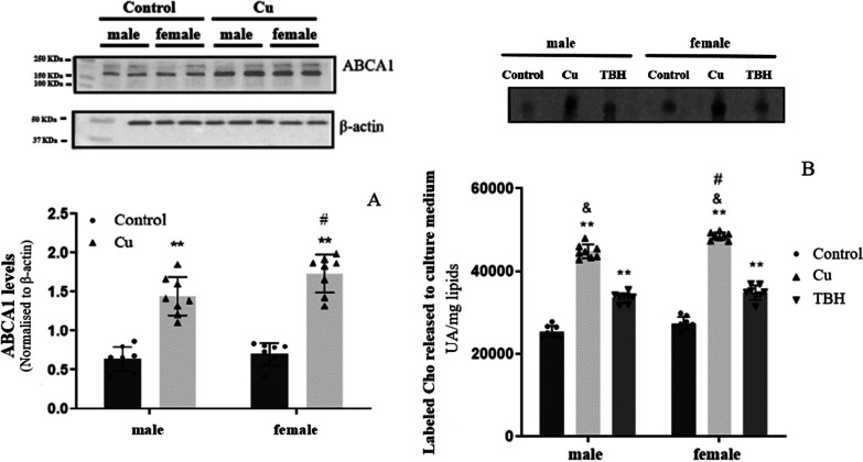
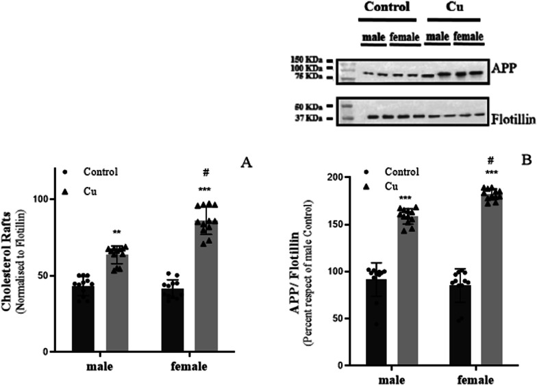
Results: Flow cytometry analysis demonstrated that Cu treatment leads to a greater increase in ROS levels in female astrocytes than in males. Furthermore, through RT-PCR analysis, we observed an upregulation of SREBP-2 and HMGCR. Consistently, we observed an increase in de novo Cho synthesis. Finally, western blot analysis indicated that the levels of ABCA1 increase after Cu treatment, accompanied by a higher release of radiolabeled Cho and an elevation in Cho and APP levels in neuronal membrane rafts. Importantly, all these results were significantly more pronounced in female astrocytes than in males.
Conclusions: Our findings confirm that Cu stimulates Cho synthesis in astrocytes, both in a ROS-dependent and -independent manner. Moreover, female astrocytes displayed elevated levels of HMGCR, and de novo Cho synthesis compared to males following TBH and Cu treatments. This corresponds with higher levels of Cho released into the culture medium and a more significant Cho and APP rise within neuronal rafts. We consider that the increased risk of AD in females partly arises from sex-specific responses to metals and/or exogenous substances, impacting key enzyme regulation in various biochemical pathways, including HMGCR.
Keywords: Astrocytes; Cholesterol; Copper; Neurons; Sexual differences.
Plain language summary
Alzheimer’s disease (AD) primarily affects the elderly and is linked to excess cholesterol (Cho) and copper (Cu). It is more prevalent and severe in women. Previous research suggested that Cu may enhance Cho synthesis in developing neurons, raising Cho levels in specialized membrane structures (rafts) and Aβ protein in the culture medium. However, the specific effects of Cu exposure on Cho synthesis in males and females are not entirely understood. We conducted experiments using astrocytes, the primary cells in the brain that produce Cho in adults, and neurons, both from male and female rats. We exposed them to non-lethal levels of Cu to explore its potential sex-related effects on (1) Cho metabolism in astrocytes, and (2) The relationship between the Cho released by astrocytes and the levels of Cho and amyloid precursor protein (APP) in neuronal membrane rafts. Our findings suggested that reactive oxygen species (ROS)-responsive sensitivity is higher in females than in male astrocytes. Cu, alongside ROS, promoted Cho synthesis, with female astrocytes being more susceptible. These released more Cho into the medium after Cu exposure, and Cho and APP levels were also higher in female neuronal rafts exposed to Cu-treated astrocyte-conditioned medium. Our results thus imply that the higher risk of AD in females may arise partly from sex-related disparities in cellular responses to external substances, impacting such crucial biochemical pathways as Cho synthesis.
© 2024. The Author(s).
Conflict of interest statement
All authors disclose any financial and personal relationships with other people or organizations that could inappropriately influence this work. The authors declare that there are no conflicts of interest.
Figures


References
MeSH terms
Substances
Grants and funding
LinkOut - more resources
Full Text Sources
Medical
