. 2024 Jan 8;14(1):828.
doi: 10.1038/s41598-023-50667-3.

Validated assays for the quantification of C9orf72 human pathology

Affiliations

Validated assays for the quantification of C9orf72 human pathology

S E Salomonsson et al. Sci Rep. .

Abstract

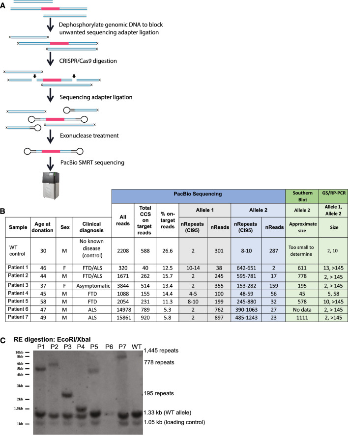
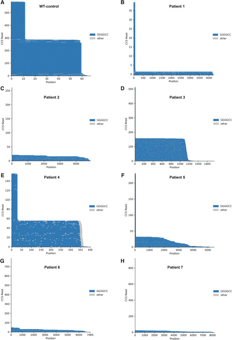
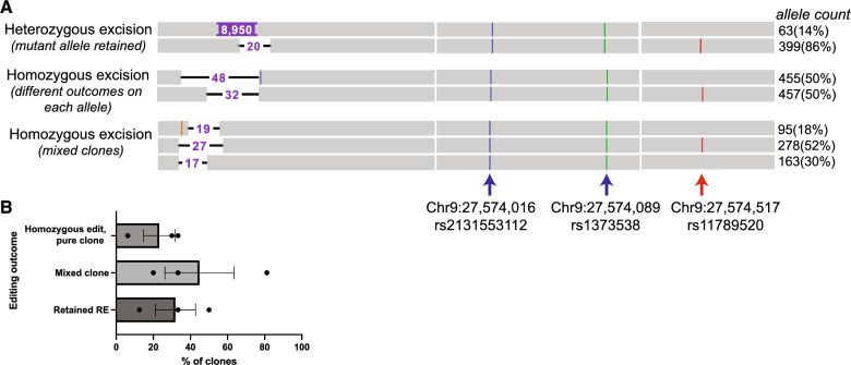
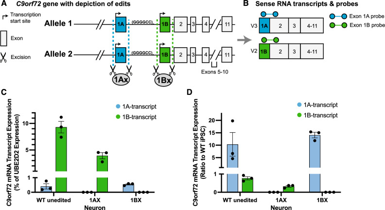
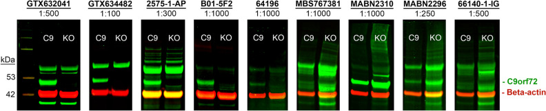
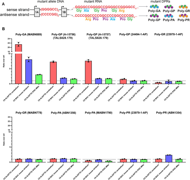
A repeat expansion mutation in the C9orf72 gene is the leading known genetic cause of FTD and ALS. The C9orf72-ALS/FTD field has been plagued by a lack of reliable tools to monitor this genomic locus and its RNA and protein products. We have validated assays that quantify C9orf72 pathobiology at the DNA, RNA and protein levels using knock-out human iPSC lines as controls. Here we show that single-molecule sequencing can accurately measure the repeat expansion and faithfully report on changes to the C9orf72 locus in what has been a traditionally hard to sequence genomic region. This is of particular value to sizing and phasing the repeat expansion and determining changes to the gene locus after gene editing. We developed ddPCR assays to quantify two major C9orf72 transcript variants, which we validated by selective excision of their distinct transcriptional start sites. Using validated knock-out human iPSC lines, we validated 4 commercially available antibodies (of 9 tested) that were specific for C9orf72 protein quantification by Western blot, but none were specific for immunocytochemistry. We tested 15 combinations of antibodies against dipeptide repeat proteins (DPRs) across 66 concentrations using MSD immunoassay, and found two (against poly-GA and poly-GP) that yielded a 1.5-fold or greater signal increase in patient iPSC-motor neurons compared to knock-out control, and validated them in human postmortem and transgenic mouse brain tissue. Our validated DNA, RNA and protein assays are applicable to discovery research as well as clinical trials.


Conflict of interest statement

B.R.C. is a founder of Tenaya Therapeutics (https://www.tenayatherapeutics.com/), a company focused on finding treatments for heart failure, including genetic cardiomyopathies. B.R.C. holds equity in Tenaya. C.D.C. is a founder, with equity, in Ciznor Co., a CNS therapeutics company. I.M and Y-C.T. are full-time employees at Pacific Biosciences (PacBio), a company developing single-molecule sequencing technologies and I.M holds stock in PacBio. K.S.B. is a consultant at Asklepios BioPharmaceutical Inc, Scribe Therapeutics and Aviado Bio. The other authors declare no competing interests.

Figures

Figure 1
Figure 1
Pacific Biosciences (PacBio) single-molecule sequencing to determine the C9orf72 repeat size in 8 iPSC lines. (A) Schematic of the pipeline used to generate the library for single-molecule sequencing. We excise the repeat region (red) from high-molecular-weight DNA using CRISPR and guide RNAs flanking the repeat regions (arrows). We then seal the CRISPR-generated double-strand breaks by ligating in sequencing adapters. Subsequent exonuclease treatment results in an enrichment for the excised repeat region, which is sealed at both ends. The enriched repeat regions are then subjected to PacBio SMRT sequencing. Because the sequenced molecules are circular, the sequencing reaction can read through them more than once, which increases the accuracy of the sequencing data. Barcoding allows us to multiplex samples to reduce sequencing costs. (B) We sequenced 3–5 µg of DNA from 1 WT-control iPSC line and 7 iPSC lines from patients harboring expansions of the C9orf72 repeat. Allele-specific SNPs allowed us to distinguish the repeat regions from the two C9orf72 alleles (Allele 1 and Allele 2) in each cell line. On-target reads are reads that sequenced the entire excised region (including the repeat region and flanking DNA) 3 times or more (> 3 pass criteria). Within these reads, we counted the number of GGCCCC repeats starting right after an anchor (CGCCC) 5′ to the repeat region. Repeat lengths and associated read counts are reported for each allele of each cell line and compared to repeat length estimated by Southern blot and GS/RP-PCR. Repeat lengths estimated by Southern blot were comparable to mean repeat lengths determined by single-molecule PacBio sequencing, while GS/RP-PCR could not determine repeat lengths > 145. (C) Southern blot of nuclear DNA from WT-control and patient iPSCs listed in (B). After EcoR/XbaI digestion, a loading control fragment (1.05 kb), WT fragment (1.33 kb) and fragments with repeat expansions of various lengths were detected. Southern blot required 20 µg of input DNA (vs. 3–5 µg input for PacBio sequencing) and a sample with 14 µg (P6) failed detection, demonstrating the insensitivity of Southern blot.
Figure 2
Figure 2
Pacific Biosciences (PacBio) single-molecule sequencing traces of the C9orf72 repeat region for patient and control cell lines. (AH) Sequencing traces showing the number of circular consensus sequencing (CCS) reads per repeat count. In the sequencing traces, each horizontal line represents one sequenced molecule of DNA. Blue color depicts on-target sequencing with GGGGCC repeat, grey color depicts sequencing error. Each molecule is anchored to an adjacent, non-repeat region (CGCCC) which is not included In the total repeat count. Y-axis = CCS count. In the WT line, repeat length has a bimodal distribution with roughly equal numbers of reads containing 2 or 10 repeats, indicating that one allele has 2 repeats and the other 10. In the patient lines, a bimodal distribution is present but not always as apparent: all lines show a peak with a low repeat number (2–10), corresponding to the unexpanded allele, and the expanded allele size can vary across cell lines from different donors.
Figure 3
Figure 3
Single molecule sequencing can detect mixed and unedited iPSC clones. (A) Examples of editing outcomes viewed in Integrative Genomics Viewer after sorting and cloning single cells from a pool of edited cells. Each horizontal grey bar represents a single sequenced molecule. Alleles can be distinguished by the phased (red arrow) SNP on the WT allele, but not by homozygous SNPs (blue and green arrow) that differ from the reference genome but are shared by both alleles. SNPs are identified by their unique chromosomal position in GRCh38 and reference SNP cluster number (rsID). We attempted to remove the repeat expansion with CRISPR editing. One clone shows a heterozygous excision with a deletion of 20 nucleotides (NTs) on the WT allele and retention of the C9orf72 repeat expansion (repeats can be variable; 8950 nucleotides or 1491 repeats shown) on the mutant allele. Another clone harbors a homozygous excision with equal read counts across two editing outcomes (48 NT excision on the mutant allele, 32 NT excision on the WT allele). An impure clone shows multiple editing outcomes: a 27 NT excision of the WT allele (52% of sequencing reads), and a 19 NT or 17 NT excision from the mutant allele. The 17 NT excision is twice as abundant as the 19 NT excision in this pool, indicating it is the dominant clone. (B) Dual gRNA excision of the repeat expansion across two cell lines with ~ 200 and ~ 1400 repeats in three independent experiments show that editing outcome detected by single-molecule sequencing of clones detects retained repeat expansions (RE) and mixed clones at a high frequency. Error bar = SEM.
Figure 4
Figure 4
Knock-out validated ddPCR probes to measure 1A- and 1B-containing C9orf72 mRNA. (A) We generated a selective excision of either exon 1A and exon 1B on both alleles to measure the specificity of exon-spanning ddPCR probes (from Table S1). We could not measure variant 1 mRNA given low abundance of this transcript in iPSC derived neurons. (B) Probes spanning exon 1A-2 (blue) and 1B-2 (green) measured 1A-containing mRNA (variant 3, V3) and 1B-containing mRNA (variant 2, V2). (C) We quantified mRNA in 2-week-old neurons from our WT, exon 1A-excised (1Ax) and exon-1B excised (1Bx) clonal cell lines. Exon 1A-2 probe is specific for 1A-transcripts and exon 1B-2 probe is specific for 1B-transcripts. Most of the transcripts in the cell derive from exon 1B. (D) We calculated the transcript expression change for motor neurons derived from each line compared to WT iPSC levels. Although exon 1B-transcripts are the most abundant, levels are equivalent between WT iPSCs and motor neurons (Dunnett’s multiple comparisons test, p = 0.13). However, 1A transcripts increased 10–15 fold compared to iPSCs (Dunnett’s multiple comparisons test, p = 0 < 0.01 for WT neurons and p < 0.001 for 1Bx neurons). Dots = biological replicates. Error bar = SEM.
Figure 5
Figure 5
Knock-out validation of commercial C9orf72 antibodies indicated for Western blot. Immunofluorescent Western blot images showing nine commercial C9orf72 antibodies tested at concentrations recommended by the manufacturer. A band of ~ 52 kDa corresponding to C9orf72 (in green) was present in 2-week-old iPSC-derived motor neurons from a C9orf72 ALS/FTD patient, but absent in the KO line only for antibodies GTX632041, GTX634482, 2575-1-AP and B01-5F2. C9 = Pooled technical replicates (wells) of 2-week-old C9 ALS/FTD patient iPSC-derived motor neurons. KO = Pooled technical replicates (wells) of 2-week-old homozygous KO iPSC-derived motor neurons. Membranes were co-incubated with beta-actin loading control (42 kDa, in red).
Figure 6
Figure 6
Specificity of commercial dipeptide repeat protein antibodies on MSD immunoassay. (A) Schematic of sense and antisense RNAs carrying the repeat expansion and of their translation through non-canonical repeat-associated non-AUG (RAN) translation. RAN translation is expected to produce 5 different dipeptide repeat proteins (DPRs): poly-GA and polyGR from the sense strand, poly-PA and poly-PR from the antisense strand and poly-GP from both the sense and antisense strands. (B) Ten DPR antibodies were tested on MSD immunoassay in 3 sample types: Human C9-ALS/FTD post-mortem cerebellum versus non-mutant neurologically unaffected post-mortem cerebellum, C9orf72 mutant transgenic mouse striatum versus WT mouse striatum, and 2-week-old C9-ALS/FTD patient iPSC-derived motor neurons (MNs) versus isogenic 2-week-old C9orf72 KO iPSC-MNs. Further information on tissues used are provided in Table S3. Electrochemiluminescence signals were normalized to background signals from negative controls of the same sample type, producing signal ratios shown on the y-axis. Raw signals are reported in Table S4. Poly-GA antibody MABN889 and lots A-I 0756 and A-I 0757 of poly-GP antibody TALS828.179 selectively recognized poly-GA and poly-GP respectively in all sample types assessed. Dotted line = ratio of 1, i.e. no change compared to control. N = 2 technical replicates. Error bars = SD.

References

    1. DeJesus-Hernandez M, et al. Expanded GGGGCC hexanucleotide repeat in noncoding region of C9ORF72 causes chromosome 9p-linked FTD and ALS. Neuron. 2011;72:245–256. doi: 10.1016/j.neuron.2011.09.011. - DOI - PMC - PubMed
    1. Renton AE, et al. A hexanucleotide repeat expansion in C9ORF72 is the cause of chromosome 9p21-linked ALS-FTD. Neuron. 2011;72:257–268. doi: 10.1016/j.neuron.2011.09.010. - DOI - PMC - PubMed
    1. Majounie E, et al. Frequency of the C9orf72 hexanucleotide repeat expansion in patients with amyotrophic lateral sclerosis and frontotemporal dementia: A cross-sectional study. Lancet Neurol. 2012;11:323–330. doi: 10.1016/S1474-4422(12)70043-1. - DOI - PMC - PubMed
    1. Ash PEA, et al. Unconventional translation of C9ORF72 GGGGCC expansion generates insoluble polypeptides specific to c9FTD/ALS. Neuron. 2013;77:639–646. doi: 10.1016/j.neuron.2013.02.004. - DOI - PMC - PubMed
    1. Gendron TF, et al. Cerebellar c9RAN proteins associate with clinical and neuropathological characteristics of C9ORF72 repeat expansion carriers. Acta Neuropathol. 2015;130:559–573. doi: 10.1007/s00401-015-1474-4. - DOI - PMC - PubMed