

Synthesis of carbon dot based Schiff bases and selective anticancer activity in glioma cells
- PMID: 38192314
- PMCID: PMC10772990
- DOI: 10.1039/d3ra06411e
Synthesis of carbon dot based Schiff bases and selective anticancer activity in glioma cells
Abstract
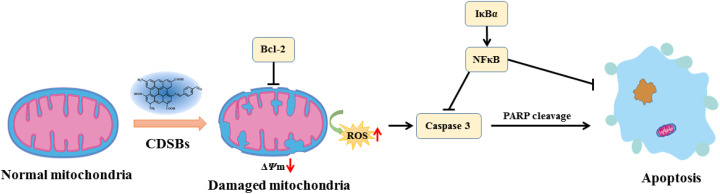
Schiff bases have remarkable anticancer activity and are used for glioma therapy. However, the poor water solubility/dispersibility limits their therapeutic potential in biological systems. To address this issue, carbon dots (CDs) have been utilized to enhance the dispersibility in water and biological efficacy of Schiff bases. The amino groups on the surface of CDs were conjugated effectively with the aldehyde group of terephthalaldehyde to form novel CD-based Schiff bases (CDSBs). The results of the MTT assays demonstrate that CDSBs have significant anticancer activity in glioma GL261 cells and U251 cells, with IC50 values of 17.9 μg mL-1 and 14.9 μg mL-1, respectively. CDSBs have also been found to have good biocompatibility with normal glial cells. The production of reactive oxygen species (ROS) in GL261 glioma cells showed that CDSBs, at a concentration of 44 μg mL-1, resulted in approximately 13 times higher intracellular ROS production than in the control group. These experiments offer evidence that CDSBs induce mitochondrial damage, leading to a reduction in mitochondrial membrane potential in GL261 cells. In particular, in this work, CDs serve not as carriers, but as an integral part of the anticancer drugs, which can expand the role of CDs in cancer treatment.
This journal is © The Royal Society of Chemistry.
Conflict of interest statement
There are no conflicts to declare.
Figures


References
-
- Tong F. Zhao J. Fang Z. et al., MUC1 promotes glioblastoma progression and TMZ resistance by stabilizing EGFRvIII. Pharmacol. Res. 2023;187:106606. - PubMed
-
- Ahamad M. N. Iman K. Raza M. K. et al., Anticancer properties, apoptosis and catecholase mimic activities of dinuclear cobalt (II) and copper (II) Schiff base complexes. Bioorg. Chem. 2020;95:103561. - PubMed
-
- Soroceanu A. Bargan A. Advanced and biomedical applications of Schiff-base ligands and their metal complexes: A review. Crystals. 2022;12:1436.
LinkOut - more resources
Full Text Sources