. 2024 Jan 5;13(1):2300882.
doi: 10.1080/2162402X.2023.2300882. eCollection 2024.

Extracellular vesicles released by cancer-associated fibroblast-induced myeloid-derived suppressor cells inhibit T-cell function

Affiliations

Extracellular vesicles released by cancer-associated fibroblast-induced myeloid-derived suppressor cells inhibit T-cell function

Carlo P Ramil et al. Oncoimmunology. .

Abstract

Myeloid cells are known to play a crucial role in creating a tumor-promoting and immune suppressive microenvironment. Our previous study demonstrated that primary human monocytes can be polarized into immunosuppressive myeloid-derived suppressor cells (MDSCs) by cancer-associated fibroblasts (CAFs) in a 3D co-culture system. However, the molecular mechanisms underlying the immunosuppressive function of MDSCs, especially CAF-induced MDSCs, remain poorly understood. Using mass spectrometry-based proteomics, we compared cell surface protein changes among monocytes, in vitro differentiated CAF-induced MDSCs, M1/M2 macrophages, and dendritic cells, and identified an extracellular vesicle (EV)-mediated secretory phenotype of MDSCs. Functional assays using an MDSC/T-cell co-culture system revealed that blocking EV generation in CAF-induced MDSCs reversed their ability to suppress T-cell proliferation, while EVs isolated from CAF-induced MDSCs directly inhibited T-cell function. Furthermore, we identified fructose bisphosphatase 1 (FBP1) as a cargo protein that is highly enriched in EVs isolated from CAF-induced MDSCs, and pharmacological inhibition of FBP1 partially reversed the suppressive phenotype of MDSCs. Our findings provide valuable insights into the cell surface proteome of different monocyte-derived myeloid subsets and uncover a novel mechanism underlying the interplay between CAFs and myeloid cells in shaping a tumor-permissive microenvironment.

Keywords: Cancer-associated fibroblasts (CAF); extracellular vesicles (EV); myeloid-derived suppressor cells (MDSC); proteomics.


Conflict of interest statement

All authors are current or previous employees of Merck Sharp & Dohme LLC, a subsidiary of Merck & Co., Inc., Rahway, NJ, USA and shareholders of Merck & Co., Inc., Rahway, NJ, USA.

Figures

Figure 1.
Figure 1.
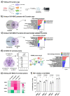
Changes in the cell surface proteome during monocyte-to-MDSC differentiation show the upregulation of vesicle-related pathways. (a) Monocytes were differentiated to CAF-MDSC and subjected to label-free proteomics. (b) Differential protein expression analysis of cell surface proteins in CAF-MDSCs vs. monocytes; cell surface proteins were filtered based on gene ontology cellular component (GOCC) terms ‘plasma membrane’ and ‘extracellular space’; colored dots represent significantly altered proteins (red = up; blue = down) (c) Enriched pathway and process terms based on differentially expressed proteins in CAF-MDSC using metascape.
Figure 2.
Figure 2.
The cell surface proteome of CAF-MDSC is enriched in proteins. (a) Monocytes were differentiated to cancer-associated fibroblast-induced myeloid derived suppressor cells (CAF-MDSC), immature dendritic cells (iDC), M1 and M2 macrophage and subjected to label-free proteomics. (b) Principal component analysis of the cell surface proteome profile of monocytes and differentiated cells – CAF-MDSC, iDC, M1 and M2 macrophage. (c) Differential protein expression analysis of cell surface proteins in CAF-MDSC vs. iDC or M1 or M2 macrophage; cell surface proteins were filtered based on GOCC terms ‘plasma membrane’ and ‘extracellular space’; significantly enriched cell surface proteins in CAF-MDSC (fold change > 2; FDR < 0.05) are enclosed in red box (dotted line on x-axis: Log2 = 1.0; y-axis: p-value (BH) = 0.05). (c) Overlap of significantly enriched cell surface proteins in CAF-MDSC vs. other cell types. (d) Gene ontology enrichment of significantly enriched cell surface proteins in CAF-MDSC (44 proteins) using STRING DB.
Figure 3.
Figure 3.
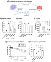
Suppression of autologous CD8 T-cell proliferation by CAF-MDSC extracellular vesicles (EV). Autologous CD8 T-cell were co-cultured with CAF-MDSC and treated with nSmase2 inhibitor (b) or treated with EVs (e-f) for 3 days then T-cell proliferation was measured by CFSE dilution using flow cytometry. (a) EVs released by CAF-MDSC can potentially suppress T-cell proliferation; this effect could be reversed by inhibiting the enzyme neutral sphingomyelinase 2 (nSmase2) that is important in EV generation. (b) Co-cultures of CAF-MDSCs and autologous CD8 T-cells were treated with an nSmase2 inhibitor (nSmase2i) or its inactive form. (c-d) EV characterization by nanoparticle tracking analysis to measure particle size and number. (e) Suppression of autologous CD8 T-cell proliferation by EVs released by CAF-MDSC using 6.25, 12.5, 25, 50, 100 µg EV or (f) 100 µg EVs isolated from myeloid cells based on protein content. Data shown is mean ± SEM from three independent experiments. Multiple group comparison by one-way ANOVA; ****= p < 0.001, ***= p < 0.001, **= p < 0.01 *= p < 0.05, ns = not significant.
Figure 4.
Figure 4.
CAF-MDSC extracellular vesicle protein cargo. (a) Proteins contained in EVs isolated from monocyte-derived cells were quantified by TMT-based LC-MS/MS. (b) Overlap of CAF-MDSC EV proteins with CAF-MDSC proteome and gene ontology enrichment of proteins uniquely identified in the EV. (c) Overlap of CAF-MDSC EV proteins with experimentally validated EV proteins in the vesiclepedia database (http://microvesicles.org/) and gene ontology enrichment of CAF-MDSC EV proteins. (d) Venn diagram showing the overlap of significantly enriched proteins in CAF-MDSC EV vs. iDC EV or M1 EV or M2 EV; box plot of the normalized intensity of the 10 enriched proteins, each box represents one replicate in the LC-Ms/MS analysis; functional protein network analysis and gene ontology enrichment of the 10 proteins using STRING DB. (e) CAF-MDSC/T-cell coculture were treated with inhibitors of CA2 (0.01 µM), MMP9 (0.1 µM), and FBP1 (10 µM) then T-cell proliferation was measured by CFSE dilution assay. The percentage of suppression was calculated using the established formula: ((Log2(y) of teff alone – Log2(y) of Teff+Myeloid cells)/Log2(y) of teff alone) x 100, where y is the mean fluorescence intensity (MFI) of CFSE on the whole population divided by the MFI of CFSE of non-proliferating cells. (f) effect of FBP1 inhibition in CAF-MDSC on T-cell proliferation. Data shown is mean ± SEM from three independent experiments. Multiple group comparison by one-way ANOVA; **= p < 0.01, *= p < 0.05, ns = not significant.

References

    1. Vesely MD, Zhang T, Chen L.. Resistance mechanisms to anti-PD cancer immunotherapy. Annu Rev Immunol. 2022;40(1):45–12. doi: 10.1146/annurev-immunol-070621-030155. - DOI - PubMed
    1. Cristescu R, Nebozhyn M, Zhang C, Albright A, Kobie J, Huang L, Zhao Q, Wang A, Ma H, Alexander Cao Z, et al. Transcriptomic determinants of response to pembrolizumab monotherapy across solid tumor types. Clin Cancer Res. 2022;28(8):1680–1689. doi: 10.1158/1078-0432.CCR-21-3329. - DOI - PMC - PubMed
    1. Singh L, Muise ES, Bhattacharya A, Grein J, Javaid S, Stivers P, Zhang J, Qu Y, Joyce-Shaikh B, Loboda A, et al. ILT3 (LILRB4) promotes the immunosuppressive function of tumor-educated human monocytic myeloid-derived suppressor cells. Mol Cancer Res. 2021;19(4):702–716. doi: 10.1158/1541-7786.MCR-20-0622. - DOI - PubMed
    1. Heim CE, Vidlak D, Kielian T. Interleukin-10 production by myeloid-derived suppressor cells contributes to bacterial persistence during staphylococcus aureus orthopedic biofilm infection. J Leukoc Biol. 2015;98(6):1003–1013. doi: 10.1189/jlb.4VMA0315-125RR. - DOI - PMC - PubMed
    1. Gillette MA, Satpathy S, Cao S, Dhanasekaran SM, Vasaikar SV, Krug K, Petralia F, Li Y, Liang W-W, Reva B, et al. Proteogenomic characterization reveals therapeutic vulnerabilities in lung adenocarcinoma. Cell. 2020;182:200–225.e35. - PMC - PubMed