

Binary semantic segmentation for detection of prostate adenocarcinoma using an ensemble with attention and residual U-Net architectures
- PMID: 38192468
- PMCID: PMC10773872
- DOI: 10.7717/peerj-cs.1767
Binary semantic segmentation for detection of prostate adenocarcinoma using an ensemble with attention and residual U-Net architectures
Abstract
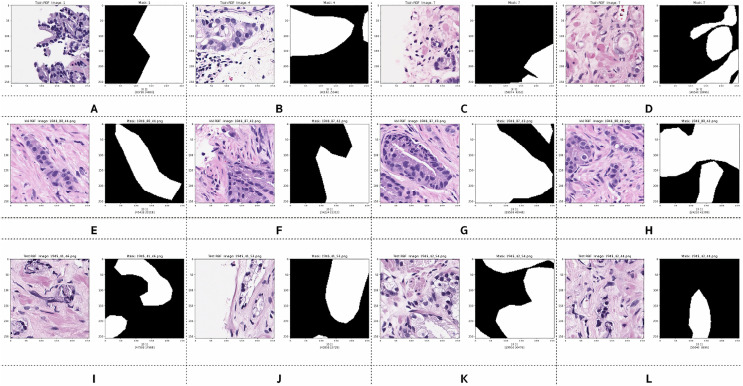
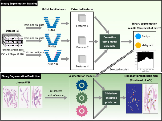
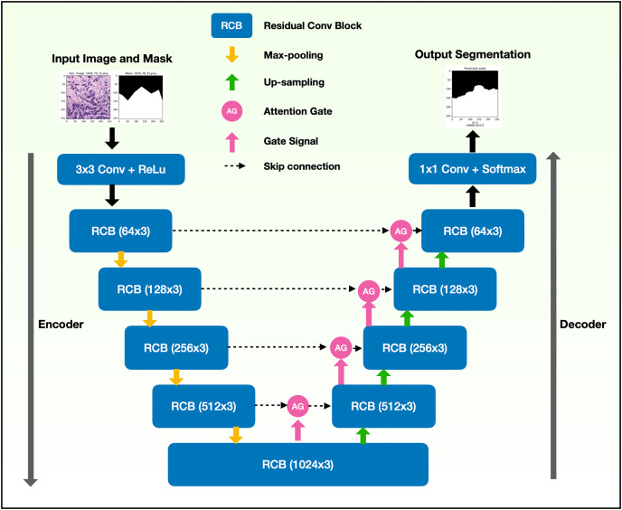
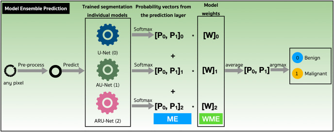
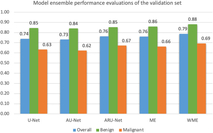
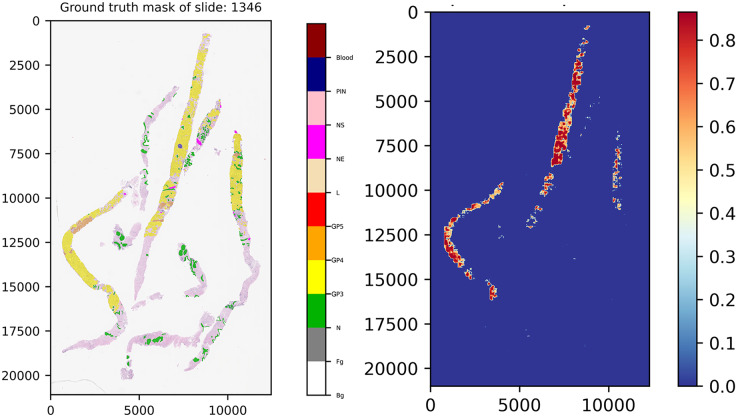
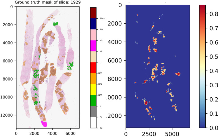
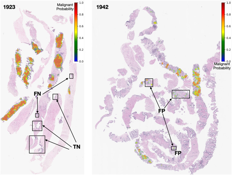
An accurate determination of the Gleason Score (GS) or Gleason Pattern (GP) is crucial in the diagnosis of prostate cancer (PCa) because it is one of the criterion used to guide treatment decisions for prognostic-risk groups. However, the manually designation of GP by a pathologist using a microscope is prone to error and subject to significant inter-observer variability. Deep learning has been used to automatically differentiate GP on digitized slides, aiding pathologists and reducing inter-observer variability, especially in the early GP of cancer. This article presents a binary semantic segmentation for the GP of prostate adenocarcinoma. The segmentation separates benign and malignant tissues, with the malignant class consisting of adenocarcinoma GP3 and GP4 tissues annotated from 50 unique digitized whole slide images (WSIs) of prostate needle core biopsy specimens stained with hematoxylin and eosin. The pyramidal digitized WSIs were extracted into image patches with a size of 256 × 256 pixels at a magnification of 20×. An ensemble approach is proposed combining U-Net-based architectures, including traditional U-Net, attention-based U-Net, and residual attention-based U-Net. This work initially considers a PCa tissue analysis using a combination of attention gate units with residual convolution units. The performance evaluation revealed a mean Intersection-over-Union of 0.79 for the two classes, 0.88 for the benign class, and 0.70 for the malignant class. The proposed method was then used to produce pixel-level segmentation maps of PCa adenocarcinoma tissue slides in the testing set. We developed a screening tool to discriminate between benign and malignant prostate tissue in digitized images of needle biopsy samples using an AI approach. We aimed to identify malignant adenocarcinoma tissues from our own collected, annotated, and organized dataset. Our approach returned the performance which was accepted by the pathologists.
Keywords: Adenocarcinoma detection; Attention gate; Binary semantic segmentation; Model-ensemble; Prostate cancer; Residual convolution.
© 2023 Damkliang et al.
Conflict of interest statement
The authors declare that they have no competing interests.
Figures

Similar articles
-
Region Segmentation of Whole-Slide Images for Analyzing Histological Differentiation of Prostate Adenocarcinoma Using Ensemble EfficientNetB2 U-Net with Transfer Learning Mechanism.Cancers (Basel). 2023 Jan 26;15(3):762. doi: 10.3390/cancers15030762. Cancers (Basel). 2023. PMID: 36765719 Free PMC article.
-
Deep learning segmentation architectures for automatic detection of pancreatic ductal adenocarcinoma in EUS-guided fine-needle biopsy samples based on whole-slide imaging.Endosc Ultrasound. 2024 Nov-Dec;13(6):335-344. doi: 10.1097/eus.0000000000000094. Epub 2024 Dec 12. Endosc Ultrasound. 2024. PMID: 39802107 Free PMC article.
-
Development of a Deep Learning Algorithm for the Histopathologic Diagnosis and Gleason Grading of Prostate Cancer Biopsies: A Pilot Study.Eur Urol Focus. 2021 Mar;7(2):347-351. doi: 10.1016/j.euf.2019.11.003. Epub 2019 Nov 22. Eur Urol Focus. 2021. PMID: 31767543 Free PMC article.
-
WeGleNet: A weakly-supervised convolutional neural network for the semantic segmentation of Gleason grades in prostate histology images.Comput Med Imaging Graph. 2021 Mar;88:101846. doi: 10.1016/j.compmedimag.2020.101846. Epub 2021 Jan 13. Comput Med Imaging Graph. 2021. PMID: 33485056
-
W-Net: Dense and diagnostic semantic segmentation of subcutaneous and breast tissue in ultrasound images by incorporating ultrasound RF waveform data.Med Image Anal. 2022 Feb;76:102326. doi: 10.1016/j.media.2021.102326. Epub 2021 Dec 5. Med Image Anal. 2022. PMID: 34936967
Cited by
-
DGCFNet: Dual Global Context Fusion Network for remote sensing image semantic segmentation.PeerJ Comput Sci. 2025 Mar 27;11:e2786. doi: 10.7717/peerj-cs.2786. eCollection 2025. PeerJ Comput Sci. 2025. PMID: 40567688 Free PMC article.
-
Artificial Intelligence Algorithms and Their Current Role in the Identification and Comparison of Gleason Patterns in Prostate Cancer Histopathology: A Comprehensive Review.Diagnostics (Basel). 2024 Sep 25;14(19):2127. doi: 10.3390/diagnostics14192127. Diagnostics (Basel). 2024. PMID: 39410530 Free PMC article. Review.
References
-
- Abadi M, Agarwal A, Barham P, Brevdo E, Chen Z, Citro C, Corrado GS, Davis A, Dean J, Devin M, Ghemawat S, Goodfellow I, Harp A, Irving G, Isard M, Jia Y, Jozefowicz R, Kaiser L, Kudlur M, Levenberg J, Mané D, Monga R, Moore S, Murray D, Olah C, Schuster M, Shlens J, Steiner B, Sutskever I, Talwar K, Tucker P, Vanhoucke V, Vasudevan V, Viégas F, Vinyals O, Warden P, Wattenberg M, Wicke M, Yu Y, Zheng X. TensorFlow: large-scale machine learning on heterogeneous systems. 2015. https://www.tensorflow.org/ https://www.tensorflow.org/
-
- Alom MZ, Hasan M, Yakopcic C, Taha TM, Asari VK. Recurrent residual convolutional neural network based on U-Net (R2U-Net) for medical image segmentation. ArXiv. 2018 doi: 10.48550/arXiv.1802.06955. - DOI
Associated data
LinkOut - more resources
Full Text Sources