

Extracellular vesicles in cardiomyopathies: A narrative review
- PMID: 38192847
- PMCID: PMC10772622
- DOI: 10.1016/j.heliyon.2023.e23765
Extracellular vesicles in cardiomyopathies: A narrative review
Abstract
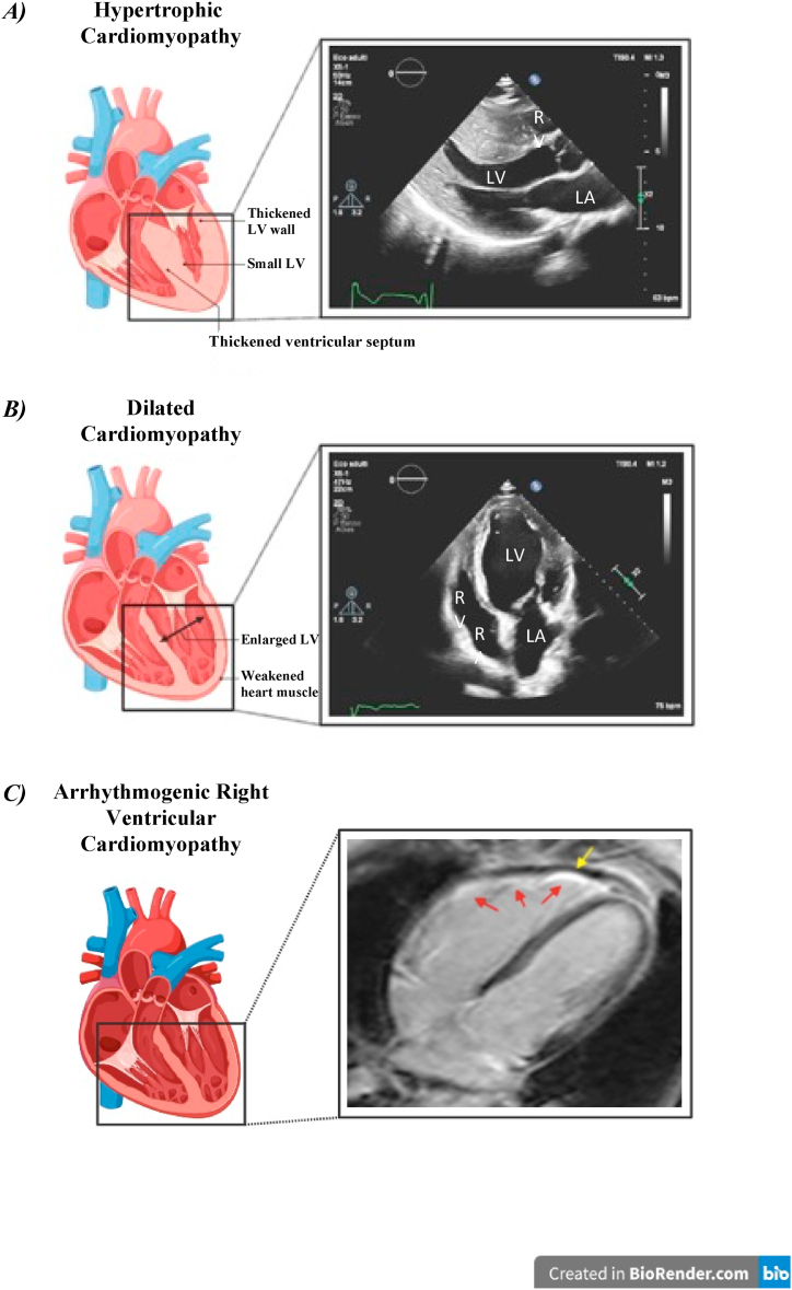
Extracellular vesicles (EVs) are membrane-bound particles released by all cells under physiological and pathological conditions. EVs constitute a potential tool to unravel cell-specific pathophysiological mechanisms at the root of disease states and retain the potential to act as biomarkers for cardiac diseases. By being able to carry bioactive cargo (such as proteins and miRNAs), EVs harness great potential as accessible "liquid biopsies", given their ability to reflect the state of their cell of origin. Cardiomyopathies encompass a variety of myocardial disorders associated with mechanical, functional and/or electric dysfunction. These diseases exhibit different phenotypes, including inappropriate ventricular hypertrophy, dilatation, scarring, fibro-fatty replacement, dysfunction, and may stem from multiple aetiologies, most often genetic. Thus, the aims of this narrative review are to summarize the current knowledge on EVs and cardiomyopathies (e.g., hypertrophic, dilated and arrhythmogenic), to elucidate the potential role of EVs in the paracrine cell-to-cell communication among cardiac tissue compartments, in aiding the diagnosis of the diverse subtypes of cardiomyopathies in a minimally invasive manner, and finally to address whether certain molecular and phenotypical characteristics of EVs may correlate with cardiomyopathy disease phenotype and severity.
Keywords: Arrhythmogenic cardiomyopathy; Cardiomyopathies; Dilated cardiomyopathy; Extracellular vesicles; Hypertrophic cardiomyopathy.
© 2023 The Authors.
Conflict of interest statement
The authors declare the following financial interests/personal relationships which may be considered as potential competing interests:Massimiliano Ruscica reports financial support was provided by Banca d’Italia. Massimiliano Ruscica reports financial support was provided by Ministry of Education and Merit. If there are other authors, they declare that they have no known competing financial interests or personal relationships that could have appeared to influence the work reported in this paper.
Figures

References
-
- Vilella-Figuerola A., et al. Platelet-released extracellular vesicle characteristics differ in chronic and in acute heart disease. Thromb. Haemostasis. 2023;123(9):892–903. - PubMed
-
- Arbelo E., et al. ESC Guidelines for the management of cardiomyopathies. Eur. Heart J. 2023;44(37):3503–3626. 2023. - PubMed
