. 2024 Jan 4:17:41-57.
doi: 10.2147/JIR.S428093. eCollection 2024.

FcγRIIb Exacerbates LPS-Induced Neuroinflammation by Binding with the Bridging Protein DAP12 and Promoting the Activation of PI3K/AKT Signaling Pathway in Microglia

Affiliations

FcγRIIb Exacerbates LPS-Induced Neuroinflammation by Binding with the Bridging Protein DAP12 and Promoting the Activation of PI3K/AKT Signaling Pathway in Microglia

YingWen Han et al. J Inflamm Res. .

Abstract

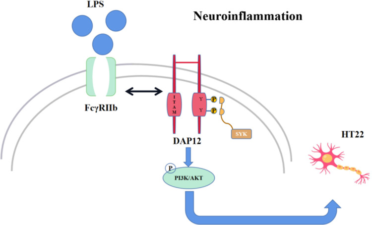
Introduction: This paper focuses on the expression and role of FcγRIIb in neuroinflammation, exploring the molecular mechanisms by which FcγRIIb interacts with the bridging protein DAP12 to regulate the PI3K-AKT signaling pathway that promote neuroinflammation and aggravate neuronal injury.

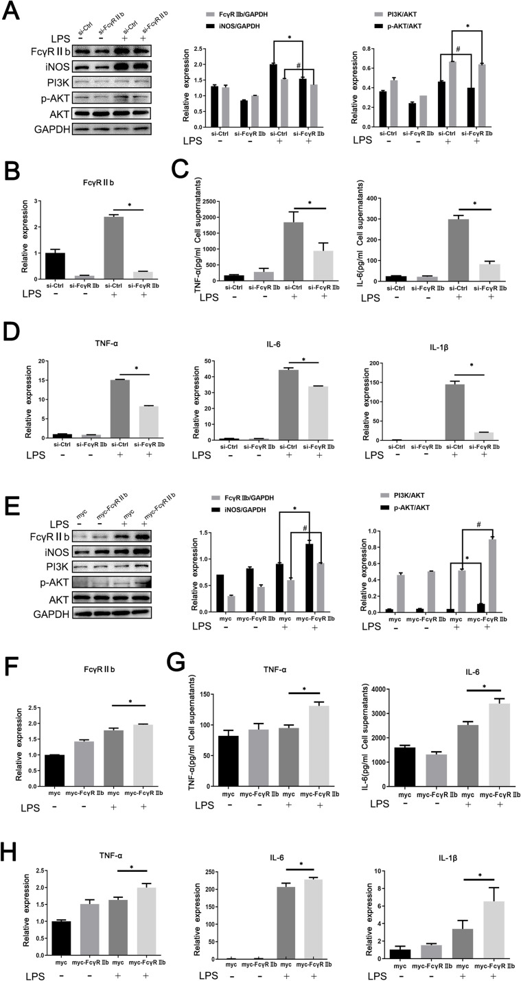
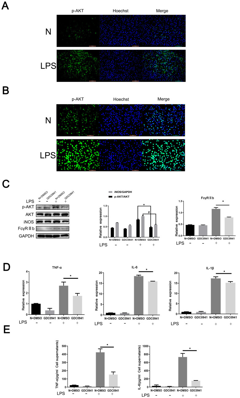
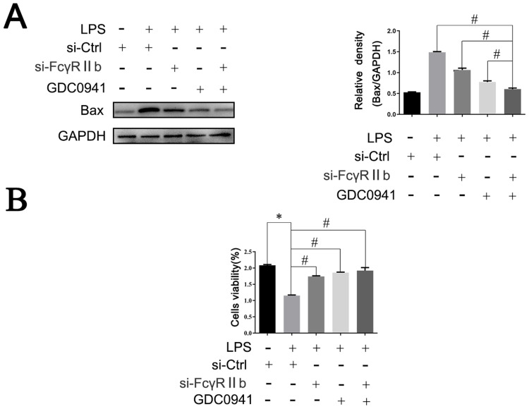
Methods: LPS-induced neuroinflammation models in vivo and in vitro were constructed to explore the role and mechanism of FcγRIIb in CNS inflammation. Subsequently, FcγRIIb was knocked down or overexpressed to observe the activation of BV2 cell and the effect on PI3K-AKT pathway. Then the PI3K-AKT pathway was blocked to observe its effect on cell activation and FcγRIIb expression. We analyzed the interaction between FcγRIIb and DAP12 by Immunoprecipitation technique. Then FcγRIIb was overexpressed while knocking down DAP12 to observe its effect on PI3K-AKT pathway. Finally, BV2 cell culture supernatant was co-cultured with neuronal cell HT22 to observe its effect on neuronal apoptosis and cell activity.

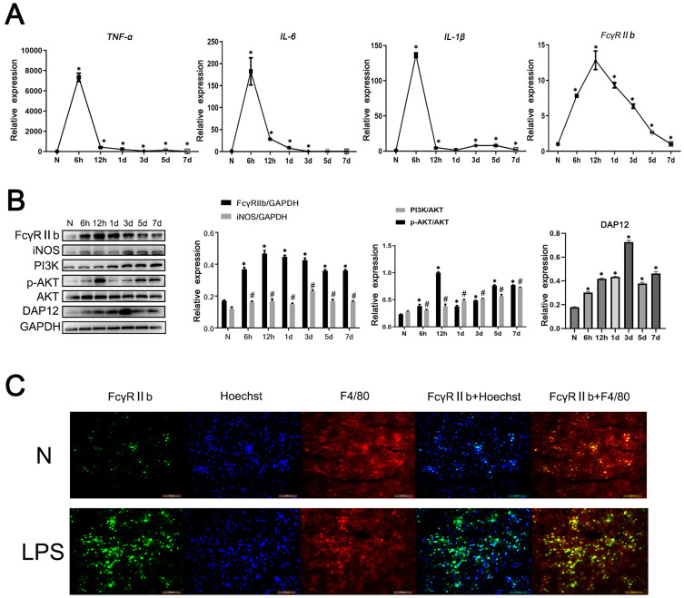
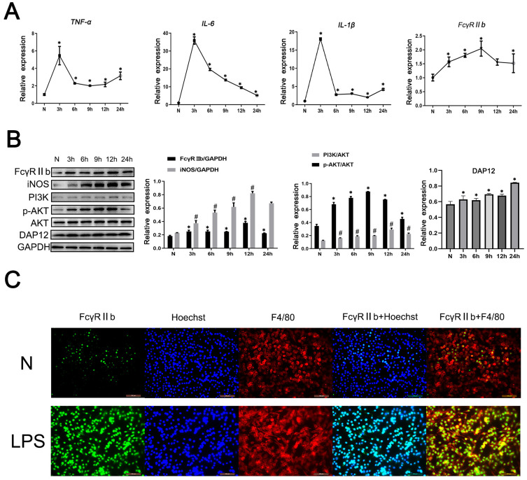
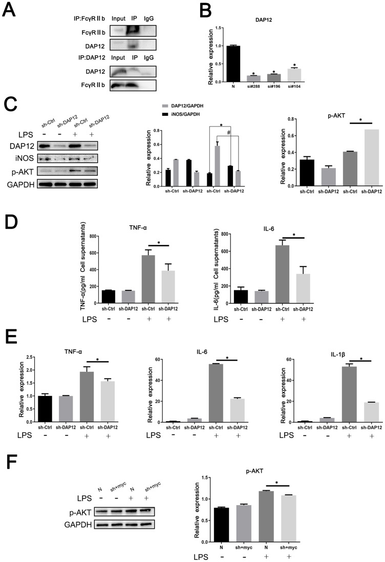
Results: In vivo and in vitro, we found that FcγRIIb expression was significantly increased and activated the PI3K-AKT pathway. Contrary to the results of overexpression of FcγRIIb, knockdown of FcγRIIb resulted in a significant low level of relevant inflammatory factors and suppressed the PI3K-AKT pathway. Furthermore, LPS stimulation induced an interaction between FcγRIIb and DAP12. Knockdown of DAP12 suppressed inflammation and activation of the PI3K-AKT pathway in BV2 cells, and meantime overexpression of FcγRIIb suppressed the level of FcγRIIb-induced AKT phosphorylation. Additionally, knockdown of FcγRIIb inhibited microglia activation, which induced neuronal apoptosis.

Discussion: Altogether, our experiments indicate that FcγRIIb interacts with DAP12 to promote microglia activation by activating the PI3K-AKT pathway while leading to neuronal apoptosis and exacerbating brain tissue injury, which may provide a new target for the treatment of inflammatory diseases in the central nervous system.

Keywords: DAP12; FcγRIIb; LPS-induced neuroinflammation; PI3K-AKT; microglia.


Conflict of interest statement

Prof. Dr. Xiaoyi Shao reports grants from Jiangsu Education Department, during the conduct of the study. The authors report no other conflicts of interest in this work.

Figures

Figure 1
Figure 1
The expression and role of FcγRIIb in a mouse model of CNS inflammation. C57BL/6 mice were injected intraperitoneally with LPS (9 mg/kg), and serum was collected at 6 h, 12 h, 1 d, 3 d, 5 d, and 7 d, while the control group (N) was injected with an equal amount of saline. (A) Q-PCR was performed to detect the mRNA expression levels of IL-6, TNF-α, IL-1β, and FcγRIIb in the mouse cerebral cortex. (B) Western blot was performed to detect the protein expression levels of FcγRIIb, iNOS, PI3K, p-AKT, AKT and DAP12 in the cerebral cortex of experimental mice. (C) Immunofluorescence staining was used to detect the expression of FcγRIIb in the cerebral cortex tissue of control and LPS mice. FcγRIIb (green), F4/80 (red), Hoechst (blue); scale bar: 100 μm (mean ± SEM represents the resultant values, n = 3, *Indicates compared to the control, #indicates compared to the control, p < 0.05).
Figure 2
Figure 2
The expression and role of FcγRIIb in an in vitro model of LPS-induced neuroinflammation. BV2 cells were stimulated with LPS (100 ng/mL) for 0, 3, 6, 9, 12, and 24 h, and then the cells and the cell culture supernatants were collected. (A) Q-PCR was performed to detect the mRNA expression levels of TNF-α, IL-6, IL-1β, and FcγRIIb in BV2 cells. (B) Western blot was used to detect the protein expression of FcγRIIb, iNOS, PI3K, p-AKT, AKT and DAP12 in BV2 cells. (C) Immunofluorescence staining was employed to detect the expression and localization of FcγRIIb in the N group and LPS-stimulated group of BV2 cells. FcγRIIb (green), F4/80 (red), Hoechst (blue); scale bar: 100 μm (mean ± SEM represents the resultant values, n = 3, *Indicates compared to the control, #indicates compared to the control, p < 0.05).
Figure 3
Figure 3
Effect of inhibition or overexpression of FcγRIIb on inflammatory activation in microglia. FcγRIIb interference knocking-down or overexpressing plasmids were transfected into BV2 cells for 48 h and then stimulated with LPS for 12 h. (A) Western blot detection of FcγRIIb, iNOS, PI3K, p-AKT and AKT protein expression levels after interference with BV2 cells. (B) Q-PCR assay of the mRNA expression levels of the inflammatory factors FcγRIIb in BV2 cells after interference with FcγRIIb. (C) ELISA the assay of the expression levels of TNF-α and IL-6 in the cell supernatants collected after interference with BV2 cells. (D) Q-PCR assay of the mRNA expression levels of the inflammatory factors TNF-α, IL-6, and IL-1β in BV2 cells after interference with FcγRIIb. (E) Western blot assay of the protein expression levels of FcγRIIb, iNOS, PI3K, p-AKT and AKT after overexpression with BV2 cells. (F) Q-PCR assay of the mRNA expression levels of FcγRIIb after overexpression with BV2 cells. (G) ELISA assay of the expression levels of inflammatory factors TNF-α and IL-6 in BV2 cells.(H) Q-PCR assay of the mRNA expression levels of TNF-α, IL-6, and IL-1β after overexpression with BV2 cells. (mean ± SEM represents the result values, n = 3, *Indicates compared to the si-Ctrl group, or myc+LPS group, #Indicates compared to the si-Ctrl+LPS group, or myc+LPS group, p < 0.05).
Figure 4
Figure 4
PI3K/AKT signaling pathway is involved in microglia-mediated LPS-induced neuroinflammation. (A) Cellular immunofluorescence staining of the expression and localization of p-AKT in control and LPS-stimulated groups in a mouse CNS inflammation model. p-AKT (green), Iba-1 (red), and Hoechst (blue); scale bar: 100 μm. (B) Cellular immunofluorescence staining of the expression and localization of p-AKT in control and LPS-stimulated groups in an in vitro model. p-AKT (green), Iba-1 (red), and Hoechst (blue); scale bar: 100 μm. (C) BV2 cells were pretreated with PI3K-AKT inhibitor GDC0941 for 1 h and then stimulated with LPS (100 ng/mL) for 9 h. Western blotting was performed to detect the protein expression levels of p-AKT, AKT, iNOS and FcγRIIb in BV2 cells. (D) Q-PCR detection of the mRNA expression levels of TNF-α, IL-6 and IL-1βin BV2 cells. (E) ELISA was performed to detect changes in expression levels of TNF-α and IL-6 in the supernatants of treated BV2 cells. (mean ± SEM represents the resultant values, n = 3, *Indicates compared to the control, or N+DMSO+LPS group, #Indicates compared to the control, or N+DMSO+LPS group, p < 0.05).
Figure 5
Figure 5
FcγRIIb and DAP12 interact to promote microglia activation-mediated LPS-induced neuroinflammation. (A) Analysis of the interaction between FcγRIIb and DAP12 by immunoprecipitation assay. (B) Si-RNA at different loci of DAP12 (si-DAP12#288, si-DAP12#196, and si-DAP12#104) were transfected into BV2 cells, before treating with LPS for 9 h. Q-PCR was performed to detect the transfection efficiency of knocking down si-RNA at different loci of DAP12. (C) Western blot detection of the changes in p-AKT, DAP12 and iNOS protein expression levels in BV2 cells after knockdown of DAP12. (D) ELISA assay of the expression levels of TNF-α and IL-6 in the supernatants of cells collected by centrifugation. (E) Q-PCR assay of the changes in NF-α, IL-6, and IL-1β expression in BV2 cells. (F) BV2 cells were transfected and then stimulated with LPS. Western blot detected the change in p-AKT protein expression in BV2 cells after overexpressed FcγRIIb while interfering with DAP12 (mean ± SEM represents the result value, n = 3, *Compared to the control, or si-Ctrl+LPS group, or N+LPS group, or si-DAP12+myc-FcγRIIb+LPS group, #Indicates compared to the sh-Ctrl+LPS group, p < 0.05).
Figure 6
Figure 6
The “FcγRIIb-PI3K-AKT” positive feedback loop exacerbates neuroinflammation-mediated neuronal injury. BV2 cells were transfected with si-Ctrl or si-FcγRIIb, pretreated with GDC0941 inhibitor, transfected with si-FcγRIIb, treated with pathway inhibitor GDC0941, and then stimulated with LPS. Subsequently, the conditioned medium was prepared with DMEM complete medium and used to culture HT22 neurons for 24 h. (A) Western blot to detect the expression level of apoptosis-related protein Bax. (n = 3, # represents p < 0.05 compared to the si-Ctrl+LPS group, si-FcγRIIb+LPS group, and GDC+LPS group). (B) CCK-8 assay of HT22 cell viability (n = 3, *Compared to the si-Ctrl group, #Compared to the si-Ctrl+LPS group, p < 0.05).
Figure 7
Figure 7
After microglia activation by LPS, FcγRIIb forms a dimer with the bridging protein DAP12, which activates the downstream PI3K/AKT signaling pathway, thereby exacerbating inflammation and causing neuronal damage.

Similar articles

References

    1. Borst K, Dumas AA, Prinz M. Microglia: immune and non-immune functions. Immunity. 2021;54(10):2194–2208. doi:10.1016/j.immuni.2021.09.014 - DOI - PubMed
    1. Lopez-Castejon G, Brough D. Understanding the mechanism of IL-1β secretion. Cytokine Growth Factor Rev. 2011;22(4):189–195. doi:10.1016/j.cytogfr.2011.10.001 - DOI - PMC - PubMed
    1. Cruceriu D, Baldasici O, Balacescu O, Berindan-Neagoe I. The dual role of tumor necrosis factor-alpha (TNF-α) in breast cancer: molecular insights and therapeutic approaches. Cell Oncol. 2020;43(1):1–18. doi:10.1007/s13402-019-00489-1 - DOI - PubMed
    1. Colonna M, Butovsky O. Microglia function in the central nervous system during health and neurodegeneration. Ann Rev Immunol. 2017;35(1):441. doi:10.1146/annurev-immunol-051116-052358 - DOI - PMC - PubMed
    1. Hickman S, Izzy S, Sen P, Morsett L, El Khoury J. Microglia in neurodegeneration. Nat Neurosci. 2018;21(10):1359–1369. doi:10.1038/s41593-018-0242-x - DOI - PMC - PubMed