. 2023 Dec 18:14:1273556.
doi: 10.3389/fimmu.2023.1273556. eCollection 2023.

Maternal consumption of a high-fat diet modulates the inflammatory response in their offspring, mediated by the M1 muscarinic receptor

Affiliations

Maternal consumption of a high-fat diet modulates the inflammatory response in their offspring, mediated by the M1 muscarinic receptor

Suleyma Oliveira Costa et al. Front Immunol. .

Abstract

Introduction: High-fat diet (HFD) consumption is associated with various metabolic disorders and diseases. Both pre-pregnancy and maternal obesity can have long-term consequences on offspring health. Furthermore, consuming an HFD in adulthood significantly increases the risk of obesity and metabolic disorders. However, an intriguing phenomenon known as the obesity paradox suggests that obesity may confer a protective effect on mortality outcomes in sepsis. In sepsis, activation of the cholinergic anti-inflammatory pathway (CAP) can help mitigate systemic inflammation. We employed a metabolic programming model to explore the relationship between maternal HFD consumption and offspring response to sepsis.

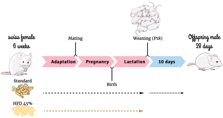
Methods: We fed female mice either a standard diet (SC) or an HFD during the pre-pregnancy, pregnancy, and lactation periods. Subsequently, we evaluated 28-day-old male offspring.

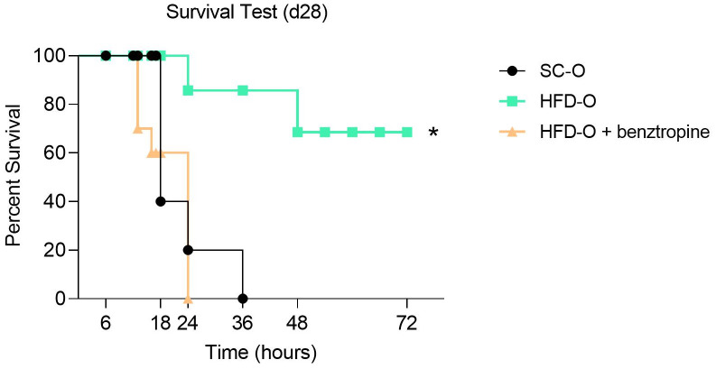
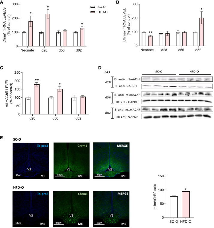
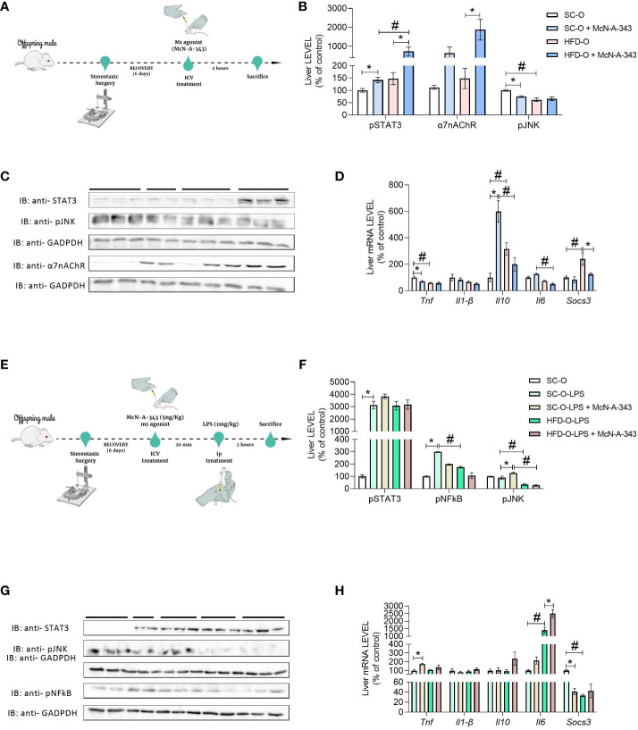
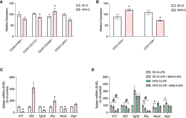
Results: Notably, we discovered that offspring from HFD-fed dams (HFD-O) exhibited a higher survival rate compared with offspring from SC-fed dams (SC-O). Importantly, inhibition of the m1 muscarinic acetylcholine receptor (m1mAChR), involved in the CAP, in the hypothalamus abolished this protection. The expression of m1mAChR in the hypothalamus was higher in HFD-O at different ages, peaking on day 28. Treatment with an m1mAChR agonist could modulate the inflammatory response in peripheral tissues. Specifically, CAP activation was greater in the liver of HFD-O following agonist treatment. Interestingly, lipopolysaccharide (LPS) challenge failed to induce a more inflammatory state in HFD-O, in contrast to SC-O, and agonist treatment had no additional effect. Analysis of spleen immune cells revealed a distinct phenotype in HFD-O, characterized by elevated levels of CD4+ lymphocytes rather than CD8+ lymphocytes. Moreover, basal Il17 messenger RNA (mRNA) levels were lower while Il22 mRNA levels were higher in HFD-O, and we observed the same pattern after LPS challenge.

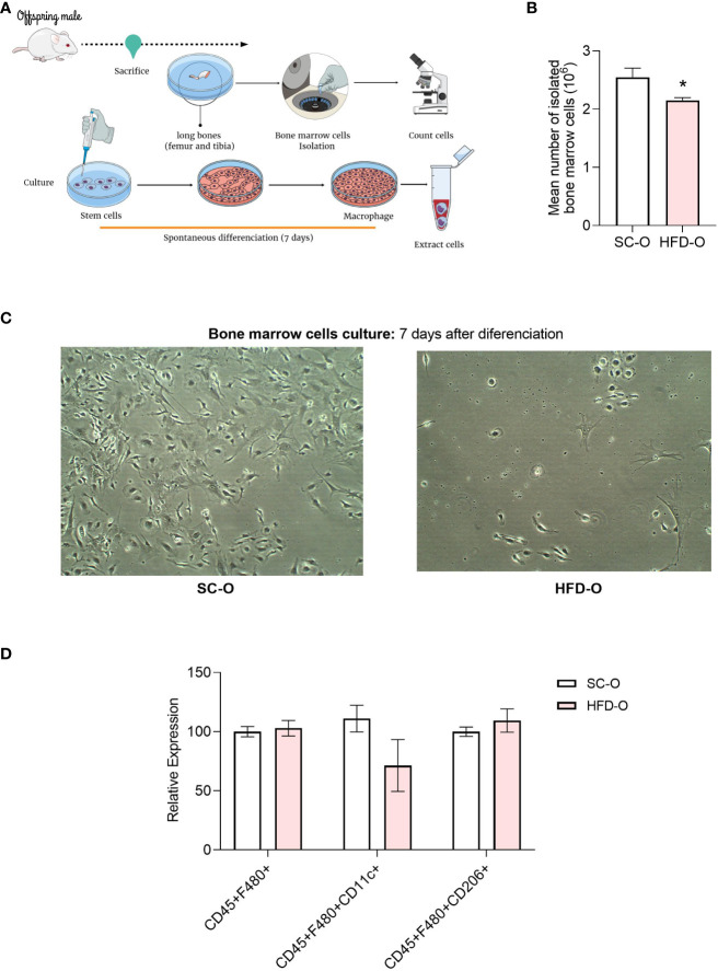
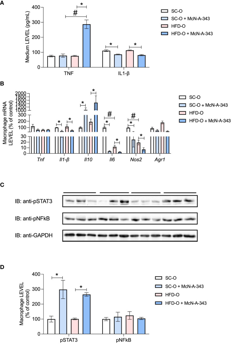
Discussion: Further examination of myeloid cells isolated from bone marrow and allowed to differentiate showed that HFD-O macrophages displayed an anti-inflammatory phenotype. Additionally, treatment with the m1mAChR agonist contributed to reducing inflammatory marker levels in both groups. In summary, our findings demonstrate that HFD-O are protected against LPS-induced sepsis, and this protection is mediated by the central m1mAChR. Moreover, the inflammatory response in the liver, spleen, and bone marrow-differentiated macrophages is diminished. However, more extensive analysis is necessary to elucidate the specific mechanisms by which m1mAChR modulates the immune response during sepsis.

Keywords: DOHaD (Developmental origins of health and disease); cholinergic; high fat diet (HFD); hypothalamus; maternal programming; muscarinic 1 acetylcholine receptors; obesity.


Conflict of interest statement

The authors declare that the research was conducted in the absence of any commercial or financial relationships that could be construed as a potential conflict of interest.

Figures

Figure 1
Figure 1
Experimental design. Mind the Graph website was used to prepare Figure 1 .
Figure 2
Figure 2
Survival analysis of the offspring. Sepsis was induced with a lethal dose of lipopolysaccharide (LPS; 30 mg LPS kg-1 body weight, intraperitoneal) administered to control offspring (SC-O, n = 10) and high-fat diet offspring (HFD-O, n = 10) at postnatal day 28. Some HFD-O were pretreated for 20 min with the m1mAChR antagonist benztropine (intracerebroventricular) (HFD-O + Benztropine, n = 10). The mice were observed for 72 h, and the survival rate was recorded every 1 h. The data represent the mean ± standard error of the mean. *p < 0.05 HFD-O versus SC-O and HFD-O + Benztropine versus HFD-O (log-rank test).
Figure 3
Figure 3
Cholinergic anti-inflammatory pathway receptor expression in the hypothalamus of the offspring. Hypothalamic m1mAChR and α7nAChR messenger RNA (mRNA) (A, B) and protein (C, D) levels were evaluated by RT-PCR and western blot, respectively, in control and high-fat diet offspring (SC-O and HFD-O, respectively). The mRNA and protein levels were evaluated at birth (neonate) and postnatal days 28, 56, and 82. Molecular weight of proteins: m1mAChR – 52KDa; α7nAChR – 55KDa; and GAPDH – 35KDa. The percent expression of control (GAPDH) is shown (mean ± standard error of the mean, n = 5 pups per group). Asterisks indicate significant differences determined by Student’s t-test (*p < 0.05 and **p < 0.01). Confocal images illustrating m1mAChR+ cells (green) and nuclear labelling with TO-PRO-3 (blue) in coronal brain sections (15 µm thick) from 28-day-old offspring (E). Scale of images 50 µm. The number of m1mAChR+ cells in the median eminence (ME) of SD-O and HFD-O (n = 3 per group) (E). V3: third ventricle.
Figure 4
Figure 4
Inflammatory markers in the liver of the offspring. Experimental design for pharmacological treatment with the m1mAChR agonist McN-A-343 (delivered intracerebroventricularly [ICV]) (A). Hepatic pSTAT3, α7nAChR, and pJNK protein expression (B, C) and Tnf, Il1b, Il10, Il6, and Socs3 messenger RNA (mRNA) expression (D) were evaluated by western blot and RT-PCR, respectively, in 28-day-old standard and high-fat diet offspring (SC-O and HFD-O, respectively). The mice were treated with the m1mAChR agonist McN-A-343 (5 ng kg-1, ICV). Experimental design for simultaneous administration of lipopolysaccharide (LPS; 1 mg kg-1, intraperitoneal) and the m1mAChR agonist (ICV) (E). Hepatic pSTAT3, pNF-κB, and pJNK protein expression (F, G) and Tnf, Il1b, Il10, Il6, and Socs3 mRNA expression (H) were evaluated by western blot and RT-PCR in 28-day-old SC-O and HFD-O. Molecular weight of proteins: pSTAT3 – 90KDa; α7nAChR – 55KDa; pJNK – 55KDa; pNFkB – 65KDa; and GAPDH – 35KDa. The data represent the mean ± standard error of the mean (n = 5 per group). The data were analysed with analysis of variance. *Significant difference (p < 0.05) between basal and agonist or LPS treatment. #Significant difference (p < 0.05) between HFD-O and SC-O. Mind the Graph website was used to prepare Figures 4A, E.
Figure 5
Figure 5
Immune response markers in the spleen of the offspring. Splenic CD45+F480+, CD45+CD11c+, CD45+CD206+, and CD45+Ly6G+ cells were evaluated with the macrophage panel (A) and CD3+CD4+ and CD3+ CD8+ cells were evaluated with the lymphocyte panel (B) by flow cytometry. The data represent expression of high-fat diet offspring (HFD-O) relative to the control offspring (SC-O). Splenic Il17, Il22, Tgfb, Ifng, Nos2, and Arg1 messenger RNA (mRNA) expression (C) was evaluated by RT-PCR in 28-day-old SC-O and HFD-O. Splenic Il17, Il22, Tgfb, Ifng, Nos2, and Arg1 mRNA expression (D) was evaluated in the offspring following lipopolysaccharide (LPS) challenge (1 mg kg-1, intraperitoneal) and treatment with the m1mAChR agonist McN-A-343 (5 ng kg-1, intracerebroventricular). The data represent the mean ± standard error of the mean. The data were analysed with analysis of variance. *Significant difference (p < 0.05) between basal and agonist or LPS treatment. #Significant difference (p < 0.05) between HFD-O and SC-O.
Figure 6
Figure 6
Profile of bone marrow cells after differentiation. Bone marrow cells were isolated from the long bones of 28-day-old control and high-fat diet offspring (SC-O and HFD-O, respectively) (A). The cells were counted before the culture (B). After 7 days of spontaneous differentiation, pictures were taken of the culture (C) to confirm that the cells had differentiated into macrophages. CD45+F480+, CD45+CD11c+, and CD45+CD206+ cells were evaluated with the macrophage panel by flow cytometry (D). The data represent the mean ± standard error of the mean. *Significant difference (p < 0.05) based on Student’s t-test. Mind the Graph website was used to prepare Figure 6A.
Figure 7
Figure 7
Inflammatory markers in bone marrow cells after differentiation. TNF and IL-1β protein expression was evaluated with enzyme-linked immunosorbent assays (A) in the culture medium after 7 days of spontaneous differentiation. Tnf, Il1b, Il10, Il6, Nos2, and Arg1 messenger RNA (mRNA) expression (B) and pSTAT3 and pNF-κB protein expression (C, D) were evaluated by RT-PCR and western blot, respectively, in macrophages differentiated from control and high-fat diet offspring (SC-O and HFD-O, respectively) bone marrow. The mice were treated with the m1mAChR agonist McN-A-343 (5 ng kg-1, intracerebroventricular). The bars at the top of blots represent the groups. The data represent the mean ± standard error of the mean. Molecular weight of proteins: pSTAT3 – 90KDa; pNFkB – 65KDa; and GAPDH – 35KDa. The data were analysed with analysis of variance. *Significant difference (p < 0.05) between basal and agonist or LPS treatment. #Significant difference (p < 0.05) between HFD-O and SC-O.

Similar articles

References

    1. Alsiö Å, Nasic S, Ljungström L, Jacobsson G. Impact of obesity on outcome of severe bacterial infections. PloS One (2021) 16:e0251887. doi: 10.1371/journal.pone.0251887 - DOI - PMC - PubMed
    1. Yeo HJ, Kim TH, Jang JH, Jeon K, Oh DK, Park MH, et al. . Obesity paradox and functional outcomes in sepsis: A multicenter prospective study. Crit Care Med (2023) 51(6):1–11. doi: 10.1097/CCM.0000000000005801 - DOI - PMC - PubMed
    1. Cichon I, Ortmann W, Kolaczkowska E. Metabolic pathways involved in formation of spontaneous and lipopolysaccharide-induced neutrophil extracellular traps (NETs) differ in obesity and systemic inflammation. Int J Mol Sci (2021) 22:7718. doi: 10.3390/ijms22147718 - DOI - PMC - PubMed
    1. Wang Q, Huang J, Chen X, Wang J, Fang F. Transcriptomic markers in pediatric septic shock prognosis: An integrative analysis of gene expression profiles. Braz J Med Biol Res (2021) 54:1–8. doi: 10.1590/1414-431X202010152 - DOI - PMC - PubMed
    1. Kalani C, Venigalla T, Bailey J, Udeani G, Surani S. Sepsis patients in critical care units with obesity: is obesity protective? Cureus (2020) 12. doi: 10.7759/cureus.6929 - DOI - PMC - PubMed

Publication types