. 2024 Feb 14;15(2):e0303923.
doi: 10.1128/mbio.03039-23. Epub 2024 Jan 9.

Genetic analysis reveals a robust and hierarchical recruitment of the LolA chaperone to the LolCDE lipoprotein transporter

Affiliations

Genetic analysis reveals a robust and hierarchical recruitment of the LolA chaperone to the LolCDE lipoprotein transporter

Kelly M Lehman et al. mBio. .

Abstract

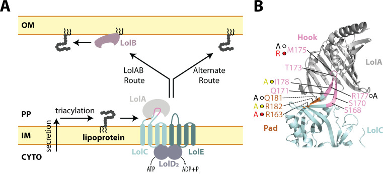
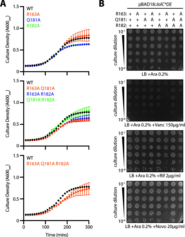
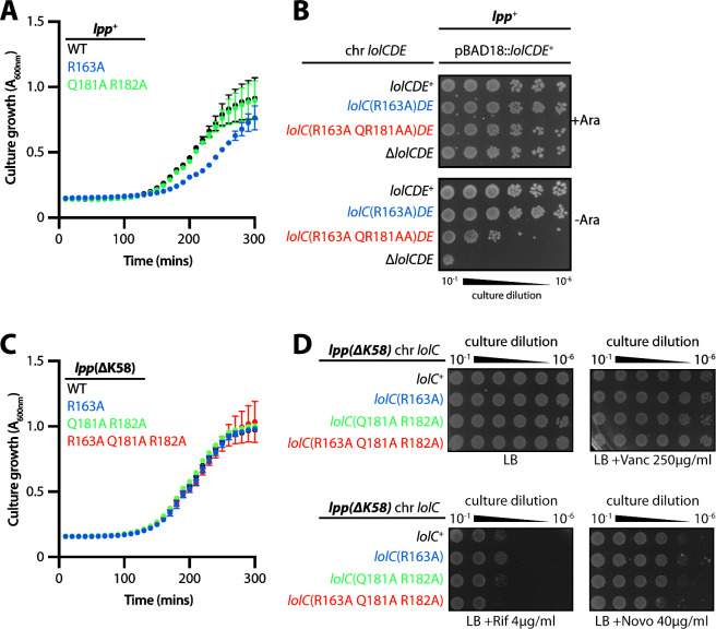
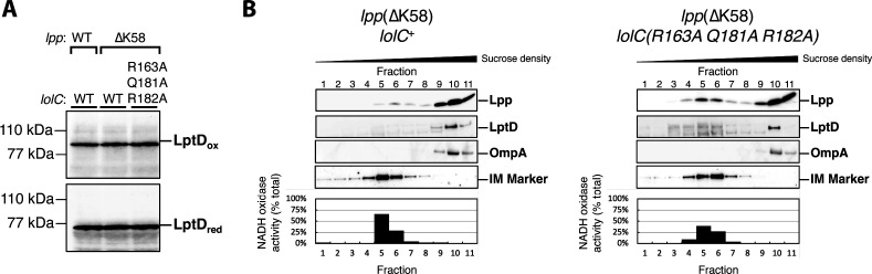
The outer membrane (OM) is an essential organelle of Gram-negative bacteria. Lipoproteins are key to building the OM, performing essential functions in several OM assembly machines. Lipoproteins mature in the inner membrane (IM) and are then trafficked to the OM. In Escherichia coli, the LolCDE transporter is needed to extract lipoproteins from the IM to begin trafficking. Lipoproteins are then transferred from LolCDE to the periplasmic chaperone LolA which ferries them to the OM for insertion by LolB. LolA recruitment by LolC is an essential trafficking step. Structural and biochemical studies suggested that two regions (termed Hook and Pad) within a periplasmic loop of LolC worked in tandem to recruit LolA, leading to a bipartite model for recruitment. Here, we genetically examine the LolC periplasmic loop in vivo using E. coli. Our findings challenge the bipartite interaction model. We show that while the Hook is essential for lipoprotein trafficking in vivo, lipoproteins are still efficiently trafficked when the Pad residues are inactivated. We show with AlphaFold2 multimer modeling that Hook:LolA interactions are likely universal among diverse Gram-negative bacteria. Conversely, Pad:LolA interactions vary across phyla. Our in vivo data redefine LolC:LolA recruitment into a hierarchical interaction model. We propose that the Hook is the major player in LolA recruitment, while the Pad plays an ancillary role that is important for efficiency but is ultimately dispensable. Our findings expand the understanding of a fundamental step in essential lipoprotein trafficking and have implications for efforts to develop new antibacterials that target LolCDE.IMPORTANCEResistance to current antibiotics is increasingly common. New antibiotics that target essential processes are needed to expand clinical options. For Gram-negative bacteria, their cell surface-the outer membrane (OM)-is an essential organelle and antibiotic barrier that is an attractive target for new antibacterials. Lipoproteins are key to building the OM. The LolCDE transporter is needed to supply the OM with lipoproteins and has been a focus of recent antibiotic discovery. In vitro evidence recently proposed a two-part interaction of LolC with LolA lipoprotein chaperone (which traffics lipoproteins to the OM) via "Hook" and "Pad" regions. We show that this model does not reflect lipoprotein trafficking in vivo. Only the Hook is essential for lipoprotein trafficking and is remarkably robust to mutational changes. The Pad is non-essential for lipoprotein trafficking but plays an ancillary role, contributing to trafficking efficiency. These insights inform ongoing efforts to drug LolCDE.

Keywords: LolA; LolC; LolCDE; lipoprotein; lipoprotein trafficking; outer membrane.


Conflict of interest statement

The authors declare no conflict of interest.

Figures

Fig 1
Fig 1
The Lol lipoprotein trafficking pathway of E. coli and LolC:LolA interaction. (A) Secreted and triacylated lipoproteins can enter the OM trafficking pathway. The LolCDE transporter uses cycles of ATP hydrolysis to extract lipoproteins from the IM, transfer them to LolA, and reset for the next lipoprotein. LolA and LolB form one trafficking route to the OM, and in their absence, an alternate route delivers lipoproteins to the OM. LolCDE is essential for all trafficking routes. (B) Summary of LolC mutations constructed in this study on the structure of LolA with the periplasmic domain of LolC (PDB 6F3Z). Direction of labeling lines indicates orientation of side chains. The Hook region (aa 167–179) and Pad residues (aa 163, 181, and 182) in LolC are colored pink and orange, respectively. Mutations that were previously tested biochemically are shown with spheres and the changed residues. Spheres indicate severity of defect that mutations cause to the biochemical interaction of LolC:LolA reported by Kaplan et al.: red spheres indicate non-binding variants, yellow spheres indicate severely defective binders, and white spheres indicate modestly defective binders (20). The amino acid substitution for each Kaplan et al. mutation is provided next to each sphere (20). Residues lacking spheres were additionally examined by mutagenesis in the current study.
Fig 2
Fig 2
Predicted interactions between LolC/LolF and LolA among diverse bacteria. (A) AlphaFold2 multimer prediction of E. coli LolA interacting with LolC aligned with the crystal structure of LolA co-crystallized with LolC periplasmic domain (PDB 6F3Z). (B) Aligned AlphaFold2 multimer predictions of LolA proteins (top) interacting with their LolC/LolF partners (bottom). Insertion of the Hook sequence into the LolA cavity occurs in each prediction. Colors indicate pLDDT quality scoring for intrachain models. Table summarizes polar contacts between Pad regions and corresponding LolA proteins. The predicted aligned error (PAE) score is a confidence prediction for an interaction between the described residue in the LolC Pad and the given LolA residue; PAE scoring ranges from 0 to 35 Å with scores <5 Å having high confidence. LolA and LolC/F sequences are from Escherichia coli MG1655 (Eco), Vibrio cholerae O1 El Tor N16961 (Vch), Pseudomonas aeruginosa PAO1 (Pae), Legionella pneumophila subsp. pneumophila Philadelphia 1 (Lpn), Bordetella pertussis Tohama I (Bpe), Burkholderia pseudomallei K96243 (Bps), Neisseria meningitidis MC58 (Nme), Caulobacter vibrioides CB15 (Ccr), Brucella suis 1330 (Bms), Campylobacter jejuni subsp. jejuni NCTC 11168 (Cje), and Helicobacter pylori 26695 (Hpy). Brackets denote Proteobacterial phyla of the organism. LolC-producing bacteria are Eco, Vch, and Pae. LolF-producing bacteria are Lpn, Bpe, Bps, Nme, Ccr, Bms, Cje, and Hpy.
Fig 3
Fig 3
The Hook is essential for LolC activity. (A) Genetic linkage between ΔycfR::kan and ΔlolCDE::cam when introduced into wild-type E. coli-carrying pBAD18::lolCDE plasmids encoding the denoted lolC mutations. Three independent transductions were measured, and 100 KanR transductants from each experiment were tested for CamR (each data point represents n = 100); statistically significant results were found by one-way ANOVA (**** indicates P < 0.0001). (B) Western immunoblotting with anti-FLAG antibodies of cell lysates from strains producing FLAG-tagged LolC proteins from pBAD18::FLAG-lolCDE plasmids. X-band is an E. coli protein that cross-reacts with anti-FLAG.
Fig 4
Fig 4
Individual Hook mutations have minimal impact on growth or OM permeability to antibiotics. (A) E. coli ΔlolCDE::cam strains carrying pBAD18::lolCDE plasmids with lolC mutations were grown in Lennox broth (LB) broth supplemented with 0.2% arabinose at 37°C. Data are average ± standard deviation (n = 3). (B) Overnight cultures of E. coli ΔlolCDE::cam strains carrying pBAD18::lolCDE plasmids with lolC mutations were 10-fold serially diluted, spotted onto indicated agar plates, and incubated overnight at 37°C.
Fig 5
Fig 5
The M175R mutation is well tolerated when introduced into the chromosomal lolC locus. (A) E. coli strains encoding either wild-type lolC+ or mutated lolC(M175R) at the native chromosome were grown in Lennox broth (LB) broth at 37°C. Data are average ± standard deviation (n = 3). (B) Overnight cultures of E. coli strains encoding either wild-type or M175R chromosomal lolC alleles were 10-fold serially diluted, spotted onto indicated agar plates, and incubated overnight at 37°C.
Fig 6
Fig 6
Multiple substitutions can inactivate the LolC Hook and impair viability. (A) Immunoblotting with anti-FLAG antibodies of cell lysates from strains producing FLAG-tagged LolC proteins from pBAD18::lolC(FLAG)DE plasmids. The lower band is an E. coli protein that cross-reacts with anti-FLAG. (B) Genetic linkage between ΔycfR::kan and ΔlolCDE::cam when introduced into wild-type E. coli carrying pBAD18::lolCDE plasmids encoding the denoted lolC mutations. Three independent transductions were measured, and 100 KanR transductants from each experiment were tested for CamR (each data point represents n = 100); statistically significant results were found by one-way ANOVA (**** indicates P < 0.0001). (C) E. coli ΔlolCDE::cam strains producing either wild-type lolC+ or mutated lolC(T173A M175R I178A) from pBAD18::lolCDE were grown in Lennox broth (LB) broth supplemented with 0.2% arabinose and incubated at 37°C. Data are average ± standard deviation (n = 3). (D) E. coli ΔlolCDE::cam strains producing either wild-type lolC+ or mutated lolC(T173A M175R I178A) from pBAD18::lolCDE were streaked onto LB agar plates supplemented with 0.2% arabinose and incubated at 37°C. Highly heterogenous colony morphology is observed in the mutant strain.
Fig 7
Fig 7
LolC Pad mutations expressed from plasmids are well tolerated in E. coli. (A) Western immunoblotting with anti-FLAG antibodies of cell lysates producing FLAG-tagged LolC proteins from pBAD18::FLAG-lolCDE plasmids. X-band is an E. coli protein that cross-reacts with anti-FLAG. (B) Genetic linkage between ΔycfR::kan and ΔlolCDE::cam when introduced into wild-type E. coli carrying pBAD18::lolCDE plasmids encoding the denoted lolC mutations. Four independent transductions were measured, and 100 KanR transductants from each experiment were tested for CamR (each data point represents n = 100); one-way ANOVA detected no statistically significant differences.
Fig 8
Fig 8
Pad mutations have minimal impact on growth or OM permeability to antibiotics. (A) E. coli ΔlolCDE::cam strains carrying pBAD18::lolCDE plasmids with lolC mutations were grown in Lennox broth (LB) broth supplemented with 0.2% arabinose at 37°C. Data are average ± standard deviation (n = 3). (B) Overnight cultures of E. coli ΔlolCDE::cam strains carrying pBAD18::lolCDE plasmids with lolC mutations were 10-fold serially diluted, spotted onto agar plates, and incubated overnight at 37°C.
Fig 9
Fig 9
The LolC Pad is non-essential, but its inactivation causes Lpp-dependent lethality. (A) E. coli strains encoding lolC mutations in the native locus were grown in Lennox broth (LB) broth at 37°C. Data are average ± standard deviation (n = 3). (B) Overnight cultures of E. coli encoding lolC mutations in the native locus were 10-fold serially diluted, spotted onto agar plates, and incubated overnight at 37°C. A chromosomal triple alanine Pad substitution lolC allele failed to support viability of lpp+ E. coli. (C) E. coli lpp(ΔK58) strains encoding lolC mutations in the native locus were grown in LB broth at 37°C. Data are average ± standard deviation (n = 3). (D) Overnight cultures of E. coli lpp(ΔK58) strains encoding lolC mutations in the native locus were 10-fold serially diluted, spotted onto indicated agar plates, and incubated overnight at 37°C.
Fig 10
Fig 10
Inactivation of the LolC Pad causes only a minor defect in OM lipoprotein localization. (A) Levels of LptD β-barrel OM protein were measured from whole-cell lysates by immunoblotting following either non-reducing SDS-PAGE (to detect LptDox) or reducing SDS-PAGE (to detect total LptD levels). (B) Inner and outer membranes from cell lysates were separated according to density using sucrose gradient centrifugation. Fractions of the gradient were collected and analyzed for composition. IM-containing fractions were identified by measuring NADH oxidase activity that only occurs in this membrane; the amount of total NADH activity per fraction is shown. An additional marker for the IM fraction was provided by immunoblotting with anti-LptD antisera and identifying the cross-reacting 55-kDa protein that was previously determined to be in the inner membrane (“IM Marker”) (35). OM-containing fractions were identified by immunoblotting against β-barrel OMPs LptD and OmpA. The localization of OM-targeted lipoprotein Lpp(ΔK58) was determined by immunoblotting fractions with anti-Lpp antisera. As expected, OM and IM fractions could be isolated from distinct fractions corresponding to high and low sucrose density, respectively.

Update of

Similar articles

Cited by

References

    1. Konovalova A, Kahne DE, Silhavy TJ. 2017. Outer membrane biogenesis. Annu Rev Microbiol 71:539–556. doi:10.1146/annurev-micro-090816-093754 - DOI - PMC - PubMed
    1. Sun J, Rutherford ST, Silhavy TJ, Huang KC. 2022. Physical properties of the bacterial outer membrane. Nat Rev Microbiol 20:236–248. doi:10.1038/s41579-021-00638-0 - DOI - PMC - PubMed
    1. Ruiz N, Wu T, Kahne D, Silhavy TJ. 2006. Probing the barrier function of the outer membrane with chemical conditionality. ACS Chem Biol 1:385–395. doi:10.1021/cb600128v - DOI - PubMed
    1. Manrique PD, López CA, Gnanakaran S, Rybenkov VV, Zgurskaya HI. 2023. New understanding of multidrug efflux and permeation in antibiotic resistance, persistence, and heteroresistance. Ann N Y Acad Sci 1519:46–62. doi:10.1111/nyas.14921 - DOI - PMC - PubMed
    1. Collaborators AR. 2022. Global burden of bacterial antimicrobial resistance in 2019: a systematic analysis. The Lancet 399:629–655. doi:10.1016/S0140-6736(21)02724-0 - DOI - PMC - PubMed

MeSH terms

LinkOut - more resources