. 2024 Mar 1;134(5):e172578.
doi: 10.1172/JCI172578.

Increased LL37 in psoriasis and other inflammatory disorders promotes LDL uptake and atherosclerosis

Affiliations

Increased LL37 in psoriasis and other inflammatory disorders promotes LDL uptake and atherosclerosis

Yoshiyuki Nakamura et al. J Clin Invest. .

Abstract

Patients with chronic inflammatory disorders such as psoriasis have an increased risk of cardiovascular disease and elevated levels of LL37, a cathelicidin host defense peptide that has both antimicrobial and proinflammatory properties. To explore whether LL37 could contribute to the risk of heart disease, we examined its effects on lipoprotein metabolism and show that LL37 enhanced LDL uptake in macrophages through the LDL receptor (LDLR), scavenger receptor class B member 1 (SR-B1), and CD36. This interaction led to increased cytosolic cholesterol in macrophages and changes in expression of lipid metabolism genes consistent with increased cholesterol uptake. Structure-function analysis and synchrotron small-angle x-ray scattering showed structural determinants of the LL37-LDL complex that underlie its ability to bind its receptors and promote uptake. This function of LDL uptake is unique to cathelicidins from humans and some primates and was not observed with cathelicidins from mice or rabbits. Notably, Apoe-/- mice expressing LL37 developed larger atheroma plaques than did control mice, and a positive correlation between plasma LL37 and oxidized phospholipid on apolipoprotein B (OxPL-apoB) levels was observed in individuals with cardiovascular disease. These findings provide evidence that LDL uptake can be increased via interaction with LL37 and may explain the increased risk of cardiovascular disease associated with chronic inflammatory disorders.

Keywords: Atherosclerosis; Cardiology; Dermatology; Innate immunity; Skin.


Conflict of interest statement

Conflict of interest: RLG is a cofounder of, a scientific advisor and consultant for, and equity holder in MatriSys Bioscience. ST is a co-inventor and receives royalties from patents (PCT/US2016/031067) owned by UCSD and is a co-founder and has an equity interest in Oxitope LLC and Kleanthi Diagnostics LLC.

Figures

Figure 1
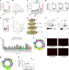
Figure 1. LL37 promotes LDL entry into cells.
(A) Visualization of pHrodo-LDL in THP-1 macrophages in the absence or presence of LL37. (B) Total fluorescence of pHrodo-LDL in THP-1 macrophages treated as in A (n = 5 per group). (C) FACS analysis and (D) proportion of CD45+ pHrodo-LDL+ THP-1 cells after treatment with LL37 (n = 6 per group). (E) Comparison of pHrodo-LDL or pHrodo-oxLDL uptake in the presence or absence of LL37 in THP-1 macrophages (n = 6–7 per group). (F) Dose-dependent uptake of pHrodo-LDL at the indicated concentrations of LL37 in THP-1 macrophages (n = 4 per concentration). (G and H) Uptake of pHrodo-LDL into HMDMs (n = 3 per group) (G), primary murine peritoneal macrophages (n = 5 per group) (H), HUVECs (n = 5 per group) (I), HAoECs (n = 5 per group) (J), and EA.hy926 endothelial cells (n = 4 per group) (K) treated with LL37. (L) Representative images of Dil-LDL uptake (red) and LL37 (green) in mouse aortas treated with LL37. White dotted lines outline the endothelial layer. (M) Proportion of positive fluorescence areas for Dil-LDL in aortic endothelium in presence and absence of LL37. Scales bars: 50 μm (A and L). Data indicate mean ± SEM. *P < 0.05, **P < 0.01, ***P < 0.001, and ****P < 0.0001, by 2-tailed Student’s t test (Student’s t test relative to no treatment in F).
Figure 2
Figure 2. Sequence elements of LL37 that promote uptake of LDL.
(A) Uptake of pHrodo-LDL into THP-1 cells treated with IL-26 or LL37 (n = 4 per group). (B) Phylogenic tree of the cathelicidin gene family. (C) pHrodo-LDL uptake into THP-1 cells treated with cathelicidin peptides from the indicated species (n = 4 in each group). (D and E) FACS analysis of pHrodo-LDL+ cells in CD45+CD11b+F4/80+ gated macrophages following peritoneal injection of pHrodo-LDL (n = 8–15 in each group). (F) SAXS profile of LDL incubated with LL37, LL34, or Cramp at a P/L ratio of 3:35. Arrows show the location of the first peak in the intensity profile, qpeak-LDL = 0.036Å–1, qpeak-LDL-Cramp = 0.032Å–1, qpeak-LDL-LL34 = 0.029Å–1, and qpeak-LDL-LL37 = 0.028Å–1. (G) Schematic of the size and structure of the LDL particle and complexes based on the fitted models of concentric core-shell ellipsoids to the SAXS spectra. The dimensions are given in angstrom. (H and I) Coimmunoprecipitation (IP) of biotinylated LDL and detection with anti-LL37 (H) or anti-Cramp (I) antibodies. (J) pHrodo-LDL uptake into THP-1 cells after addition of LL37, LL34, or LL34 with alanine substitutions at positions 1–34 (LL34 L1A-R34A) (n = 6 per group). (K) Helical wheel plot of LL34 with green circles indicating substitutions resulting in more than a 30% decrease in LDL uptake compared with parent LL34 peptide. (L) Representative immunofluorescence study of Dil-LDL aggregate cultured with LL37, LL34, or LL34-I13A. Scale bars: 20 μm. (M) Helical wheel plot of LL34, in which green circles indicate the position where alanine substitution resulted in more than a 50% decrease in aggregate fluorescence. (N) Linear regression analysis for associations between LDL uptake and fluorescence of LDL aggregate induced by the LL34 mutant peptides. Data indicate the mean ± SEM; *P < 0.05, **P < 0.01, and ****P < 0.0001, by Dunnett’s test (C) or 1-way ANOVA multiple-comparison test (A and E).
Figure 3
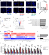
Figure 3. LL37 enhances the binding of LDL to its receptors.
(AD) pHrodo-LDL uptake into THP-1 cells ± LL37 after pretreatment with Pitstop or Genistein (A), anti-LDLR antibody (B), anti–SR-B1 antibody (C), or anti-CD36 antibody (D) (n = 4–7 per group). (EG) PLA between LDL and LDL receptors of THP-1 cells treated with biotinylated LDL with or without LL37. Schema (E), representative PLA images detecting an association between LDL and LDLR (F), and fluorescence quantification of positive signal (n = 4 per group) (G). (HJ) PLA between LL37 and LDL receptors of THP-1 cells treated with LDL with or without LL37. Schema (H), representative images detecting association between LL37 and LDLR (I), and fluorescence quantification of positive signals (n = 4 per group) (J). Scale bars: 10 μm. Data indicate the mean ± SEM. *P < 0.05, **P < 0.01, ***P < 0.001, and ****P < 0.001, by Dunnett’s test (A), 2-tailed Student’s t test (BD), or 1-way ANOVA multiple-comparison test (G and J).
Figure 4
Figure 4. LL37 and LDL increase intracellular lipid and alter macrophage gene expression.
(AC) Representative images of THP-1 cells treated with LDL with or without LL37 after staining with filipin (blue) to detect free cholesterol (A), or with Nile red (red) to detect lipids, and with DAPI (blue) to detect DNA (B), or with Bodipy (green) to detect lipids and DAPI (blue) to detect DNA (C). Scale bars: 50 μm (A) and 20 μm (B and C). (D) Quantitative analysis of signal intensity in THP-1 cells after Bodipy staining as in C (n = 4 per group). (EI) RNA-Seq analyses of THP-1 cells treated with LDL with or without LL37 for 24 hours (n = 3 per group). (E) PCA plot of the transcriptional profile. (F) Volcano plot of differentially expressed genes between no treatment and LDL plus LL37. (G) GO term analysis and (H) heatmap visualization of selected genes downregulated by LDL plus LL37 treatment compared with LDL or LL37 monotherapy. (I) Transcription factors predicted to influence the expression of genes shown in H. (J) qPCR quantification of mRNA expression for the indicated genes in THP-1 cells treated with LDL with or without LL37 (n = 4 per group). Data indicate the mean ± SEM. *P < 0.05, **P < 0.01, ***P < 0.001, and ****P < 0.001, by 1-way ANOVA multiple-comparison test.
Figure 5
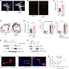
Figure 5. Transgenic expression of CAMP enhances the development of atherosclerosis.
(AF) Apoe–/– and LL37tg/tg Apoe–/– mice were fed a HFD for 10 weeks. (A) Representative images of the aortic arch. (B and C) Representative en face images of aortas stained with oil red (B) to detect atherosclerotic plaques and quantitation of lesion surface area (C) (n = 13 in Apoe–/– mice, n = 12 in LL37tg/tg Apoe–/– mice). (D and E) Representative images of oil red/hematoxylin-stained aortic sinus sections (D) and quantitation of plaque area (E) (n = 13 in Apoe–/– mice, n = 11 in LL37tg/tg Apoe–/– mice). Scale bar: 500 μm (D). (F) Mouse serum concentrations of total cholesterol and LDL cholesterol (n = 4 per group fed a ND, n = 8 per group fed a HFD, respectively). (G) Coimmunoprecipitation of serum from Apoe–/– mice or LL37tg/tg Apoe–/– mice fed a ND with anti-LL37 and detection with anti-LL37 and anti-apoB. (H) Coimmunoprecipitation of human serum from healthy blood donors with anti-LL37 and detection with anti-LL37 and anti-apoB. (I and J) Representative images of Nile red/LL37–stained plaques (I) and CD68/ LL37-stained plaques (J) in LL37tg/tg Apoe–/– mice. Scale bars: 50 μm (I and J). Original magnification, ×3 (enlarged inset on right in J). (K) Linear regression analysis of human plasma LL37 and PC-oxPL in patients with atherosclerosis (n = 20). Data indicate the mean ± SEM. **P < 0.01 and ****P < 0.0001, by 2 tailed Student’s t test (C and E) or linear regression analysis (K).

Similar articles

Cited by

References

    1. Wu Q, et al. Natural compounds from botanical drugs targeting mTOR signaling pathway as promising therapeutics for atherosclerosis: A review. Front Pharmacol. 2023;14:1083875. doi: 10.3389/fphar.2023.1083875. - DOI - PMC - PubMed
    1. Moore KJ, et al. Macrophages in atherosclerosis: a dynamic balance. Nat Rev Immunol. 2013;13(10):709–721. doi: 10.1038/nri3520. - DOI - PMC - PubMed
    1. Hua TC, et al. Cardiovascular comorbidities in patients with rosacea: A nationwide case-control study from Taiwan. J Am Acad Dermatol. 2015;73(2):249–254. doi: 10.1016/j.jaad.2015.04.028. - DOI - PubMed
    1. Tsai TY, et al. Cardiovascular risk and comorbidities in patients with rosacea: a systematic review and meta-analysis. Acta Derm Venereol. 2020;100(17):adv00300. doi: 10.2340/00015555-3668. - DOI - PMC - PubMed
    1. Prodanovich S, et al. Association of psoriasis with coronary artery, cerebrovascular, and peripheral vascular diseases and mortality. Arch Dermatol. 2009;145(6):700–703. doi: 10.1001/archdermatol.2009.94. - DOI - PubMed