. 2024 Jan 16;121(3):e2317668121.
doi: 10.1073/pnas.2317668121. Epub 2024 Jan 9.

Disruption of DNA methylation-mediated cranial neural crest proliferation and differentiation causes orofacial clefts in mice

Affiliations

Disruption of DNA methylation-mediated cranial neural crest proliferation and differentiation causes orofacial clefts in mice

Caden M Ulschmid et al. Proc Natl Acad Sci U S A. .

Abstract

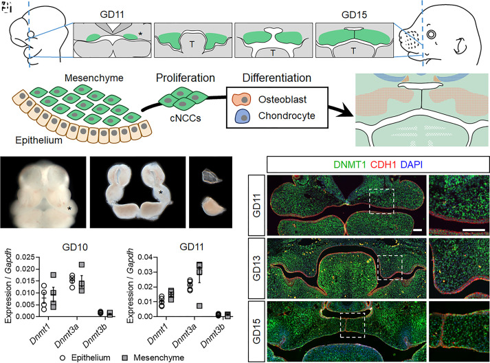
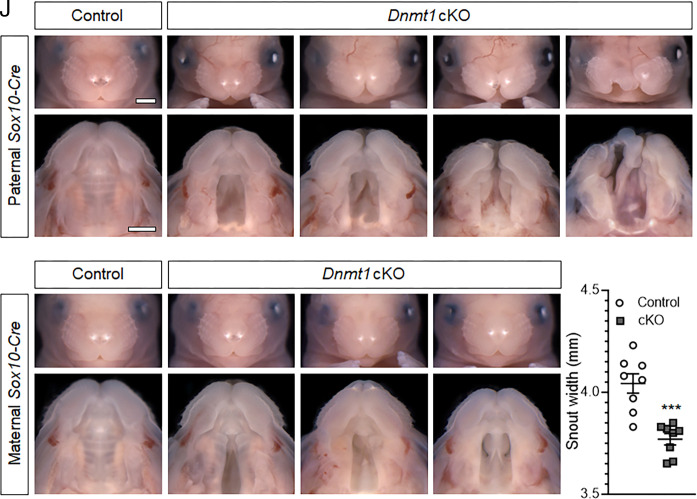
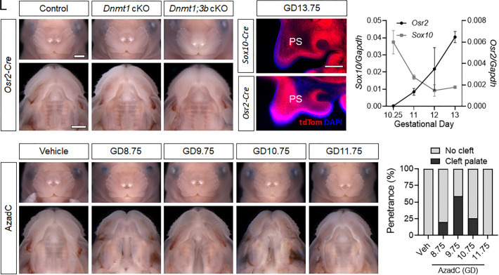
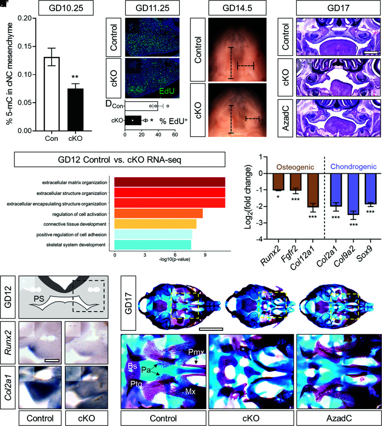
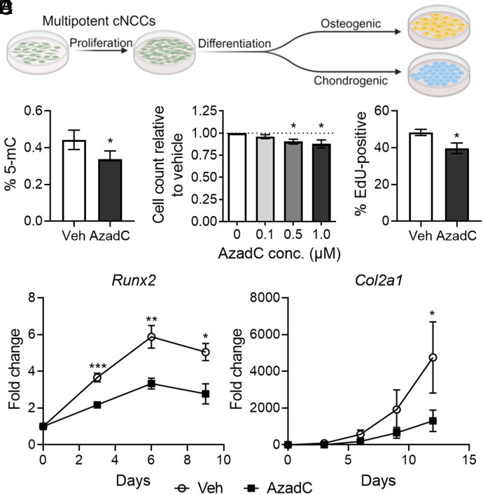
Orofacial clefts of the lip and palate are widely recognized to result from complex gene-environment interactions, but inadequate understanding of environmental risk factors has stymied development of prevention strategies. We interrogated the role of DNA methylation, an environmentally malleable epigenetic mechanism, in orofacial development. Expression of the key DNA methyltransferase enzyme DNMT1 was detected throughout palate morphogenesis in the epithelium and underlying cranial neural crest cell (cNCC) mesenchyme, a highly proliferative multipotent stem cell population that forms orofacial connective tissue. Genetic and pharmacologic manipulations of DNMT activity were then applied to define the tissue- and timing-dependent requirement of DNA methylation in orofacial development. cNCC-specific Dnmt1 inactivation targeting initial palate outgrowth resulted in OFCs, while later targeting during palatal shelf elevation and elongation did not. Conditional Dnmt1 deletion reduced cNCC proliferation and subsequent differentiation trajectory, resulting in attenuated outgrowth of the palatal shelves and altered development of cNCC-derived skeletal elements. Finally, we found that the cellular mechanisms of cleft pathogenesis observed in vivo can be recapitulated by pharmacologically reducing DNA methylation in multipotent cNCCs cultured in vitro. These findings demonstrate that DNA methylation is a crucial epigenetic regulator of cNCC biology, define a critical period of development in which its disruption directly causes OFCs, and provide opportunities to identify environmental influences that contribute to OFC risk.

Keywords: birth defects; epigenetics; neural crest; orofacial morphogenesis.


Conflict of interest statement

Competing interests statement:The authors declare no competing interest.

Figures

Fig. 1.
Fig. 1.
DNMT1 is expressed in the epithelium and mesenchyme throughout palate morphogenesis. (A) A schematic of palatal shelf morphogenesis depicting their emergence as expansions of the maxillary processes (*), vertical growth along the sides of the tongue (T), and subsequent elevation, approximation, and fusion at the midline. (B) A cartoon of the maxillary process showing surface epithelium covering dense mesenchyme composed of multipotent cNCCs that rapidly proliferate and then differentiate into osteoblasts and chondrocytes. (CE) Facial growth centers, including the maxillary processes (*), in an intact GD11 embryo (C), after microdissection (D), and after enzymatic separation of the epithelium and mesenchyme (E). (F and G) Expression of the three Dnmt genes in the epithelium and mesenchyme of gestational day (GD)10 (F) and GD11 (G) maxillary tissue was assessed by qRT-PCR. Data points representing pooled tissue from independent litters are shown along with mean ± SEM. (HJ’) Immunohistochemical staining for DNMT1 (green) on sections through the median aspect of the developing palate. Sections are also stained for CDH1 (red) to mark epithelium. DAPI (blue) was used to stain nuclei. (Scale bars: 100 µm.)
Fig. 2.
Fig. 2.
Dnmt1 in the cranial neural crest mesenchyme is required for orofacial morphogenesis (AE’) Representative face and palate images of targeted Dnmt1 deletion from paternally inherited Sox10-Cre of control (Sox10-Cre;Dnmt1fl/fl) and conditional knockout (cKO, Sox10-Cre+;Dnmt1fl/fl) fetuses at GD17. (Scale bar: 1 mm.) (FI’) Representative face and palate images of targeted Dnmt1 deletion from maternally inherited Sox10-Cre of control (Sox10-Cre;Dnmt1fl/fl) and conditional knockout (cKO, Sox10-Cre+;Dnmt1fl/fl) fetuses at GD17. (J) Linear measurements of snout width of control (Sox10-Cre;Dnmt1fl/fl) and cKO (Sox10-Cre+;Dnmt1fl/fl) fetuses. Data points represent individual fetuses and are shown with mean ± SEM. ***P < 0.001 by an unpaired t test.
Fig. 3.
Fig. 3.
The temporal requirement for Dnmt1 is restricted to initial palate morphogenesis. (AC’) Representative face and palate images of targeted Dnmt1 deletion or Dnmt1 and Dnmt3b (Dnmt1;3b) double deletion from paternally inherited Osr2-Cre of control (Osr2-Cre;Dnmt1fl/fl), conditional Dnmt1 knockout (cKO, Osr2-Cre+;Dnmt1fl/fl), and conditional Dnmt1;Dnmt3b double knockout (Osr2-Cre+;Dnmt1fl/fl;Dnmt3b fl/fl) fetuses at GD17. (Scale bar: 1 mm.) (D and E) Representative fluorescent microscopy images of tdTomato (tdTom, red) reporter expression from Sox10-Cre or Osr2-Cre in palatal shelves (PS) at GD13.75. Nuclear staining shown with DAPI (blue). (Scale bar: 0.2 mm.) (F) Expression of Sox10 and Osr2 in isolated maxillary/palatal shelf mesenchyme at the indicated stage of gestation as assessed by qRT-PCR. Data points represent the mean ± SEM of n = 3 samples per time point. (GK’) Face and palate images of wild-type fetuses at GD17 exposed to vehicle (PBS) or 5-aza-2′-deoxycytidine (AzadC) at the indicated gestational day. (L) Penetrance of cleft palate in wild-type GD17 fetuses exposed to vehicle (veh) or AzadC between GD8.75 and 11.75.
Fig. 4.
Fig. 4.
DNA methylation regulates cNCC proliferation and differentiation. (A) Global DNA methylation as assessed by 5-mC ELISA in maxillary process mesenchyme tissue of control (Con, Sox10-Cre+;Dnmt1fl/+) and conditional knockout (cKO, Sox10-Cre+;Dnmt1fl/fl) embryos at GD10.25. N = 7 embryos were used for each genotype, and means ± SEM are shown. **P < 0.01 compared to control by unpaired t test. (B and C) Representative examples of EdU incorporation in sections through the maxillary processes of control (Sox10-Cre+;Dnmt1fl/+) and conditional knockout (cKO, Sox10-Cre+;Dnmt1fl/fl) embryos. (D) Quantification of percent EdU-positive cells. N = 3 embryos were used for each genotype, with data points representing individual embryos shown along with mean ± SEM. *P < 0.05 by the t test. (E and F) Light images of the palatal shelves of GD14.5 control (Sox10-Cre;Dnmt1fl/+) and cKO (Sox10-Cre+;Dnmt1fl/fl) fetuses showing representative linear measurements of palatal shelf width and length. (GI) Representative H&E-stained coronal sections showing the region of the palatal shelves in control, cKO (Sox10-Cre+;Dnmt1fl/fl), or AzadC-treated (GD9.75) fetuses. (Scale bar: 0.5 mm.) (J) GO analysis from RNA sequencing conducted on palatal shelves isolated from control (Sox10-Cre+;Dnmt1fl/+) and cKO (Sox10-Cre+;Dnmt1fl/fl) embryos at GD12. (K) Expression of established cNC osteogenic and chondrogenic markers from RNA sequencing conducted on palatal shelves isolated from control (Sox10-Cre+;Dnmt1fl/+) and cKO (Sox10-Cre+;Dnmt1fl/fl) embryos at GD12. *P < 0.05 and ***P < 0.001. (L) Schematic of a GD12 section showing the palatal shelves (PS) with the dashed box indicating field of view of (MP). (MP) In situ hybridization for Runx2 and Col2a1 was performed on 100 µm vibratome sections of GD12 control (Sox10-Cre;Dnmt1fl/fl) and cKO (Sox10-Cre+;Dnmt1fl/fl) embryos. (Scale bar = 0.2 mm.) (QS’) Whole-mount bone and cartilage staining was performed on control (Sox10-Cre;Dnmt1fl/+), cKO (Sox10-Cre+;Dnmt1fl/fl), or AzadC-treated (GD9.75) GD17 fetuses with Insets (dashed boxes) showing higher magnification of the palatine bone region. (Scale bar = 2 mm.) Bs, basisphenoid; Ptg, pterygoid process; Pa, palatine bones; Pmx, premaxilla; Mx, maxilla.
Fig. 5.
Fig. 5.
DNA methylation regulates cNCC proliferation and differentiation in cultured cNCCs. (A) Schematic depicting the proliferative and differentiation capacity of cNCCs cultured in vitro and experimental conditions to evaluate the impact of pharmacological DNMT inhibition. (B) Global DNA methylation as assessed by 5-mC ELISA in cultured cNCCs treated with PBS (vehicle, veh) or 1.0 µM AzadC for 48 h. N = 6 for each group with mean ± SEM shown. *P < 0.05 by the t test. (C) Cell counts were performed after 48-h treatments with the indicated concentrations of AzadC. N = 5 for each concentration and means ± SEM are shown. *P < 0.05 compared to the 0 µM AzadC group by ANOVA with Dunnett’s post hoc test for multiple comparisons. (D) EdU-positive cells were counted after 12 h of PBS (vehicle, veh) of 1.0 µM AzadC treatment. N = 5 biological replicates for each group and means ± SEM are shown. *P < 0.05 by the t test. (E and F) Cultured cNCCs were treated with PBS (vehicle, veh) or 1.0 µM AzadC for 48 h before undergoing differentiation conditions for osteogenic (E) or chondrogenic (F) differentiation. Expression of Runx2 or Col2a1 was assayed at the indicated days after the start of differentiation. N = 5 for each group at each time point and means ± SEM are shown. *P < 0.05, **P < 0.01, or ***P < 0.001 compared to the vehicle group at the same time point by t test.

References

    1. Rahimov F., Jugessur A., Murray J. C., Genetics of nonsyndromic orofacial clefts. Cleft Palate Craniofac J. 49, 73–91 (2012). - PMC - PubMed
    1. Boulet S. L., Grosse S. D., Honein M. A., Correa-Villaseñor A., Children with orofacial clefts: Health-care use and costs among a privately insured population. Public Health Rep. 124, 447–453 (2009). - PMC - PubMed
    1. Semb G., Brattström V., Mølsted K., Prahl-Andersen B., Shaw W. C., The Eurocleft study: Intercenter study of treatment outcome in patients with complete cleft lip and palate. Part 1: Introduction and treatment experience. Cleft Palate Craniofac J. 42, 64–68 (2005). - PubMed
    1. Wehby G. L., et al. , Academic achievement of children and adolescents with oral clefts. Pediatrics 133, 785–792 (2014). - PMC - PubMed
    1. Wehby G. L., Cassell C. H., The impact of orofacial clefts on quality of life and healthcare use and costs. Oral Dis. 16, 3–10 (2010). - PMC - PubMed

Substances

Supplementary concepts