. 2024 Jan 16;121(3):e2315354120.
doi: 10.1073/pnas.2315354120. Epub 2024 Jan 9.

Conversion of monoclonal IgG to dimeric and secretory IgA restores neutralizing ability and prevents infection of Omicron lineages

Affiliations

Conversion of monoclonal IgG to dimeric and secretory IgA restores neutralizing ability and prevents infection of Omicron lineages

Harold Marcotte et al. Proc Natl Acad Sci U S A. .

Abstract

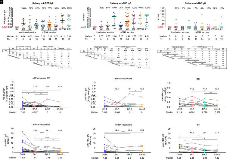
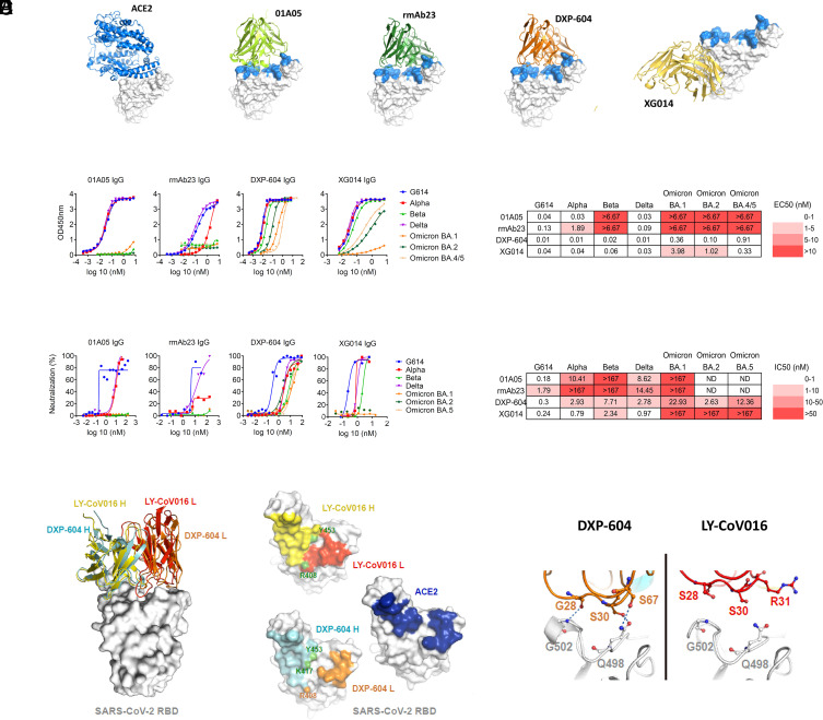
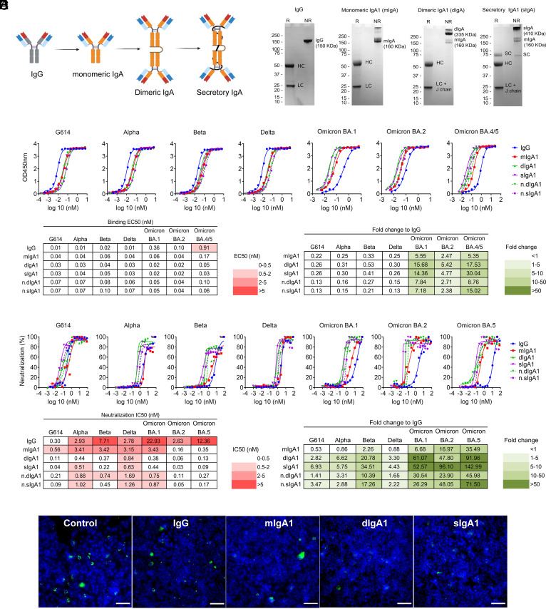
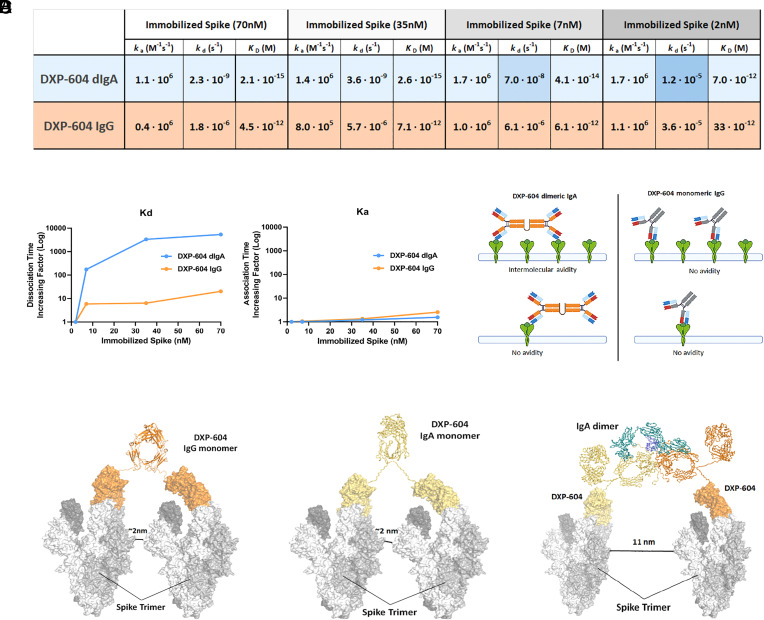
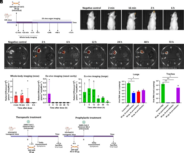
The emergence of Omicron lineages and descendent subvariants continues to present a severe threat to the effectiveness of vaccines and therapeutic antibodies. We have previously suggested that an insufficient mucosal immunoglobulin A (IgA) response induced by the mRNA vaccines is associated with a surge in breakthrough infections. Here, we further show that the intramuscular mRNA and/or inactivated vaccines cannot sufficiently boost the mucosal secretory IgA response in uninfected individuals, particularly against the Omicron variant. We thus engineered and characterized recombinant monomeric, dimeric, and secretory IgA1 antibodies derived from four neutralizing IgG monoclonal antibodies (mAbs 01A05, rmAb23, DXP-604, and XG014) targeting the receptor-binding domain of the spike protein. Compared to their parental IgG antibodies, dimeric and secretory IgA1 antibodies showed a higher neutralizing activity against different variants of concern (VOCs), in part due to an increased avidity. Importantly, the dimeric or secretory IgA1 form of the DXP-604 antibody significantly outperformed its parental IgG antibody, and neutralized the Omicron lineages BA.1, BA.2, and BA.4/5 with a 25- to 75-fold increase in potency. In human angiotensin converting enzyme 2 (ACE2) transgenic mice, a single intranasal dose of the dimeric IgA DXP-604 conferred prophylactic and therapeutic protection against Omicron BA.5. Thus, dimeric or secretory IgA delivered by nasal administration may potentially be exploited for the treatment and prevention of Omicron infection, thereby providing an alternative tool for combating immune evasion by the current circulating subvariants and, potentially, future VOCs.

Keywords: IgA; Omicron; SARS-CoV-2; antibody engineering; antibody therapy.


Conflict of interest statement

Competing interests statement:Y.C. and X.S.X. are listed as inventors on a patent on DXP-604 antibody (PCT/CN2021/093305) for Peking University. H.M., Y.C., L.H., X.S.X., and Q.P.-H. have filed a patent on DXP-604 IgA antibodies. All other authors declare that they have no competing interests.

Figures

Fig. 1.
Fig. 1.
Salivary anti-RBD IgA antibodies are produced at low levels following vaccination. (AC) Salivary anti-RBD IgA (A), IgG (B), and IgM (C) antibodies in different vaccination groups. For each group, the number of samples (n=) and median antibody titers are shown below the x-axis. Whiskers indicate the interquartile range. The results of anti-RBD antibodies are presented as arbitrary units (AU)/µg total IgA (salivary IgA), binding antibody units (BAU/mL) (salivary and plasma IgG) or arbitrary units (AU)/mL (salivary IgM). HV: heterologous vaccination (two doses of inactivated vaccine followed by a heterologous mRNA boost), Inf+Vac: one or two doses of mRNA vaccine after SARS-CoV-2 infection (during the G614 wave), BTI: breakthrough infection (during the BA.1, BA.2, and BA.5 waves) after inactivated and/or mRNA vaccines. A two-sided Mann–Whitney U test was used. (D and E) Salivary anti-RBD IgA (D) and IgG (E) antibodies against G614 and Omicron variants BA.1, BA.2, and BA.4/5 after the second (D2) and third (D3) doses of mRNA vaccine and following BTI in mRNA-vaccinated individuals. In AE, samples were collected 5 to 59 d (median day 20) after each mRNA dose including after mRNA heterologous boost, 6 to 92 d (median day 51) after doses 2 and 3 of inactivated vaccine, and 8 to 43 d (median day 19) after BTI. The number of fold differences of the median compared to G614 are indicated. A Wilcoxon paired-sample signed-rank test was used. *P < 0.05, **P < 0.01, ***P < 0.001, and ****P < 0.0001. ns, not significant. See also SI Appendix, Fig. S1.
Fig. 2.
Fig. 2.
Characterization of neutralizing antibodies 01A05, rmAb23, DXP-604, and XG014. (A) In silico binding of ACE2 and IgG antibodies to RBD. The ACE2 receptor binding motif is indicated (blue). (B and C) Binding to RBD (B) and neutralization (C) of G614 and VOCs by IgG antibodies. The EC50 and IC50 and fold-change differences between IgG and IgA antibody forms are indicated. (D) Overlaid crystal structures of LY-CoV016 Fab (PDB ID: 7C01) and DXP-604 Fab 473 (PDB ID: 7CH4) in complex with SARS-CoV-2 RBD (Left picture) and the footprints of LY-CoV016, DXP-604, and ACE2 (PDB ID: 6M0J) on SARS-CoV-2 RBD. Atoms of the RBD within 5.0 Å of the antibodies or ACE2 are colored yellow (LY-CoV016 H), red (LY-CoV016 L), cyan (DXP-604 H), orange (DXP-604 L), or blue (ACE2) (Right picture). (E) Hydrogen bonds were formed between S30/S67 in the light chain of DXP-604 and RBD Q498, which is a key ACE2-binding site, and between the main chain groups of G28 and RBD G502. See also SI Appendix, Figs. S2–S4.
Fig. 3.
Fig. 3.
Dimeric and secretory IgA1 showed enhanced binding and neutralization activity against VOCs. (A) Illustration showing antibodies engineered from IgG into monomeric, dimeric, and secretory IgA1. (B) SDS–PAGE under reducing (R) and nonreducing (NR) conditions showing the assembly and purity of DXP-604 IgG and IgA1 antibodies. HC, heavy chain; LC, light chain; SC, secretory component; J chain, joining chain. The J chain migrates at the same molecular weight as the light chain. (C and D) Binding to RBD (C) and neutralization (D) of G614 and VOCs by DXP-604 IgG and IgA [monomeric (mIgA1), dimeric (dIgA1), and secretory IgA1 (sIgA1)] antibodies. The EC50 and IC50 and fold-change differences between IgG and IgA1 antibody forms are indicated. n.dIgA1 and n.sIgA1 represent normalized values according to the number of binding sites. (E) Staining of virus following infection of Vero E6 cells with SARS-CoV-2 Omicron BA.1 preincubated with 3.3 nM DXP-604 IgG or IgA1 forms. Omicron BA.1-infected cells were used as a negative control (control). SARS-CoV-2 virus was visualized using Alexa 488 (green)-conjugated antibody, and the nucleus was stained with DAPI (blue). (Scale bar, 100 μM.) See also SI Appendix, Figs. S5 and S6.
Fig. 4.
Fig. 4.
Increased neutralization potency of DXP-604 dimeric IgA is associated with increased avidity. (A) ka/kd values obtained at different concentrations of immobilized antigen (Spike) for DXP-604 dimeric IgA1 and IgG forms. ka remains equal across concentrations whereas kd becomes ~1,000 times slower for the dIgA1, indicative of intermolecular avidity available only to the dIgA1. Shades of blue indicate the difference in kd value for DXP-604 dIgA1. (B) Plots of kd (Left) and ka (Right) at different concentrations of immobilized spike, highlighting intermolecular avidity effects (slower dissociation, same association) for the dIgA1 (blue) in comparison to the IgG (orange). (C) DXP-604 dIgA1 and monomeric IgG have different binding modes that are available when high or low quantities of S-trimers are immobilized on the surface of the SPR chip. (D) Computational simulation showing inter-Spike linking by DXP-604 monomeric IgG and IgA1, and dimeric IgA1 antibodies. The predicted distance between S-trimers necessary for interlinking is indicated.
Fig. 5.
Fig. 5.
Intranasal administration of dimeric IgA in hACE2 mice is protective against Omicron BA.5. (A) Experimental design for the evaluation of antibody biodistribution after administration of 60 µg DXP-604 dIgA1 (labeled with Alexa Fluor 647). (B) Representative whole-body images. (C) Representative ex vivo images. N, nasal cavity; Lu, lungs and section of the trachea; H, heart; Li, liver; S, spleen; K, kidney. In (B) and (C), the negative control mice received PBS only. (D) Quantification of fluorescence signals. Data are presented as mean ± SD of five mice (whole-body imaging) or three mice (ex vivo nasal cavity and lung imaging). (E) Experimental design for the evaluation of DXP-604 dIgA1 using therapeutic and prophylactic intranasal administration. (F) Viral loads in the lung and tracheal tissues at 3 d post-infection of Omicron BA.5-infected mice after administration of a single dose of DXP-604 dIgA1 in a therapeutic (60 µg, 2 h post-infection) and prophylactic (40 or 60 µg, 4 h preinfection) setting. Viral loads are expressed as the mean ± SD for three mice. A two-sided unpaired t test was used. *P < 0.05 and **P < 0.01.

References

    1. Chen Y., et al. , Emerging SARS-CoV-2 variants: Why, how, and what’s next? Cell Insight 1, 100029 (2022). - PMC - PubMed
    1. Cao Y., et al. , Omicron escapes the majority of existing SARS-CoV-2 neutralizing antibodies. Nature 602, 657–663 (2022). - PMC - PubMed
    1. Viana R., et al. , Rapid epidemic expansion of the SARS-CoV-2 Omicron variant in southern Africa. Nature 603, 679–686 (2022). - PMC - PubMed
    1. Wang Q., et al. , Alarming antibody evasion properties of rising SARS-CoV-2 BQ and XBB subvariants. Cell 186, 279–286 (2022). - PMC - PubMed
    1. Mannar D., et al. , SARS-CoV-2 Omicron variant: Antibody evasion and cryo-EM structure of spike protein–ACE2 complex. Science 375, 760–764 (2022). - PMC - PubMed