Comparison of posttreatment stability after total mandibular arch distalization with mini-implants and mandibular setback surgery
- PMID: 38195065
- PMCID: PMC10893925
- DOI: 10.2319/062723-447.1
Comparison of posttreatment stability after total mandibular arch distalization with mini-implants and mandibular setback surgery
Abstract
Objectives: To compare posttreatment stability in skeletal Class III patients between those treated by total mandibular arch distalization (TMAD) with buccal mini-implants and those by mandibular setback surgery (MSS).
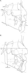
Materials and methods: The samples included 40 Class III adults, 20 treated by TMAD using buccal interradicular mini-implants and 20 treated with MSS. Lateral cephalograms were taken at pretreatment, posttreatment, and at least 1-year follow-up, and 24 variables were compared using statistical analysis.
Results: Mandibular first molars moved distally 1.9 mm with intrusion of 1.1 mm after treatment in the TMAD group. The mandibular incisors moved distally by 2.3 mm. The MSS group exhibited a significant skeletal change of the mandible, whereas the TMAD group did not. During retention, there were no skeletal or dental changes other than 0.6 mm labial movement of the mandibular incisors (P < .05) in the MSS group. There was 1.4° of mesial tipping (P < .01) and 0.4 mm of mesial movement of the mandibular molars and 1.9° of labial tipping (P < .001) and 0.8 mm of mesial movement of the mandibular incisors in the TMAD group. These dental changes were not significantly different between the two groups.
Conclusions: The TMAD group showed a slightly decreased overjet with labial tipping of the mandibular incisors and mesial tipping of the first molars during retention. Posttreatment stability of the mandibular dentition was not significantly different between the groups. It can be useful to plan camouflage treatment by TMAD with mini-implants in mild-to-moderate Class III patients.
Keywords: Mandibular arch distalization; Mandibular setback surgery; Mini-implants; Stability.
© 2024 by The EH Angle Education and Research Foundation, Inc.
Figures


Similar articles
-
Comparison of treatment effects after total mandibular arch distalization with miniscrews vs ramal plates in patients with Class III malocclusion.Am J Orthod Dentofacial Orthop. 2022 Apr;161(4):529-536. doi: 10.1016/j.ajodo.2020.09.040. Epub 2021 Dec 23. Am J Orthod Dentofacial Orthop. 2022. PMID: 34953658
-
Effects of mandibular incisor extraction on anterior occlusion in adults with Class III malocclusion and reduced overbite.Am J Orthod Dentofacial Orthop. 1999 Feb;115(2):113-24. doi: 10.1016/s0889-5406(99)70337-9. Am J Orthod Dentofacial Orthop. 1999. PMID: 9971920
-
Camouflage treatment of skeletal Class III malocclusion with multiloop edgewise arch wire and modified Class III elastics by maxillary mini-implant anchorage.Angle Orthod. 2013 Jul;83(4):630-40. doi: 10.2319/091312-730.1. Epub 2013 Jan 11. Angle Orthod. 2013. PMID: 23311602 Free PMC article.
-
The effectiveness of pendulum, K-loop, and distal jet distalization techniques in growing children and its effects on anchor unit: A comparative study.J Indian Soc Pedod Prev Dent. 2016 Oct-Dec;34(4):331-40. doi: 10.4103/0970-4388.191411. J Indian Soc Pedod Prev Dent. 2016. PMID: 27681396
-
Comparison of treatment outcomes between skeletal anchorage and extraoral anchorage in adults with maxillary dentoalveolar protrusion.Am J Orthod Dentofacial Orthop. 2008 Nov;134(5):615-24. doi: 10.1016/j.ajodo.2006.12.022. Am J Orthod Dentofacial Orthop. 2008. PMID: 18984393 Clinical Trial.
Cited by
-
Camouflage treatment of severe skeletal class III malocclusion with effective torque control in an adolescent combined with forward functional shift and hypodivergent.BMC Oral Health. 2025 May 22;25(1):762. doi: 10.1186/s12903-025-06113-z. BMC Oral Health. 2025. PMID: 40405176 Free PMC article.
References
-
- Saito I, Yamaki M, Hanada K. Nonsurgical treatment of adult open bite using edgewise appliance combined with high-pull headgear and class III elastics Angle Orthod 2005. 75 277–283 - PubMed
-
- Kim YH, Han UK, Lim DD, Serraon ML. Stability of anterior openbite correction with multiloop edgewise archwire therapy: a cephalometric follow-up study Am J Orthod Dentofacial Orthop 2000. 118 43–54 - PubMed
-
- Janson G, de Souza JE, Alves Fde A, Andrade P, Jr, Nakamura A, de Freitas MR, Henriques JF. Extreme dentoalveolar compensation in the treatment of Class III malocclusion Am J Orthod Dentofacial Orthop 2005. 128 787–794 - PubMed
-
- Mora DR, Oberti G, Ealo M, Baccetti T. Camouflage of moderate Class III malocclusions with extraction of lower second molars and mandibular cervical headgear Prog Orthod 2007. 8 300–307 - PubMed
MeSH terms
LinkOut - more resources
Full Text Sources

