Review
. 2024 Apr;21(3):e00313.
doi: 10.1016/j.neurot.2023.e00313. Epub 2024 Jan 8.

The role of the motor thalamus in deep brain stimulation for essential tremor

Affiliations
Review

The role of the motor thalamus in deep brain stimulation for essential tremor

Clemens Neudorfer et al. Neurotherapeutics. 2024 Apr.

Abstract

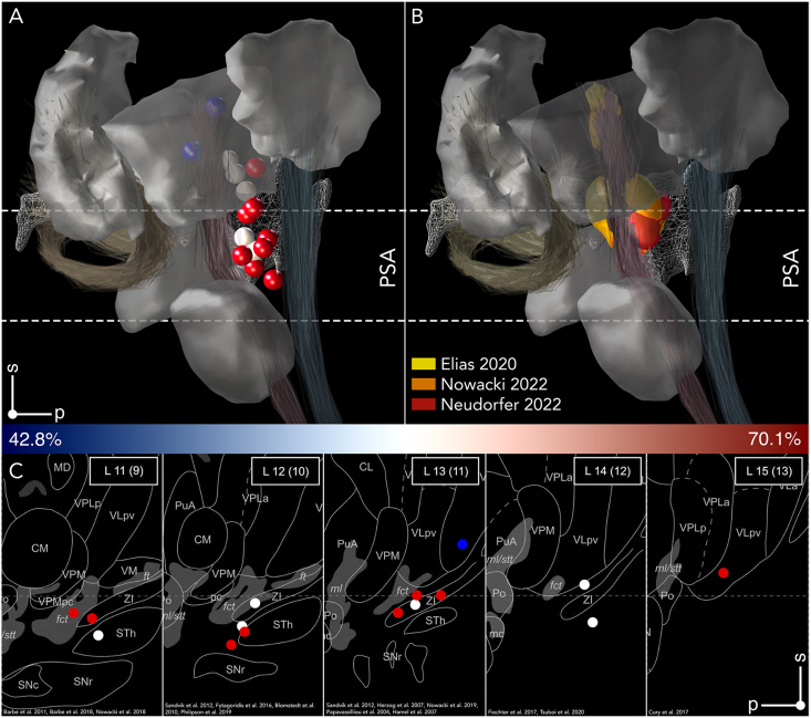
The advent of next-generation technology has significantly advanced the implementation and delivery of Deep Brain Stimulation (DBS) for Essential Tremor (ET), yet controversies persist regarding optimal targets and networks responsible for tremor genesis and suppression. This review consolidates key insights from anatomy, neurology, electrophysiology, and radiology to summarize the current state-of-the-art in DBS for ET. We explore the role of the thalamus in motor function and describe how differences in parcellations and nomenclature have shaped our understanding of the neuroanatomical substrates associated with optimal outcomes. Subsequently, we discuss how seminal studies have propagated the ventral intermediate nucleus (Vim)-centric view of DBS effects and shaped the ongoing debate over thalamic DBS versus stimulation in the posterior subthalamic area (PSA) in ET. We then describe probabilistic- and network-mapping studies instrumental in identifying the local and network substrates subserving tremor control, which suggest that the PSA is the optimal DBS target for tremor suppression in ET. Taken together, DBS offers promising outcomes for ET, with the PSA emerging as a better target for suppression of tremor symptoms. While advanced imaging techniques have substantially improved the identification of anatomical targets within this region, uncertainties persist regarding the distinct anatomical substrates involved in optimal tremor control. Inconsistent subdivisions and nomenclature of motor areas and other subdivisions in the thalamus further obfuscate the interpretation of stimulation results. While loss of benefit and habituation to DBS remain challenging in some patients, refined DBS techniques and closed-loop paradigms may eventually overcome these limitations.

Keywords: Deep brain stimulation; Human thalamic nomenclature; Motor thalamus; Parcellation; Tremor; Vim.


Conflict of interest statement

Declaration of competing interest The authors declare the following financial interests/personal relationships which may be considered as potential competing interests: Guenther Deuschl reports a relationship with Boston Scientific Corp that includes: consulting or advisory. Guenther Deuschl reports a relationship with Cavion that includes: consulting or advisory. Guenther Deuschl reports a relationship with Functional Neuromodulation that includes: consulting or advisory. Guenther Deuschl reports a relationship with Thieme Medical Publishers that includes: consulting or advisory. Andreas Horn reports a relationship with German Research Foundation that includes: funding grants. Andreas Horn reports a relationship with Deutsches Zentrum für Luft-und Raumfahrt that includes: funding grants.

Figures

Fig. 1
Fig. 1
Early depictions of the thalamus and its nuclear parcellation according. (A) Diagram of the brain in the sagittal plane featuring the principal cellular centers (labeled 4, 9, 14, and 19) of the thalamus as hypothesized by Luys. Note the significant functional emphasis on the processing of sensory information as illustrated by the input from the olfactory bulb (20), retina (13), spinal cord (8), and the inner ear (3). Information from the thalamus is relayed to the cortex before being transmitted to the striatum (1), from where it descends to the spinal cord. Arrows represent the presumed direction of information flow. (B) Three-dimensional representation of the thalamus and its input and output structures, viewed in the axial plane. (C) Drawing of a frontal section through the human thalamus displaying the centers moyens (9, 9’) and médians (10, 10’). Sagittal (D), axial (E), and coronal (F) transparent sections by Meynert suggesting poor differentiation of individual thalamic nuclei. (GI) Depiction of coronal transparent sections by Forel. (J,K) Coronal sections of a postmortem brain as reproduced by von Monakow, showing retrograde thalamic degeneration within thalamus (K) secondary to lesioning of the orbital and anterior insular cortex (J). Adopted from Luys [3] (A–C), Meynert [14] (D–F), Forel [15] (G–I), and von Monakow [16] (J,K).
Fig. 2
Fig. 2
Heterogeneity in the delineation of the motor thalamus across atlases. The figure illustrates different parcellation schemes of the thalamus (in gray) and the motor thalamic nuclei (red), as defined by different anatomical atlases. The variability can in part be attributed to the diverse methods used to identify the boundaries of the thalamic nuclei and the translation of findings from non-human primates to human anatomy, in part by different levels of granularity in their segregations. The reconstruction by Ewert uses the Hassler nomenclature and features the motor thalamus as assigned by Hassler [31] capturing only the ventral portion of the thalamus. Notably, while the term ‘motor thalamus' typically refers to the territory occupied by cerebellar, pallidal, and nigral. Contemporary pathway tracing studies reveal that both pallidal and cerebellar projection zones span up to the dorsal edge of the thalamus, encompassing Hassler's dorsal nuclei. Historically, the emphasis on the ventral part is due lack of experimental data at the time and its significance as a neurosurgical intervention target. Atlases adopted from Ilinsky et al. 2018 [32], Ewert et al. 2017 [33] which is based on histological data from Chakravarty et al. 2006 [34], Morel 2007 [35], Saranathan et al. 2019 [36] (based on the Morel atlas), Ding et al. 2020 [37] and Iglesias et al. 2018 [38] (based on the Morel atlas) as available in Lead-DBS.
Fig. 3
Fig. 3
Evolution of the understanding of subcortical afferent termination zones in the motor thalamus. The figure features the chronological progression in understanding the topography of termination zones within the motor thalamus, focusing on the cytoarchitectonic confines outlined by Hassler (A-C, E) and Morel (D). Outlines are shown in the axial plane 2 ​mm above the intercommissural line. Colors code for afferent termination zones of cerebellothalamic (dark red), pallidothalamic (orange), and nigrothalamic (green) projections as identified and revised by different groups. Hassler's description of thalamic termination zones additionally incorporates vestibulothalamic afferents (blue) projecting to Vim.i, ascending pathways for muscle spindle excitations originating in Clark's column (dark orange) that reach Vim.e, and oculomotor afferents projecting from the interstitial nucleus of Cajal to Voi (A,B) Nomenclature and zones as originally described by Hassler [16]. I Afferent termination zones extrapolated from tracing data in monkeys by Ilinsky et al. [39]. (D) Outlines from Morel's atlas, nomenclature introduced by Walker, which was revised and translated to humans by Hirai and Jones and adopted by Morel et al [25]. I Revised nomenclature based on immunocytochemical staining specific for afferent zones in human thalamus [32,40]. Adopted from Schaltenbrand and Wahren [1] (A-C,E) and Morel et al [35]. (D).
Fig. 4
Fig. 4
Variability in the delineation of V.im/VL and the cerebellothalamic afferent system. (A) N-map of Vim/VL parcellations, derived from the atlases in Fig. 2 is shown in axial, coronal, and sagittal planes, superimposed on a 100 ​μm resolution, 7T brain scan in MNI 152 space. The maps were generated by summing the binary atlas parcellations in a voxel-wise manner. (B) Passage of the cerebellothalamic tract as derived from different anatomical atlases. Tracts are featured exhibit significantly greater variability in their passage in the sagittal plane (right), as compared to the coronal plane (left) at the subthalamic level. Note that tracts converge onto V.im and are constrained with respect to their termination zones (refer also to Table 2). Adopted from Petersen et al. 2019 [64] and Middlebrooks et al. 2020 [65].
Fig. 5
Fig. 5
Anatomical description of the posterior subthalamic area (PSA). (A) Inferior (from below), (B) medial, and (C) sagittal views of PSA and its anatomical confines. Pallidothalamic fibers constrain the PSA anteriorly and traverse the internal capsule through ansa lenticularis (al, orange) and fasciculus lenticularis (fl, orange) forming the fasciculus thalamicus (ft, orange) medial to the STN, which ascends into the anterior nuclei of the motor thalamus. PSA adjoins the posterior border of STN. The caudal border of PSA is formed by the medial lemniscus (ml, blue) and spinothalamic tract (stt, blue), which ascend into the sensory thalamic nuclei. The main structures comprised within the PSA are zona incerta (ZI), prelemniscal radiation, which consists of fibers ascending from the mesencephalic reticular formation (not shown), as well as the cerebellothalamic tract (fct). Abbreviations: bic brachium of the inferior colliculus; CM centromedian thalamic nucleus; fct fasciculus cerebellothalamicus; fl fasciculus lenticularis; ft fasciculus thalamicus; ic internal capsule; MGN medial geniculate nucleus; ml medial lemniscus; PuM, medial pulvinar; PSA posterior subthalamic area; pt, pallidothalamic tract; SG suprageniculate nucleus; SNc substantia nigra, pars compacta; SNr substantia nigra, pars reticulata; STN nucleus subthalamicus; stt spinothalamic tract; VLa ventral lateral anterior thalamic nucleus; VLpv ventral lateral posterior nucleus, ventral portion; VM ventral medial nucleus; VPM ventral posteromedial nucleus. (C) adapted from Morel et al [35].
Fig. 6
Fig. 6
The ‘electrophysiologically defined V.im’ as pioneered by Albe-Fessard and Guiot. (A) Sagittal section through the thalamus, showing two of the most commonly chosen trajectories for stereotactic lesioning by the group using a parietal approach. At the thalamic level, the trajectory typically traversed pulvinar (Pulv.), nucleus lateralis posterior (L.P.), the superior part of the nuvleus ventralis posterior (V.P.), and nucleus ventralis lateralis (defined as V.O.p by Albe-Fessard et al.). (B) Differentiation of thalamic nuclei based on oscillographic recordings. During thalamic exploration, spike activity intensifies in the nucleus lateralis posterior (LP) and is especially prominent in the nucleus ventralis posterior (VP). This activity diminishes in the nucleus reticularis and is significantly reduced in the internal capsule. Entering the globus pallidus (G.P.i.) leads to another spike activity surge. Adopted from Albe-Fessard et al [75].
Fig. 7
Fig. 7
Spatial relationship between anatomical structures and previously reported DBS targets in the literature. (A) Medial view of the thalamus and posterior subthalamic area (PSA) features previously reported target coordinates associated with symptom improvements following DBS of the V.im and PSA. Targets associated with improved clinical outcome are closely related to the cerebellothalamic outflow tract (dark red) and the zona incerta (white mesh) at the level of the PSA. For a comprehensive overview of study characteristics, MNI coordinates, and clinical outcomes, see Table 3. (B) Relationship of publicly available DBS hotspots to anatomical substrates. Probabilistic maps were derived from large cohorts of ET-patients undergoing DBS and are featured in 3D. (C) Sagittal sections feature previously reported target coordinates within PSA and motor thalamus. Mediolateral planes, expressed in millimeters relative to the intercommissural line and estimated from the margin of the third ventricle (indicated in parentheses), are specified in the upper right corner of each section. Sections are aligned with the intercommissural line (horizontal dashed line).
Fig. 8
Fig. 8
Comparison of conventional and advanced imaging sequences used for surgical planning in V.im-DBS. Top row: Conventional imaging sequences, including T1w-imaging, T2w-imaging, and inversion recovery sequences have been clinically effective, but lack of contrast at the thalamic level, which hinders the direct identification of the DBS target and surrounding anatomical substrates during surgical planning. Bottom row: Advanced imaging sequences such as quantitative susceptibility mapping (QSM) and white-matter nulled (WMnulled) sequences (e.g., fast gray-matter acquisition T1 inversion recovery (FGATIR) sequences), provide superior contrast at the thalamic level allowing the identification of individual thalamic nuclei and white matter tracts, which can be used to tailor the target towards the patient's anatomy. Increases in magnetic field strength enhance the signal-to-noise-ratio and yield improved resolutions, however, they also have distinct disadvantages including increased sensitivity to motion and susceptibility artifacts. Green circles denote a hypointensity at the level of the PSA as identifiable on FGATIR and WMnulled sequences, considered to be the imaging equivalent of the cerebellothalamic tract (CTT). Overlap of stimulation volumes with the hypointensity was shown to predict clinical outcome in patients undergoing DBS for ET.
Fig. 9
Fig. 9
Efficacy data available to the FDA for the approval of Vim-DBS of the first multicenter, uncontrolled trials for therapy for ET and PD in Europe and North America. (A, B) Reduction of mean tremor scores (0–4) contralateral to the stimulated side in a randomized ON/OFF design as the main outcome parameter of the two studies at 3 months after implantation. This outcome was measured for 16 ​ET-patients and 13 PD-patients in the US-study and for 6 Patients with ET and 9 patients with PD in the EU study, respectively. (C, D) Open one-year follow-up of tremor improvement during open ON- and OFF-assessments for essential tremor (EU n ​= ​28 patients; USA n ​= ​45) and PD (EU: n ​= ​57; USA n ​= ​39). The graphs illustrate how the criteria for the approval of such therapies was handled at the end of the last century. Adopted from Food and Drug Administration (FDA) [81].
Fig. 10
Fig. 10
Main results of the Abbott/St. Jude study. (A) Extend and persistence of the main effect on lateralized symptoms. Unilateral stimulation in the whole study-cohort of 122 patients showed a profound effect on the lateralized tremor score which persisted over 90, 180 and 365 days [167]. The stim-OFF score is 21 ​% lower than the baseline score, which is most likely attributable to a microlesion effect induced during electrode placement intraoperatively. Notably for this study this main result was confirmed by a randomized blinded evaluation of the video-recorded tremor assessment of a subgroup (n ​= ​76) at baseline and 90 days [170]. (B) Bilateral stimulation has a stronger effect on axial symptoms than unilateral stimulation. The axial manifestations of essential tremor are captured at 6 months after first side and 3 months after second side surgery by the axial score and differ for uni- and bilateral stimulation for the subgroup of 38 patients with bilateral stimulation (All bars represent mean ​± ​SEM in percent of the respective baseline scores).

References

    1. Schaltenbrand G., Wahren W., Hassler R.G. Atlas for Stereotaxy of the Human Brain. 2d, rev.Enl. Thieme; Stuttgart: 1977.
    1. Jones E.G., editor. The thalamus [Internet] Springer US; Boston, MA: 1985. http://link.springer.com/10.1007/978-1-4615-1749-8 [cited 2023 Jul 28]. Available from: - DOI
    1. Luys J.B. J.B. Baillière et fils; 1865. Recherches sur le système nerveux cérébro-spinal: sa structure, ses fonctions et ses maladies.
    1. Broca P. Perte de la parole, ramouissement chronique et destruction partielle du lobe antérieur gauche du cerveau | Max Planck Institute. Bulletin de la Société Anthropologique. 1861:235–238.
    1. Nissl F. Die Grosshirnanteile des Kaninchens. Archiv f Psychiatrie. 1913;52:867–953.

Publication types

MeSH terms