Review
. 2024 Feb 20;11(2):ENEURO.0435-23.2023.
doi: 10.1523/ENEURO.0435-23.2023. Print 2024 Feb.

Deprivation-Induced Plasticity in the Early Central Circuits of the Rodent Visual, Auditory, and Olfactory Systems

Affiliations
Review

Deprivation-Induced Plasticity in the Early Central Circuits of the Rodent Visual, Auditory, and Olfactory Systems

Li Huang et al. eNeuro. .

Abstract

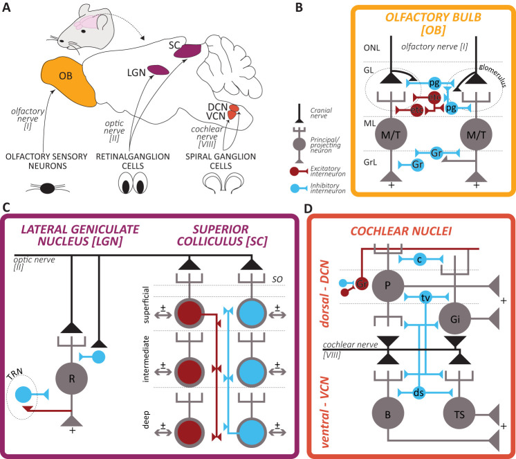
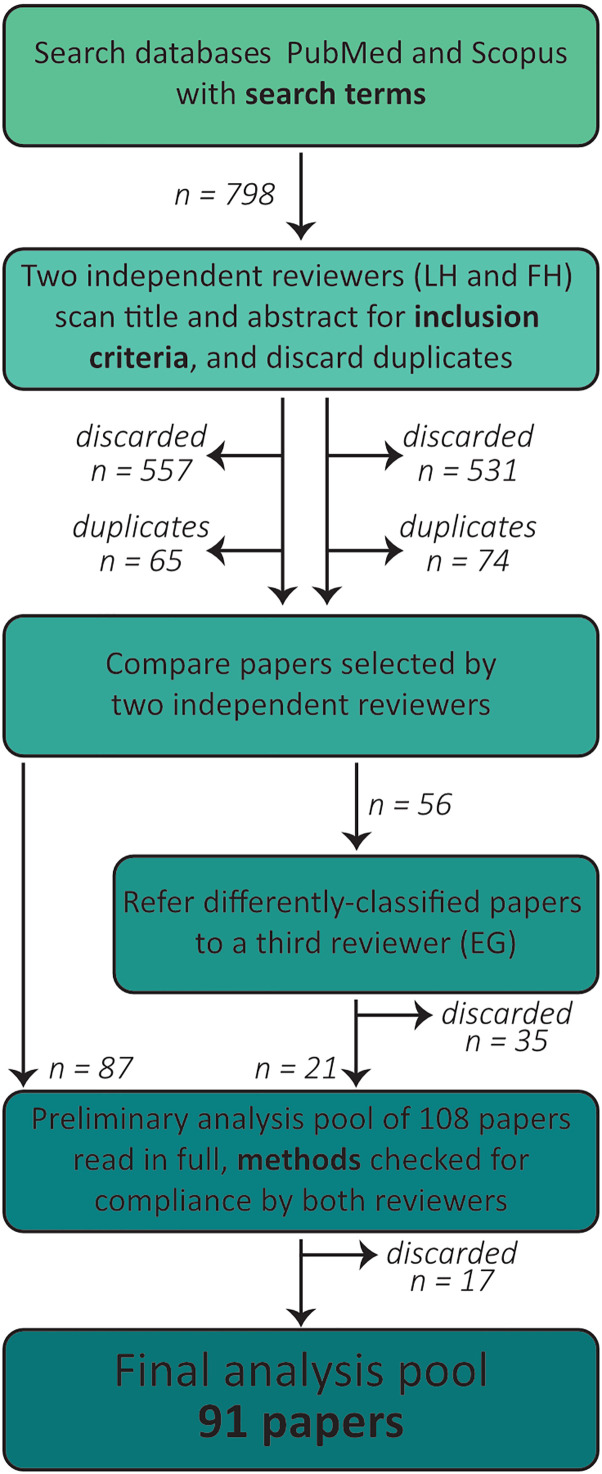
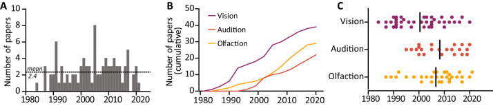
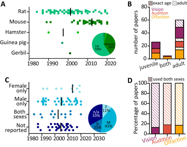
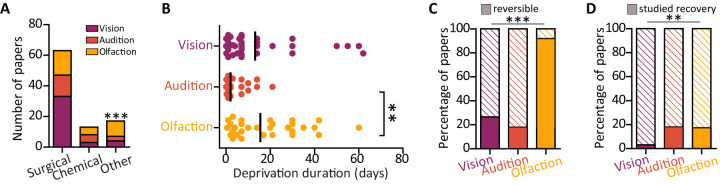
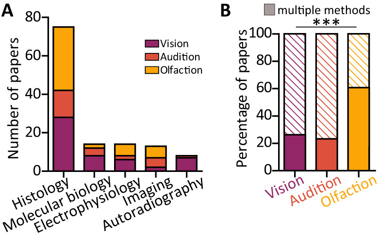
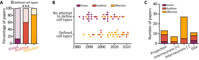
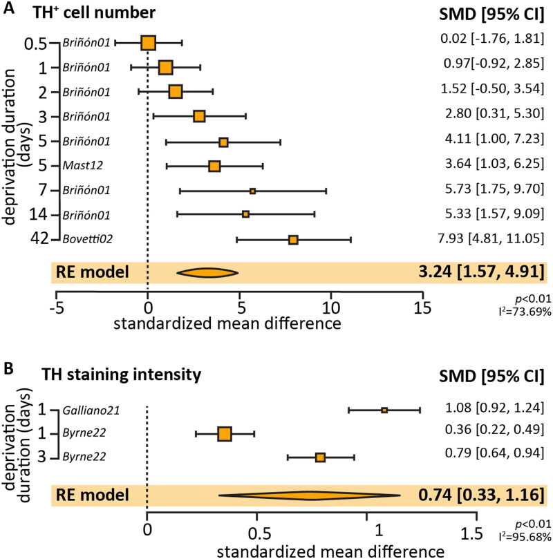
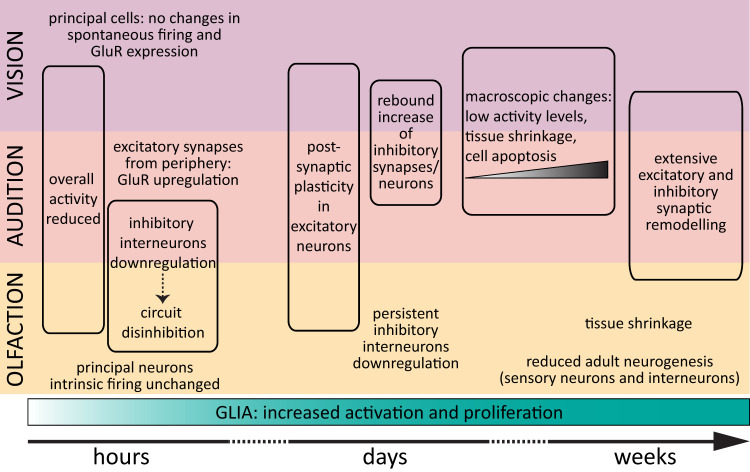
Activity-dependent neuronal plasticity is crucial for animals to adapt to dynamic sensory environments. Traditionally, it has been investigated using deprivation approaches in animal models primarily in sensory cortices. Nevertheless, emerging evidence emphasizes its significance in sensory organs and in subcortical regions where cranial nerves relay information to the brain. Additionally, critical questions started to arise. Do different sensory modalities share common cellular mechanisms for deprivation-induced plasticity at these central entry points? Does the deprivation duration correlate with specific plasticity mechanisms? This study systematically reviews and meta-analyzes research papers that investigated visual, auditory, or olfactory deprivation in rodents of both sexes. It examines the consequences of sensory deprivation in homologous regions at the first central synapse following cranial nerve transmission (vision - lateral geniculate nucleus and superior colliculus; audition - ventral and dorsal cochlear nucleus; olfaction - olfactory bulb). The systematic search yielded 91 papers (39 vision, 22 audition, 30 olfaction), revealing substantial heterogeneity in publication trends, experimental methods, measures of plasticity, and reporting across the sensory modalities. Despite these differences, commonalities emerged when correlating plasticity mechanisms with the duration of sensory deprivation. Short-term deprivation (up to 1 d) reduced activity and increased disinhibition, medium-term deprivation (1 d to a week) involved glial changes and synaptic remodeling, and long-term deprivation (over a week) primarily led to structural alterations. These findings underscore the importance of standardizing methodologies and reporting practices. Additionally, they highlight the value of cross-modal synthesis for understanding how the nervous system, including peripheral, precortical, and cortical areas, respond to and compensate for sensory inputs loss.

Keywords: audition; olfaction; plasticity; rodent; sensory deprivation; vision.


Figures

Figure 1.
Figure 1.
Architecture of the early olfactory, visual, and auditory pathways. A, Schematic representation of the mouse brain and location of the olfactory bulb (OB), lateral geniculate nucleus (LGN; dorsal, dLGN; and ventral, vLGN, combined), superior colliculus (SC), dorsal and ventral cochlear nuclei (DCN and VCN), and their respective cranial nerves input from the sensory organs. B–D, Simplified circuitry of the early central circuits processing olfactory, visual, and auditory information (summarized and adapted from Oertel and Young, 2004; Shepherd, 2004; Campagnola and Manis, 2014; Kosaka and Kosaka, 2016; Weyand, 2016; Duménieu et al., 2021; Liu et al., 2022). Black line and triangle indicate the cranial nerve ending, gray cells are principal neurons projecting outside these early circuits to higher processing areas, red and blue cells are local interneurons, respectively, excitatory and inhibitory. For ease of representation, the many central inputs to these circuits are not depicted. In the bulbar circuit: ONL, olfactory nerve layer; GL, glomerular layer; ML, mitral layer; GrC, granule cells layer; M/T, mitral/tufted cell; pg, periglomerular cell; etc, external tufted cell; gr, granule cell. In the geniculate circuit: R, relay neuron; TRN, thalamic reticular nucleus. In the collicular circuit: SO, stratum opticum; note that the exact circuitry has not been fully resolved, and that all layers send projections outside the SC. In the cochlear nuclei circuit: P, pyramidal (or fusiform); Gi, giant cell; B, bushy cell; TS, T-stellate cell; ds, d-stellate cell; tv, tubercoloventral (or vertical) cell; c, cartwheel cell; Gr, granule cell with its axon called parallel fiber.
Figure 2.
Figure 2.
Strategy for literature search and papers selection. Flowchart indicating the number of articles returned by the parallel searches in the databases PubMed and Scopus using the search terms detailed in the text and their subsequent selection by two independent scrutineers.
Figure 3.
Figure 3.
Publication trends over time. A, No significant trend in the volume of published primary research articles over the 1984–2021 time period. Dotted line represents mean number of publications per year (2.4 mean, 1.7 standard deviation). B, Cumulative distribution of papers published over the 1984–2021 period for each investigated sensory modality. C, Distribution of papers over time for each investigated sensory modality. Dots are individual studies, line indicates median year. Purple, vision; orange, audition; yellow, olfaction.
Figure 4.
Figure 4.
Animal models. A, Distribution of rodent species used over time (1984–2021). Dots are individual studies; line indicates median publication year. Inset: pie chart reporting the percentages of the 91 selected studies using rats (59%), mice (36%), and other rodents (5% split among hamsters, guinea pigs, and gerbils). B, Number of papers using only juvenile animals (postweaning to P40; 26 papers), both juvenile and adult animals (5 papers), and only adult animals older than 40 postnatal days (filled rectangles) or stated “adult” without specifying the age (striped rectangles) across vision (purple), audition (orange), and olfaction (yellow). C, Distribution of rodent sex used over time (1984–2021). Dots are individual studies; line indicates median publication year. Inset: pie chart reporting the percentages of the 91 selected studies using females only (11%), males only (41%), both sexes (15%), or failing to report the sex of the used animals (33%). D, Proportion of papers using both sexes (filled rectangles) across the three sensory modalities: purple, vision, 13%; orange, audition, 18%; yellow, olfaction, 17%. Striped rectangles include studies which used only one sex or failed to report the sex used.
Figure 5.
Figure 5.
Deprivation method. A, Number of papers using surgical, chemical, or other (plugs, patches) deprivation methods. B, Minimum deprivation duration in days used in each study (individual dots) and mean duration (black line). C, Proportion of papers using reversible deprivation methods (filled rectangles) across the three sensory modalities: purple, vision, 26%; orange, audition, 18%; yellow, olfaction, 90%. D, Proportion of papers which used a reversible method to induce deprivation and investigated recovery (filled rectangles) across the three sensory modalities: purple, vision, 3%; orange, audition, 18%; yellow, olfaction, 17%. **p < 0.01, ***p < 0.001.
Figure 6.
Figure 6.
Plasticity readouts. A, Number of papers across the three sensory modalities using each investigative methodology. B, Proportion of papers using more than one method to investigate the effects of deprivation (filled rectangles) across the three sensory modalities: purple, vision, 26%; orange, audition, 23%; yellow, olfaction, 60%. ***p < 0.001.
Figure 7.
Figure 7.
Definition of cell types. A, Proportion of papers defining the cell types where the deprivation-induced plasticity was investigated (filled rectangles). ***p < 0.001. B, Distribution of papers over time split by the lack/presence of cell type definition. Dots are individual studies; purple, vision; orange, audition; yellow, olfaction. C, Among the studies which defined cell types, number of papers investigating deprivation-induced plasticity in projection/principal neurons, excitatory (+) or inhibitory (−) interneurons, and glia across the three sensory modalities. Note the lack of vision papers focusing on interneurons.
Figure 8.
Figure 8.
Meta-analysis of TH expression after olfactory deprivation of various durations. A, Effect size of olfactory deprivation on the number of TH-positive DA neurons in the OB. Note that 7/11 data points originated from the same study (Briñón et al., 2001). B, Effect size of olfactory deprivation duration on the TH staining intensity in bulbar DA neurons. Note that 2/3 data points originated from the same study (Byrne et al., 2022).
Figure 9.
Figure 9.
Deprivation-induced plasticity. Graphical representation of the cellular and circuit effects of sensory deprivations of increasing durations in early visual, auditory, and olfactory areas. See Table 1 for details and references.

Similar articles

Cited by

References

    1. Gudden (1870) Experimentaluntersuchungen über das peripherische und centrale Nervensystem. Archiv f Psychiatrie 2:693–723. 10.1007/BF02046772 - DOI
    1. Akol I, Kalogeraki E, Pielecka-Fortuna J, Fricke M, Löwel S (2022) MMP2 and MMP9 activity is crucial for adult visual cortex plasticity in healthy and stroke-affected mice. J Neurosci 42:16–32. 10.1523/JNEUROSCI.0902-21.2021 - DOI - PMC - PubMed
    1. Allen NJ, Lyons DA (2018) Glia as architects of central nervous system formation and function. Science 362:181–185. 10.1126/science.aat0473 - DOI - PMC - PubMed
    1. Andrade-Talavera Y, Pérez-Rodríguez M, Prius-Mengual J, Rodríguez-Moreno A (2023) Neuronal and astrocyte determinants of critical periods of plasticity. Trends Neurosci 46:566–580. 10.1016/j.tins.2023.04.005 - DOI - PubMed
    1. Andrade C (2020) Understanding the basics of meta-analysis and how to read a forest plot: as simple as it gets. J Clin Psychiatry 81:20f13698. 10.4088/JCP.20f13698 - DOI - PubMed

LinkOut - more resources