. 2024 Jan 10;43(1):15.
doi: 10.1186/s13046-023-02942-4.

EMID2 is a novel biotherapeutic for aggressive cancers identified by in vivo screening

Affiliations

EMID2 is a novel biotherapeutic for aggressive cancers identified by in vivo screening

Ambra Cappelletto et al. J Exp Clin Cancer Res. .

Abstract

Background: New drugs to tackle the next pathway or mutation fueling cancer are constantly proposed, but 97% of them are doomed to fail in clinical trials, largely because they are identified by cellular or in silico screens that cannot predict their in vivo effect.

Methods: We screened an Adeno-Associated Vector secretome library (> 1000 clones) directly in vivo in a mouse model of cancer and validated the therapeutic effect of the first hit, EMID2, in both orthotopic and genetic models of lung and pancreatic cancer.

Results: EMID2 overexpression inhibited both tumor growth and metastatic dissemination, consistent with prolonged survival of patients with high levels of EMID2 expression in the most aggressive human cancers. Mechanistically, EMID2 inhibited TGFβ maturation and activation of cancer-associated fibroblasts, resulting in more elastic ECM and reduced levels of YAP in the nuclei of cancer cells.

Conclusion: This is the first in vivo screening, precisely designed to identify proteins able to interfere with cancer cell invasiveness. EMID2 was selected as the most potent protein, in line with the emerging relevance of the tumor extracellular matrix in controlling cancer cell invasiveness and dissemination, which kills most of cancer patients.

Keywords: AAV vectors; Biotherapeutics; Cancer; Cell invasiveness; Gene therapy; In vivo screening.


Conflict of interest statement

MG is a founder, member of the board, equity holder, and consultant for Forcefield Therapeutics, London, FB is a consultant of the same company. The AAV secretome library and the FunSel technology are owned by Purespring Therapeutics, for which MG is a cofounder, observer to the board, equity holder, and consultant.

Figures
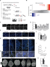
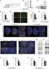
Fig. 1
Fig. 1
CanSel identifies EMID2 as the most potent factor inhibiting cancer cell invasiveness. A. Outline of the CanSel procedure. B. Results of the competitive screening ranking 1028 secreted factors. Each dot shows the relative abundance of each transgene in muscles that were injected with both AAV and cancer cells over muscles injected with AAV only. The grey area covers factors that were not enriched. The red area contains factors that were positively selected (enrichment > 1 SD). C. Representative images of migrated LLC cells in response to FBS in combination with the indicated factors. D. Quantification of LLC migration. E. Representative images of LLC cancers, positive for the proliferation marker Ki67, growing in muscles injected with AAV9 vectors expressing the indicated factors. F. Percentage of the area covered by Ki67+ LLC cells. G. Representative images of LG tumor borders with front of invasion indicated in yellow (n = 2 muscles/group). H. Higher magnification of peripheral branches showing LG cells migrating on collagen fibers. I. Quantification of tumor circularity, a quantitative shape factor of roundness (range, 0–1; 1 = a true circle). J. Representative images of LG tumors in muscles injected with AAV9-CTR or AAV9-EMID2. K. Quantification of LG tumor size. Scale bar in C, E, G 50 μm, in H 100 μm, in J 2 mm. In D, F, K data are shown as mean ± SEM and were analyzed by one-way ANOVA with Student-Newman-Keuls correction. *p < 0.05, **p < 0.01, ***p < 0.001
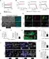
Fig. 2
Fig. 2
EMID2 inhibits CAF activation and normalizes cancer ECM. A Western blot of total cell lysates and supernatants of HEK293T cells transfected with the indicated plasmids. B Quantification of active-TGFβ, relative to pro-TGFβ. C Western blot of whole muscle tissue injected with LG cells and the indicated vectors. D Quantification of active-TGFβ, relative to pro-TGFβ. Tubulin was used as a loading control. E Primary fibroblasts from COLL-EGFP/αSMA-RFP mice cultured in absence or presence of rEMID2. F Quantification of the percentage of red activated fibroblasts. G Quantification of the area covered by EGFP+/α-SMA+ CAFs in muscles co-injected with LG cells and AAV9-CTR or AAV9-EMID2. H Representative images of muscles co-injected with LG cells and AAV9-CTR or AAV9-EMID2. I Representative images of LG tumors in muscles injected with AAV9-CTR and AAV9-EMID2 stained for collagen I. J Representative images of LG tumors in muscles injected with AAV9-CTR and AAV9-EMID2 stained for fibronectin. K. Quantification of collagen I+ area as in H. L. Quantification of fibronectin+ area as in J. M. Western blots showing collagen I (Col I) content in muscles co-injected with LG cells and AAV9-CTR or AAV9-EMID2. N. Western blots showing fibronectin (Fn) content in muscles co-injected with LG cells and AAV9-CTR or AAV9-EMID2. Tubulin (Tub) was used as loading control. O. Quantification of collagen I as in M. P. Quantification of fibronectin as in N. Scale bar in E 50 μm, in H 1 mm, in I, J 100 μm. In B, D, F, G, K, L, O, P data are shown as mean ± SEM and were analyzed by one-way ANOVA with Student-Newman-Keuls correction. *p < 0.05, **p < 0.01
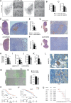
Fig. 3
Fig. 3
EMID2 incorporation in the extracellular matrix modifies its mechanical properties with consequent cytoskeletal re-organization. A. Rheological analysis of Matrigel polymerization in presence of recombinant EMID2 (rEMID2, red) compared to control Matrigel (black). G’, elastic modulus; G’’, viscous modulus. B. Strain sweep of Matrigel in the presence of rEMID2 (red) or control (black). Arrows indicate breaking points. C. Decellularized ECM secreted by primary fibroblasts cultured in a medium containing rEMID2 or in a control medium. D. Representative images of LG cancer cells seeded on Matrigel either in the absence or in the presence of rEMID2. E. Quantification of the number of stress fibers per cell, identified by F-actin, in cancer cells seeded on Matrigel either in the absence or in the presence of rEMID2. F. Quantification of the length of focal adhesions, identified by paxillin, in cancer cells seeded on Matrigel either in the absence or in the presence of rEMID2. G. NIH-3T3 fibroblasts cultured on the decellularized ECM. H, I. Quantification of the number and length of filopodia protruding from NIH-3T3 fibroblasts. J. Representative images of LG cancer cells seeded on Matrigel either in the absence or in the presence of rEMID2. K. Quantification of the ratio of YAP integrated density in the nucleus and cytoplasm of LG cells as in J. Scale bar in C, G, J 50 μm, in D 100 μm. In E, F, H, I, K, data are shown as mean ± SEM and were analyzed by Student t-test. *p < 0.05, **p < 0.01
Fig. 4
Fig. 4
EMID2 overexpression inhibits the growth and dissemination of murine and human cancers. A Representative hematoxylin/eosin staining of total lung and zoomed fields of LG tumor mass. B Number of LG tumor foci. C Area covered by LG cells. D Representative images of total pancreas and zoomed fields injected with KPC cells. KPC cells are stained for Ki67. Nuclei are counterstained with hematoxylin. E Number of KPC tumor foci per pancreas. F Area covered by Ki67+ KPC cells. G Representative images of total pancreas and zoomed fields of KC mice. Cancer cells are stained for Ki67. Nuclei are counterstained with hematoxylin. H Pancreas/body weight for each mouse. I Number of tumor foci per pancreas. J Area covered by Ki67+ cancer cells. K. Area covered by YAP+ cancer cells on tumor area. L. Representative images of the pancreas of KC mice stained for YAP. M. Representative images of wound healing assay with EGFP+ Panc1 cells co-cultured human fibroblasts overexpressing EMID2 or control fibroblasts, at the indicated time points. N. Wound closure at T20 relative to T0. O, P. Kaplan-Meier curves of patients affected by pancreatic adenocarcinoma (PAAD) and lung adenocarcinoma (LUAD), according to the expression levels of EMID2, at the indicated time points from diagnosis. Q. Forest plot indicating the Hazard Ratio (HR) for the most aggressive human solid cancers. GBMLGG: glioblastoma multiforme and low-grade glioma, LGG: low grade glioma, PAAD: pancreatic adenocarcinoma, LIHC: liver hepatocellular carcinoma, BLCA: bladder urothelial carcinoma, LUAD: lung adenocarcinoma, LUSC: lung squamous cell carcinoma, CHOL: cholangiocarcinoma, ESCA: esophageal carcinoma, MESO: mesothelioma. Square size reflects the weight of the significance. Source: TCGA database. Scale bar in A (whole lung), D, G (whole pancreas) 1 mm, in A, D, G (magnification) 200 μm, in L, M 50 μm. In B, C, E, F, H, I, J, K, N data are shown as mean ± SEM and were analyzed by one-way ANOVA with Student-Newman-Keuls correction. *p < 0.05, ***p < 0.001

References

    1. Sun D, Gao W, Hu H, Zhou S. Why 90% of clinical drug development fails and how to improve it? Acta Pharm Sin B. 2022;12(7):3049–62. doi: 10.1016/j.apsb.2022.02.002. - DOI - PMC - PubMed
    1. Lin A, Giuliano CJ, Palladino A, John KM, Abramowicz C, Yuan ML, et al. Off-target toxicity is a common mechanism of action of cancer Drugs undergoing clinical trials. Sci Transl Med. 2019;11:509. doi: 10.1126/scitranslmed.aaw8412. - DOI - PMC - PubMed
    1. Ruozi G, Bortolotti F, Falcione A, Dal Ferro M, Ukovich L, Macedo A, et al. AAV-mediated in vivo functional selection of tissue-protective factors against ischaemia. Nat Commun. 2015;6:7388. doi: 10.1038/ncomms8388. - DOI - PMC - PubMed
    1. Ruozi G, Bortolotti F, Mura A, Tomczyk M, Falcione A, Martinelli V, et al. Cardioprotective factors against Myocardial Infarction selected in vivo from an AAV secretome library. Sci Transl Med. 2022;14(660):eabo0699. doi: 10.1126/scitranslmed.abo0699. - DOI - PubMed
    1. Bortolotti F, Ruozi G, Falcione A, Doimo S, Dal Ferro M, Lesizza P, et al. In vivo functional selection identifies Cardiotrophin-1 as a Cardiac Engraftment factor for mesenchymal stromal cells. Circulation. 2017;136(16):1509–24. doi: 10.1161/CIRCULATIONAHA.117.029003. - DOI - PubMed