. 2024 Jan;21(210):20230521.
doi: 10.1098/rsif.2023.0521. Epub 2024 Jan 10.

Adhesive properties of Aphrophoridae spittlebug foam

Affiliations

Adhesive properties of Aphrophoridae spittlebug foam

Hannelore Hoch et al. J R Soc Interface. 2024 Jan.

Abstract

Aphrophora alni spittlebug nymphs produce a wet foam from anal excrement fluid, covering and protecting themselves against numerous impacts. Foam fluid contact angles on normal (26°) and silanized glass (37°) suggest that the foam wets various substrates, including plant and arthropod surfaces. The pull-off force depends on the hydration state and is higher the more dry the fluid. Because the foam desiccates as fast as water, predators once captured struggle to free from drying foam, becoming stickier. The present study confirms that adhesion is one of the numerous foam characteristics resulting in multifunctional effects, which promote spittlebugs' survival and render the foam a smart, biocompatible material of biological, biomimetic and biomedical interest. The sustainable 'reuse' of large amounts of excrement for foam production and protection of the thin nymph integument suggests energetic and evolutionary advantages. Probably, that is why foam nests have evolved in different groups of organisms, such as spittlebugs, frogs and fish.

Keywords: cryo-SEM; defence; foam nest; insect cuticle; pull-off force; surfactant.


Conflict of interest statement

The authors declare no competing or financial interests.

Figures

Figure 1.
Figure 1.
Video stills of Aphrophora alni nymphs, predators and commensals in spittlebug foam. (ac) A nymph moves in the foam without hindrance, pulling bulks (b) and filaments of foam (c; arrow). Note the glossy appearance of the body surface, which is totally covered with anal fluid. (d,e) The anal tip with the ventral channel is initially opening (d) and fully opening (e). Note the pulled bead-on-string structure of foam fluid in (e) (arrow tip). (fj,k–o) Sequences of foam bubble release. The abdominal tip raises over the foam (f,g,k,l,m) to take air into the open ventral channel. Then, the abdominal tip with the closed channel moves into the foam (h,n) to release a bubble (i,o) before raising again (j). (pt) A sequence of foam bubble aggregation, which starts ventrally before covering all the body after about 2–3 min. The ventral tip alternately extends (p) and bends (q) before releasing a bubble (r). The bubbles aggregate (s), forming the foam (t). (uw) An ant trapped in spittlebug foam struggles to free itself (u,v) and grooms intensively after escaping from foam (w). (x) A spider entangled in the foam. (y) Ciliates belonging to Colpodea move actively in the foam (arrows).
Figure 2.
Figure 2.
The outcome of behavioural assays with Lasius niger ant workers and spittlebug foam. (a) The number of contacts with tap water and foam droplets during 30 min in a 13 cm diameter glass Petri dish (χ2 = 70.0, p = 0.3, n = 12 per fluid); means and standard deviations. (b) The time required to escape and groom after submerging all legs in the spittlebug foam and tap water. Asterisks indicate statistical differences: time to escape, Mann–Whitney rank sum test, T = 75.0, p = 0.029; time to groom: T = 83.0, p ≤ 0.001; n = 8 per fluid and observation; box-and-whisker diagrams with the ends of the boxes defining the 25th and 75th percentiles, with a line at the median and error bars depicting the 10th and 90th percentiles. (c) Relationship between time to escape and groom; linear regressions; regression equations, coefficients, F statistics and probability values.
Figure 3.
Figure 3.
(ad) Photographs of Aphrophora alni foam nests on the abaxial leaf lamina of ribwort Plantago lanceolata (a) and cinquefoil Potentilla sp. stems (bd), occupied with single larva (a,c) and groups of larvae (b,d). The foam is built of regular, tightly packed circular and pentagonal bubbles surrounded by fluid lamellae. Note the foam iridescence in (a,b). (eg) Stereomicroscopic images showing different aspects of the foam fluid. (e) A fluid film includes few single gas bubbles, fluid foam consisting of larger tightly packed gas bubbles, and a fluid filament pulled between two cinquefoil leaflets. (f) Dried foam residues on a cinquefoil stem. Dried lamellae (arrow) form the pentagonal shape established in drying/draining foam bubbles (dotted line). (g) Foam dried in a frame, leaving very thin, stable filaments (dried lamellae). The pentagonal bubble shape and nodes between the lamellae of different bubbles are still recognizable.
Figure 4.
Figure 4.
Cryo-SEM images of Aphrophora alni third-instar nymphs and foam fluid (residues) on insect and plant surfaces. (a) Lateral view of a nymph attached upside down on a Potentilla sp. stem. The concavities between body segments are covered with foam fluid. (b) Back view of the nymph showing the anal tip and seven proximal tergites with broadened lobes forming a channel. Note the foam fluid trapped between leg segments. (c) Close-up of the ventral abdominal tip and anal valves with a matt coverage (asterisks). The arrow points to the opening of the channel formed by tergites' lobes. (df). Surface close-ups of the anal valves and channel (asterisks in c): cone-shaped outgrowths covered with protruded thin wax filaments which are partly coalesced; top view (d,e), cross-section (f). (g,h) Dorsal abdominal nymph surface after washing with water (g) and untreated covered with foam, appearing like a meshed network (h). (i,j) Cross sections of abdominal cuticle after washing (i) and untreated, covered with foam fluid (j). (k) A nymph femur covered with a meshed network of foam fluid. (l,m) A Potentilla sp. stem covered with foam fluid pulled into bulk and thin filaments under tension (l) and foam bubbles indicated by Plateau borders (dotted line), fluid lamellae and nodes (m). (n) For example, a Myrmica rubra worker entangled in spittlebug foam fluid, embedding the mouthparts and legs. (o,p) Round and pentagonal bubbles surrounded by fluid lamellae forming the so-called Plateau borders (dotted lines) and converging nodes in a dense foam (o) and foam residues on spittlebug nymph cuticle (p). (q) Under tension, the foam fluid lamellae may be pulled into very thin filaments. Note the bead-on-string structures (arrow tips). Note that the watery foam content is evaporated in all SEM micrographs. b, bubble; e, epicuticle; endo, endocuticle; exo, exocuticle; f, foam fluid; fl, fluid lamella; n, nodes between fluid lamellae of different bubbles; g, epicuticular grease; t, tergites' lobes.
Figure 5.
Figure 5.
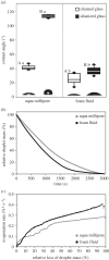
(a) Contact angles of Aqua Millipore water and spittlebug foam fluid. Box-and-whisker diagrams: the ends of the boxes define the 25th and 75th percentiles, with a line at the median and error bars depicting the 10th and 90th percentiles. Difference between surfaces for foam fluid: t-test, t = −4.1, p ≤ 0.001 (uppercase letters). Difference between surfaces for Aqua Millipore water: t-test, t = −66.0, p ≤ 0.001 (uppercase letters). Difference between fluids on hydrophobic (silanized) and hydrophilic (cleaned) glass for each substrate: Mann–Whitney rank sum test, T = 155.0, p ≤ 0.001 (lowercase letters); n = 10 per fluid and substrate. The insets show droplet images obtained during contact angle measurements. (b) Relative loss of fluid drop mass due to evaporation; ‘means and standard deviations per 5th second'. (c) Temporary evaporation rate [5 s × ((Mn − Mn + 1)/5)] obtained from relative values. See table 1 for the half-life period (t50) and time point of evaporation of 80% (t80) of Aqua Millipore and cicada foam fluid.
Figure 6.
Figure 6.
Adhesive properties of the foam. Force–distance diagrams obtained between the clean glass surface and the sapphire ball in the presence of the foam under an initial load of 6.3 mN. The blue curve was obtained after 10 min of foam preparation. The red curve was obtained after 30 min of foam preparation. The area under the negative part of the curve corresponds to the energy of the separation. The inset illustrates the experimental design. The arrow indicates the direction of the pull-off. FL, foam filament; GL, glass surface; SB, sapphire ball; SE, force sensor.
Figure 7.
Figure 7.
Adhesive properties of the foam fluid. Data are based on the force–distance diagrams (figure 1). (a,b) Pull-off force Fa (a) and area under the pull-off curve A (separation energy) (b) measured at an individual sample over time (n = 36 single measurements). (c,d) Box-and-whisker diagrams of the pull-off force (c) and area under the pull-off curve (separation energy) (d) of the fluid compared to the control experiments (dry sapphire on glass). The ends of the boxes define the 25th and 75th percentiles, with a line at the median and error bars representing the 10th and 90th percentiles. A statistically significant difference exists between data obtained on the fluid and the control experiment (Mann–Whitney rank sum test; force: T = 2027.0, p ≤ 0.001; area: T = 2053.0, p ≤ 0.001).
Figure 8.
Figure 8.
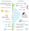
Spittlebug foam characteristics and facilitated integrative effects support nymph protection and survival in multiple ways (schematic chart). The present study is set in context with previous outcomes [,,,,,–,–,,–75]. The smart multifunctional foam material may inspire innovative biomimetic and biomedical approaches.

References

    1. Cantat I, Cohen-Addad S, Elias F, Graner F, Höhler R, Pitois O, Rouyer F, Saint-Jalmes A. 2013. Foams. Structure and dynamics. Oxford, UK: Oxford University Press.
    1. Mecozzi M, Pietroletti M. 2016. Chemical composition and surfactant characteristics of marine foams investigated by means of UV-vis, FTIR and FTNIR spectroscopy. Environ. Sci. Poll. Res. 23, 22 418-22 432. (10.1007/s11356-016-7423-5) - DOI - PubMed
    1. Hill C, Eastoe J. 2017. Foams: from nature to industry. Adv. Coll. Interface Sci. 247, 496-513. (10.1016/j.cis.2017.05.013) - DOI - PubMed
    1. Cooper A, Kennedy MW. 2010. Biofoams and natural protein surfactants. Biophys. Chem. 151, 96-104. (10.1016/j.bpc.2010.06.006) - DOI - PMC - PubMed
    1. Seymour RS, Loveridge JP. 1994. Embryonic and larval respiration in the arboreal foam nests of the African frog Chiromantis xerampelina. J. Exp. Biol. 197, 31-46. (10.1242/jeb.197.1.31) - DOI - PubMed

Publication types

Substances

LinkOut - more resources