[Preprint]. 2023 Dec 19:rs.3.rs-3716881.
doi: 10.21203/rs.3.rs-3716881/v1.

Sex differences in mitochondrial gene expression during viral myocarditis

Affiliations

Sex differences in mitochondrial gene expression during viral myocarditis

Damian Di Florio et al. Res Sq. .

Update in

  • Sex differences in mitochondrial gene expression during viral myocarditis.
    Di Florio DN, Weigel GJ, Gorelov DJ, McCabe EJ, Beetler DJ, Shapiro KA, Bruno KA, Chekuri I, Jain A, Whelan ER, Salomon GR, Khatib S, Bonvie-Hill NE, Fliess JJ, Giresi PG, Hamilton C, Hartmoyer CJ, Balamurugan V, Darakjian AA, Edenfield BH, Kocsis SC, McLeod CJ, Cooper LT Jr, Audet-Walsh É, Coronado MJ, Sin J, Fairweather D. Di Florio DN, et al. Biol Sex Differ. 2024 Dec 18;15(1):104. doi: 10.1186/s13293-024-00678-0. Biol Sex Differ. 2024. PMID: 39696682 Free PMC article.

Abstract

Background: Myocarditis is an inflammation of the heart muscle most often caused by an immune response to viral infections. Sex differences in the immune response during myocarditis have been well described but upstream mechanisms in the heart that might influence sex differences in disease are not completely understood.

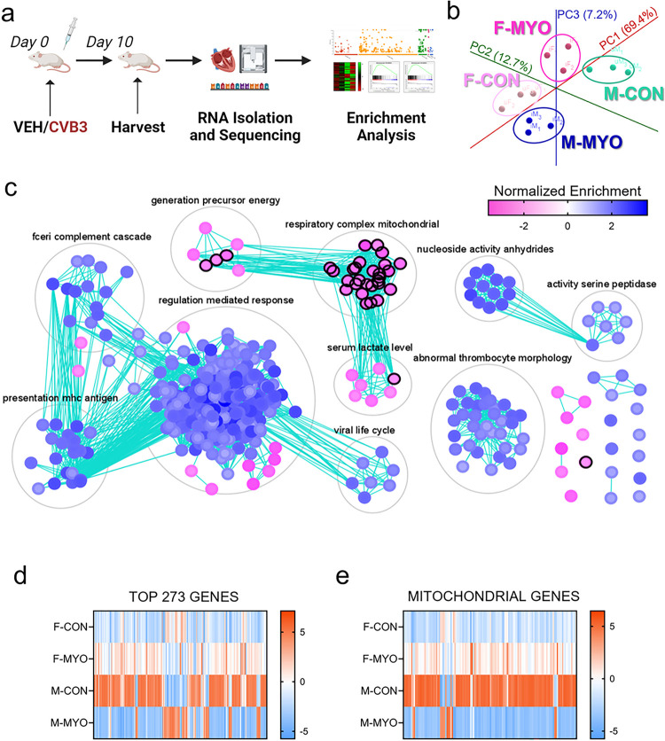
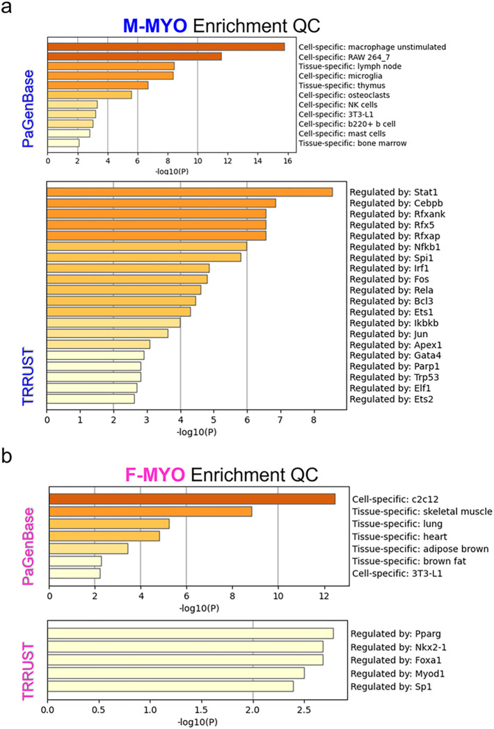
Methods: Male and female BALB/c wild type mice received an intraperitoneal injection of heart-passaged coxsackievirus B3 (CVB3) or vehicle control. Bulk-tissue RNA-sequencing was conducted to better understand sex differences in CVB3 myocarditis. We performed enrichment analysis to understand sex differences in the transcriptional landscape of myocarditis and identify candidate transcription factors that might drive sex differences in myocarditis.

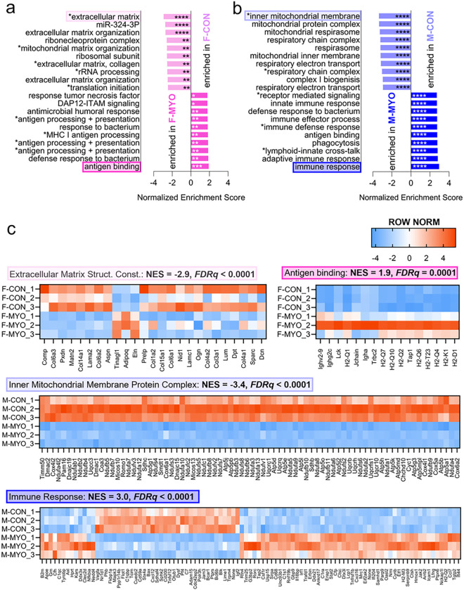
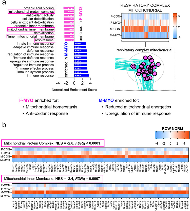
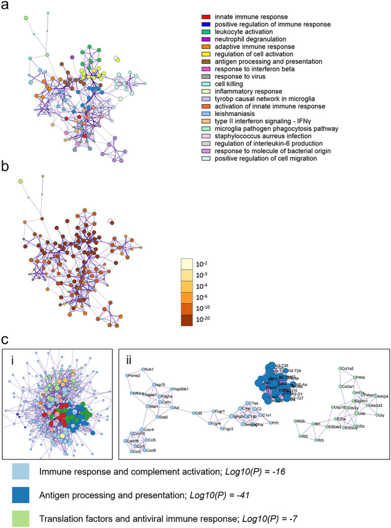
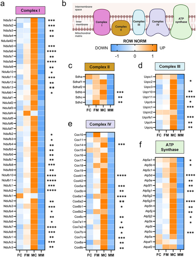
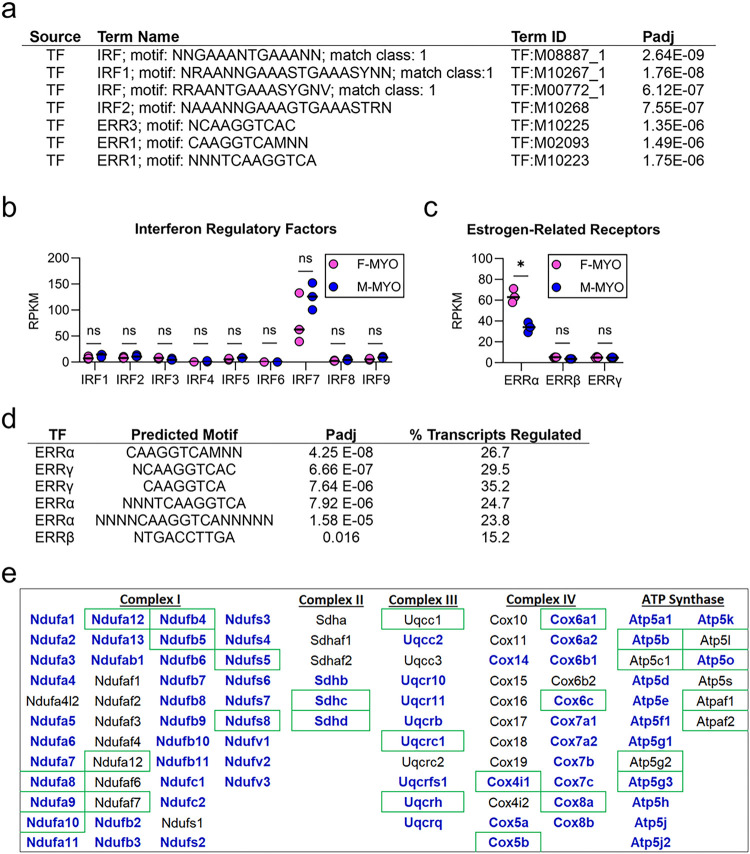
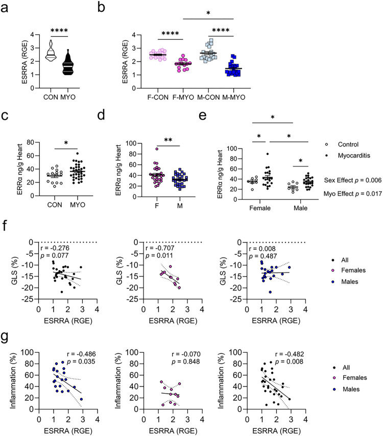
Results: The hearts of male and female mice with myocarditis were significantly enriched for pathways related to an innate and adaptive immune response compared to uninfected controls. When comparing females to males with myocarditis, males were enriched for inflammatory pathways and gene changes that suggested worse mitochondrial transcriptional support (e.g., mitochondrial electron transport genes). In contrast, females were enriched for pathways related to mitochondrial respiration and bioenergetics, which were confirmed by higher transcript levels of master regulators of mitochondrial function including peroxisome proliferator-activated receptor gamma coactivator 1 (PGC1α), nuclear respiratory factor 1 (NRF1) and estrogen-related receptor alpha (ERRα). TRANSFAC analysis identified ERRa as a transcription factor that may mediate sex differences in mitochondrial function during myocarditis.

Conclusions: Master regulators of mitochondrial function were elevated in females with myocarditis compared to males and may promote sex differences in mitochondrial respiratory transcript expression during viral myocarditis resulting in less severe myocarditis in females following viral infection.

Keywords: autoimmune disease; coxsackievirus B3; estrogen-related receptor alpha estrogen/ERRa; inflammation; innate immunity; interleukin-1 beta; mitochondria; nuclear respiratory factor 1/NRF1; peroxisome proliferator-activated receptor gamma coactivator 1/PGC1a.


Conflict of interest statement

Competing Interests The authors declare that they have no competing interests.

Figures

Figure 1
Figure 1. Myocardial inflammation is increased in males compared to females
a, Myocarditis severity (% inflammation) between female controls (F-CON, n= 27), females with myocarditis (F-MYO, n = 41), male controls (M-CON, n= 30), and males with myocarditis (M-MYO, n = 40); b, representative heart sections (scale bars = 80μm); c-h, relative gene expression (RGE) for controls (CON, n = 31–34) versus mice with myocarditis (MYO, n = 32–41) and for F-CON (n = 15–16), F-MYO (n = 15–21), M-CON (n = 18), and M-MYO (n = 15–20).
Figure 2
Figure 2. RNA sequencing reveals sex differences in immune and mitochondrial genes
a, RNA-sequencing experimental pipeline; b, principal component analysis plot showing female controls (F-CON), females with myocarditis (F-MYO), male controls (M-CON), and males with myocarditis (M-MYO); c, results from GSEA pre-ranked plotted on Cytoscape with Enrichment Map and AutoAnnotate, pink = F-MYO and blue = M-MYO, nodes circled in black are mitochondrial-related pathways; heat map for d, the top 273 most differentially expressed genes and e, mitochondrial genes between all four groups.
Figure 3
Figure 3. Sex-specific gene expression pathways during myocarditis
The top ten most enriched pathways from GSEA ranked by normalized enrichment score comparing a, female controls (F-CON) to females with myocarditis (F-MYO), b, male controls (M-CON) to males with myocarditis (M-MYO) (pathway text marked with astricts indicate abbreviated pathway names, see supplement for abbreviations and full pathway names) c, Row normalized heatmaps for the most enriched pathways from F-CON vs F-MYO and M-CON vs M-MYO, respective to colors highlighting pathways in (a) and (b). NES = normalized enrichment score, Extracellular Matrix Structural (Struct.) Constituent (Const.). *FDRq<0.05,**FDRq<0.01, *** FDRq<0.001, **** FDRq<0.00001
Figure 4
Figure 4. Sex differences in differential gene expression pathways during myocarditis
The top ten most enriched pathways from GSEA ranked by normalized enrichment score comparing a, females with myocarditis (F-MYO) and males with myocarditis (M-MYO) and heatmap of auto-annotated cluster of pathways/nodes describing the mitochondrial respiratory complex, b, Row normalized heatmaps for pathways highlighted in pink for F-MYO (from F-MYO vs M-MYO comparison; other 2 pathways share common genes with those in (b) and are available in Supplemental Figure 7). *FDRq<0.05, ** FDRq<0.01, *** FDRq<0.001, **** FDRq<0.00001
Figure 5
Figure 5. Females with myocarditis are enriched for pathways related to mitochondrial respiration compared to males with myocarditis
Metascape enrichment results for females with myocarditis (comparing females and males with myocarditis) a, the top enriched pathways colored by cluster/pathway and b, by p-value. Top enriched pathways c, Protein-protein interaction analysis clustered by interaction outside of pathways (i) and interaction inside pathways (ii). Log10(P) vals are derived by averaging the Log10(P) vals for the 3 MCODE annotations, rounded to whole number with colors indicating respective pathways. Images in (i) are cropped to show the bulk of pathways and interactors and the top 3 pathways only are shown in (ii).
Figure 6
Figure 6. Males with myocarditis are enriched for pathways related immune activation compared to females with myocarditis
Metascape enrichment results for males with myocarditis (comparing females and males with myocarditis) a, the top enriched pathways colored by cluster/pathway and b, by p-value. Top enriched pathways c, Protein-protein interaction analysis clustered by interaction outside of pathways (i) and interaction inside pathways (ii). Log10(P) vals are derived by averaging the Log10(P) vals for the 3 MCODE annotations, rounded to whole number with colors indicating respective pathways. Images in (i) are cropped to show the bulk of pathways and interactors and the top 3 pathways only are shown in (ii).
Figure 7
Figure 7. Metascape Enrichment QC shows cell specific signals and identifies potential transcriptional regulators of sex differences in myocarditis
a, Enrichment quality control (QC) for males with myocarditis (M-MYO) shows cell specific enrichment signals from Pattern Gene Database (PaGenBase) and suggested transcription factors from Transcriptional Regulatory Relationships Unraveled by Sentence-based Text mining (TRRUST) and for b, females with myocarditis (F-MYO)
Figure 8
Figure 8. Sex differences in mitochondrial electron transport genes during myocarditis
Row normalized RPKM comparing female controls (FC), females with myocarditis (FM), male controls (MC) and males with myocarditis (MM) for nuclear encoded genes for a, complex I, b, color-coded illustration of the mitochondrial electron transport chain; c, complex II, d, complex III, e, complex IV, and f, ATP synthase. *p<0.05, ** p<0.01, *** p<0.001,****p<0.0001
Figure 9
Figure 9. TRANSFAC analysis identifies interferon regulatory factors and estrogen-related receptors as potential mediators of sex difference during myocarditis
a, TRANSFAC results comparing females with myocarditis (F-MYO) and males with myocarditis (M-MYO); RPKM (reads per kilobase per million) using false discovery rate to compare F-MYO and M-MYO for b, interferon regulatory factors (IRFs) and c, estrogen-related receptors (ERRs); d, predicted binding capacity of ERRs for electron transport chain transcripts; e, significantly different transcripts by sex in electron transport genes are indicated by bold blue lettering, green boxes indicate genes that ERRa predicted to bind to.
Figure 10
Figure 10. Females with myocarditis express higher levels of mitochondrial master regulators PGC1a and NRF1
Relative gene expression (RGE) for controls (CON, n = 33–35) versus mice with myocarditis (MYO, n = 35–39) and for F-CON (n = 15), F-MYO (n = 20–21), M-CON (n = 17–18), and M-MYO n = 19–20) for a-b, PGC1a; and c,d, NRF1.
Figure 11
Figure 11. Females with myocarditis express higher levels of ERRαcompared to males
Relative gene expression (RGE) for controls (CON, n = 33–35) versus mice with myocarditis (MYO, n = 35–39) and for F-CON (n = 15), F-MYO (n = 20–21), M-CON (n = 17–18), and M-MYO n = 19–20) for a,b, ERRa ELISA of ERRa protein from whole heart homogenate supernatant comparing c, CON (n = 14) to MYO (n= 42); d, all females (F, n = 27) to all males (M, n = 29) regardless of disease state; e, two-way ANOVA F-CON (n = 7), F-MYO (n = 20), M-CON (n = 7), and M-MYO (n = 22). Two-tailed assessment of correlations for, f ERRα gene expression and global longitudinal strain (GLS (%)), and g for ERRα gene expression and myocarditis severity (Inflammation %) scored by H+E stain.
Figure 12
Figure 12. In situ expression of ERRα demonstrates sex differences
Representative heart sections (based on H&E scores) for F-CON, M-CON, F-MYO, and M-MYO stained for ERRα. Images of the myocardium were taken at the base, middle (mid) and apex of the heart for each sample. Sale bars = 70 μm.

References

    1. Coronado MJ, Bruno KA, Blauwet LA, Tschope C, Cunningham MW, Pankuweit S, van Linthout S, Jeon ES, McNamara DM, Krejci J, et al. Elevated Sera sST2 Is Associated With Heart Failure in Men =50 Years Old With Myocarditis</at. J Am Heart Assoc. 2019;8(2):e008968. - PMC - PubMed
    1. Roth GA, Mensah GA, Johnson CO, Addolorato G, Ammirati E, Baddour LM, Barengo NC, Beaton AZ, Benjamin EJ, Benziger CP, et al. Global Burden of Cardiovascular Diseases and Risk Factors, 1990–2019: Update From the GBD 2019 Study. J Am Coll Cardiol. 2020;76(25):2982–3021. - PMC - PubMed
    1. Fu M, Kontogeorgos S, Thunstrom E, Zverkova Sandstrom T, Kroon C, Bollano E, Schaufelberger M, Rosengren A. Trends in myocarditis incidence, complications and mortality in Sweden from 2000 to 2014. Sci Rep. 2022;12(1):1810. - PMC - PubMed
    1. Boehmer TK, Kompaniyets L, Lavery AM, Hsu J, Ko JY, Yusuf H, Romano SD, Gundlapalli AV, Oster ME, Harris AM. Association Between COVID-19 and Myocarditis Using Hospital-Based Administrative Data - United States, March 2020-January 2021. MMWR Morb Mortal Wkly Rep. 2021;70(35):1228–32. - PMC - PubMed
    1. Fairweather D, Beetler DJ, Di Florio DN, Musigk N, Heidecker B, Cooper LT Jr.. COVID-19, Myocarditis and Pericarditis. Circ Res. 2023;132(10):1302–19. - PMC - PubMed

Publication types

LinkOut - more resources