[Preprint]. 2023 Dec 22:rs.3.rs-3722563.
doi: 10.21203/rs.3.rs-3722563/v1.

Anti-Ebola virus mAb 3A6 with unprecedented potency protects highly viremic animals from fatal outcome and physically lifts its glycoprotein target from the virion membrane

Affiliations

Anti-Ebola virus mAb 3A6 with unprecedented potency protects highly viremic animals from fatal outcome and physically lifts its glycoprotein target from the virion membrane

Erica Saphire et al. Res Sq. .

Update in

Abstract

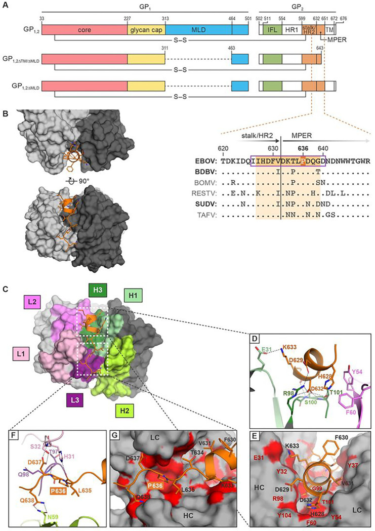
Monoclonal antibodies (mAbs) against Ebola virus (EBOV) glycoprotein (GP1,2) are the standard of care for Ebola virus disease (EVD). Anti-GP1,2 mAbs targeting the stalk and membrane proximal external region (MPER) potently neutralize EBOV in vitro. However, their neutralization mechanism is poorly understood because they target a GP1,2 epitope that has evaded structural characterization. Moreover, their in vivo efficacy has only been evaluated in the mouse model of EVD. Using x-ray crystallography and cryo-electron tomography of 3A6 complexed with its stalk- GP1,2 MPER epitope we reveal a novel mechanism in which 3A6 elevates the stalk or stabilizes a conformation of GP1,2 that is lifted from the virion membrane. In domestic guinea pig and rhesus monkey EVD models, 3A6 provides therapeutic benefit at high viremia levels, advanced disease stages, and at the lowest dose yet demonstrated for any anti-EBOV mAb-based monotherapy. These findings can guide design of next-generation, highly potent anti-EBOV mAbs.

Keywords: 3A6; Animal study; EBOV; Ebola virus; MPER; antibody; efficacy study; mAb; membrane proximal external region; monoclonal antibody; stalk.


Conflict of interest statement

Competing interests A.G., E.D., and B.J.D. are employees of Integral Molecular, and B.J.D.is a shareholder in that company. Additional Declarations: There is NO Competing Interest.

Figures

Figure 1
Figure 1
Crystal structures of unbound human mAb 3A6 Fab and mAb 3A6 Fab bound to the Ebola virus glycoprotein stalk–MPER (A) Top: Schematic of proteolytically processed mature EBOV GP1,2 using the amino acid residue numbering of its uncleaved precursor minus signal peptide residues. Middle and bottom: EBOV GP1,2 constructs used in this study. Inset: Sequence alignment of GP2 stalk–MPER region amino-acid sequences. Aligned are the stalk–MPER transition areas (with the two regions separated by a vertical black line) of all six known orthoebolaviruses. The predicted linear epitope of 3A6 is indicated by a purple box. The EBOV residues observed to interact with 3A6 in the crystal structure highlighted in dark orange and the corresponding regions in glycoproteins of the other orthoebolaviruses highlighted in light orange. Orthoebolaviruses associated with fatal human disease are indicated in bold. (B) Top and front view of the 3A6 Fab fragment (grey) bound to the EBOV stalk–MPER peptide (orange). The heavy and light chains of 3A6 are highlighted in dark and light grey, respectively. (C) View of 3A6 highlighting the molecular surface contributed by the heavy (H1, H2, H3) and light chain (L1, L2, L3) CDRs in the paratope. Polar interactions (D, E) and hydrophobic interactions (F, G) (atoms within 4 Å) are shown as black dotted lines and red surfaces. P636, associated with escape from 3A6 after change P636S, is boxed in red in (A, F, G). BDBV, Bundibugyo virus; BOMV, Bombali virus; CDR, complementarity determining region; EBOV, Ebola virus; GP1,2, glycoprotein; GP2, glycoprotein subunit 2; HR, heptad repeat regions; IFL, internal fusion loop; MLD, mucin-like domain; MPER, membrane proximal external region; RESTV, Reston virus; SUDV, Sudan virus; TAFV, Taï Forest virus; TM, transmembrane domain.
Figure 2
Figure 2
Residues D632 and P636 of the Ebola virus glycoprotein MPER are key for mAb 3A6 binding (A) Cells expressing WT EBOV GP1,2 or recombinant EBOV GP1,2 with the indicated changes to cognate amino-acid residues found in SUDV GP2 were incubated with 3A6 IgG and then stained with DyLight 488 anti-human IgG for detection by fluorescence microscopy, followed by quantification of antibody-positive cells. (B) ELISA binding curves for 3A6 IgG to purified EBOV GP1,2ΔTM/ΔMLD or variants thereof containing the indicated amino-acid residue changes. (C) Flow cytometry analysis of mAb binding to cell-surface expressed EBOV GP bearing a D632A or P636A mutation. Error bars represent the mean ± standard deviation of triplicates (A and B) and duplicates (C); black dots in (A) indicate the values for the individual experiments. EBOV, Ebola virus; ELISA, enzyme-linked immunosorbent assay; Fab, fragment antigen binding; GP1,2, glycoprotein; GP2, glycoprotein subunit 2; Ig, immunoglobulin; SUDV, Sudan virus; WT, wild-type.
Figure 3
Figure 3
Binding of mAb 3A6 lifts Ebola virus glycoprotein relative to the membrane surface (A) Alignment of the EBOV stalk–MPER peptide (orange) to the full-length EBOV GP1,2 structure (PDB ID: 5JQ7; GP1 in blue, GP2 in yellow) illustrates anchoring of 3A6 Fab to the C-terminus of the ectodomain of the GP2 stalk. (B) The close trimeric bundle arrangement of GP2 is incompatible with the GP2–3A6 complex structure, as the bound antibody sterically clashes with neighboring monomers. (C) Negative-stain EM reference-free two-dimensional class averages of 3A6 Fab in complex with trimeric EBOV GP1,2ΔTM/ΔMLD, showing representative side and tilted views. Scale bar, 20 nm. EBOV, Ebola virus; EM, electron microscopy; GP1,2, glycoprotein; GP1, glycoprotein subunit 1; GP2, glycoprotein subunit 2; ID, identification number; MPER, membrane proximal external region; PDB, Protein Data Bank; VLP, virion-like particle; VP40, EBOV matrix protein.
Figure 4
Figure 4
Binding of mAb 3A6 lifts Ebola virus glycoprotein relative to the membrane surface (A) Representative tomographic slices of filamentous EBOV VLPs bound to mAb Fabs. VP40 of VLPs is visible as a dotted layer underneath the lipid bilayer. The bottom row corresponds to magnified view of areas enclosed by red boxes in the top row. (B–D) VLP GP1,2-Fab complexes were imaged by cryogenic electron microscopy, followed by subtomogram averaging. (B) Isosurface representations of reconstructions of GP1,2 on the surface of VLPs bound to 3A6 Fab (gold) or KZ52 Fab (blue) superimposed to align with GP1,2 (top) or the outer layer of the VLP membrane (bottom). Density corresponding to the VP40 layer has been removed for clarity. (C) Reconstructions of GP1,2 on the surface of VLPs bound to 3A6 and KZ52 Fabs (magenta) and KZ52 Fab alone (blue) were superimposed to align with GP1,2 (top) or the outer layer of the VLP membrane (bottom). (D) GP1,2 on the surface of VLPs bound to 3A6 Fab and bound to KZ52 and 3A6 Fabs overlap well both in GP1,2 and membrane positions. EBOV, Ebola virus; VLP, virion-like particles; mAb, monoclonal antibody; VP40, EBOV matrix protein.
Figure 5
Figure 5
Low-dose mAb 3A6 monotherapy is efficacious in domesticated guinea pigs and rhesus monkeys exposed to Ebola virus (A) Domesticated guinea pigs (n=6 per group) were exposed to a typically lethal 1,000-PFU dose of domesticated guinea-pig-adapted EBOV on Day 0. On Day 3, the indicated mAbs were administered at 5 mg each in DPBS. Control guinea pigs were either given a FLUAV-specific human immunoglobulin G1 or were untreated. (B) Rhesus monkeys (n=3) were exposed to a typically lethal 1,000-PFU dose of EBOV on Day 0. On Day 4 and Day 7, 25 mg/kg of 3A6 was administered in PBS. One control monkey was given PBS. Treatment days are indicated by dotted lines. mAb, monoclonal antibody; FLUAV, influenza A virus; PBS, phosphate-buffered saline.

References

    1. Kuhn J.H., Amarasinghe G.K. & Perry D.L. Filoviridae. in Fields Virology Vol. 1 (Emerging Viruses) (eds. Howley P.M., Knipe D.M. & Whelan S.P.J.) 449–503 (Wolters Kluwer/Lippincott Williams & Wilkins, Philadelphia, Pennsylvania, USA, 2020).
    1. Yu X. & Saphire E.O. Development and structural analysis of antibody therapeutics for filoviruses. Pathogens 11, 374 (2022). - PMC - PubMed
    1. Sanchez A., et al. Biochemical analysis of the secreted and virion glycoproteins of Ebola virus. J Virol 72, 6442–6447 (1998). - PMC - PubMed
    1. Volchkov V.E., Feldmann H., Volchkova V.A. & Klenk H.-D. Processing of the Ebola virus glycoprotein by the proprotein convertase furin. Proc Natl Acad Sci U S A 95, 5762–5767 (1998). - PMC - PubMed
    1. Wang H., et al. Ebola viral glycoprotein bound to its endosomal receptor Niemann-Pick C1. Cell 164, 258–268 (2016). - PMC - PubMed

Publication types

LinkOut - more resources