External Rotation Strength After TSA in Osteoarthritic Shoulders with Eccentric Deformity Is Not Impacted by Posterior Rotator Cuff Deficiency
- PMID: 38196852
- PMCID: PMC10773797
- DOI: 10.2106/JBJS.OA.23.00053
External Rotation Strength After TSA in Osteoarthritic Shoulders with Eccentric Deformity Is Not Impacted by Posterior Rotator Cuff Deficiency
Abstract
Background: Patients with persistent glenohumeral osteoarthritis symptoms despite nonoperative management may pursue anatomic total shoulder arthroplasty (TSA). TSA revision rates are higher in patients with preoperative eccentric (asymmetric posterior erosion) compared with concentric (symmetric) glenoid deformity. If posterior rotator cuff deficiency demonstrated preoperatively in patients with eccentric deformity persists after TSA, it may manifest as relative weakness in external compared with internal rotation secondary to deficient activity of the shoulder external rotator muscles. Persistent posterior rotator cuff deficiency is hypothesized to contribute to TSA failures. However, it remains unknown whether rotational strength is impaired after TSA in patients with eccentric deformity. Our goal was to determine if patients with eccentric deformity exhibit relative external rotation weakness that may be explained by posterior rotator cuff deficiency after TSA.
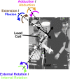
Methods: Patients who were >1 year after TSA for primary glenohumeral osteoarthritis and had had preoperative eccentric or concentric deformity were prospectively recruited. Torque was measured and electromyography was performed during maximal isometric contractions in 26 three-dimensional direction combinations. Relative strength in opposing directions (strength balance) and muscle activity of 6 shoulder rotators were compared between groups.
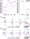
Results: The internal (+) and external (-) rotation component of strength balance did not differ in patients with eccentric (mean internal-external rotation component of strength balance: -7.6% ± 7.4%) compared with concentric deformity (-10.3% ± 6.8%) (mean difference: 2.7% [95% confidence interval (CI), -1.3% to 6.7%]; p = 0.59), suggesting no relative external rotation weakness. Infraspinatus activity was reduced in patients with eccentric (43.9% ± 10.4% of maximum voluntary contraction [MVC]) compared with concentric (51.3% ± 10.4% of MVC) deformity (mean difference: -7.4% [95% CI, -13.4% to -1.4%] of MVC; p = 0.04).
Conclusions: A relative external rotation strength deficit following TSA was not found, despite evidence of reduced infraspinatus activity, in the eccentric-deformity group. Reduced infraspinatus activity suggests that posterior rotator cuff deficiencies may persist following TSA in patients with eccentric deformities. Longitudinal study is necessary to evaluate muscle imbalance as a contributor to higher TSA failure rates.
Level of evidence: Prognostic Level III. See Instructions for Authors for a complete description of levels of evidence.
Copyright © 2024 The Authors. Published by The Journal of Bone and Joint Surgery, Incorporated. All rights reserved.
Conflict of interest statement
Disclosure: The Disclosure of Potential Conflicts of Interest forms are provided with the online version of the article (http://links.lww.com/JBJSOA/A587).
Figures


References
-
- Bercik MJ, Kruse K, 2nd, Yalizis M, Gauci MO, Chaoui J, Walch G. A modification to the Walch classification of the glenoid in primary glenohumeral osteoarthritis using three-dimensional imaging. J Shoulder Elbow Surg. 2016. Oct;25(10):1601-6. - PubMed
-
- Kiet TK, Feeley BT, Naimark M, Gajiu T, Hall SL, Chung TT, Ma CB. Outcomes after shoulder replacement: comparison between reverse and anatomic total shoulder arthroplasty. J Shoulder Elbow Surg. 2015. Feb;24(2):179-85. - PubMed
-
- Walch G, Moraga C, Young A, Castellanos-Rosas J. Results of anatomic nonconstrained prosthesis in primary osteoarthritis with biconcave glenoid. J Shoulder Elbow Surg. 2012. Nov;21(11):1526-33. - PubMed
-
- Donohue KW, Ricchetti ET, Ho JC, Iannotti JP. The association between rotator cuff muscle fatty infiltration and glenoid morphology in glenohumeral osteoarthritis. J Bone Joint Surg Am. 2018. Mar 7;100(5):381-7. - PubMed
-
- Domos P, Checchia CS, Walch G. Walch B0 glenoid: pre-osteoarthritic posterior subluxation of the humeral head. J Shoulder Elbow Surg. 2018. Jan;27(1):181-8. - PubMed
LinkOut - more resources
Full Text Sources
Research Materials
