

A Canal of Nuck Cyst Presented as a Femoral Hernia: A Rare Case Report With Diagnostic Dilemma
- PMID: 38196983
- PMCID: PMC10775744
- DOI: 10.7759/cureus.51908
A Canal of Nuck Cyst Presented as a Femoral Hernia: A Rare Case Report With Diagnostic Dilemma
Abstract
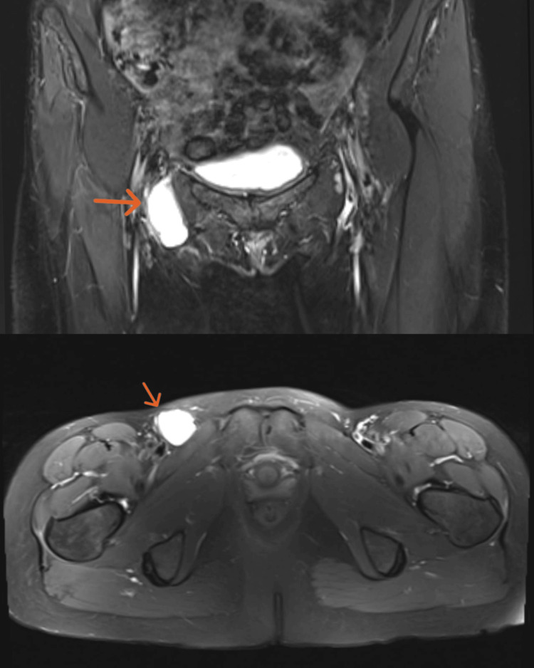
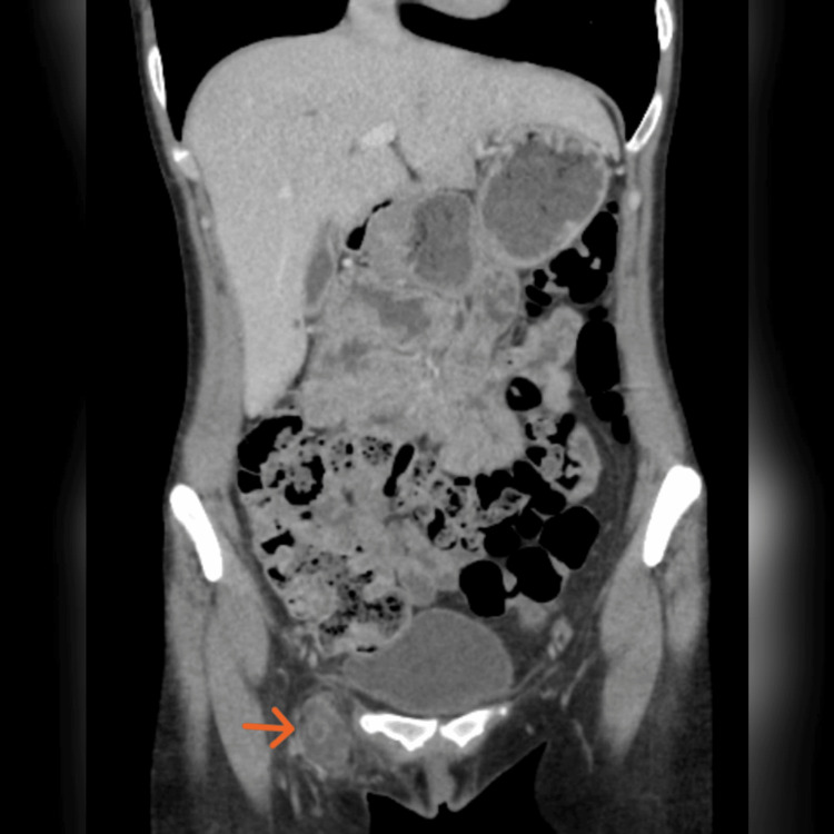
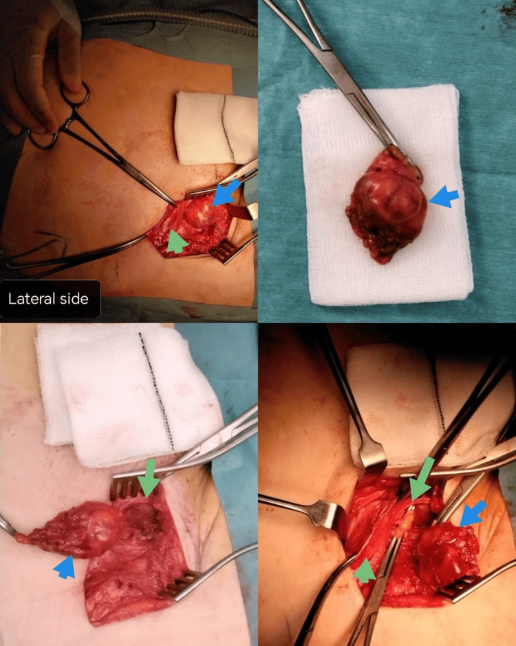
A cyst of the canal of Nuck is an uncommon disorder in females. It results from the failure of obliteration of the peritoneal fold that runs along the round ligament. This case report details a unique and rare presentation of a 38-year-old female who presented with a right groin swelling. Although her preoperative images showed only the right canal of the Nuck cyst, the intraoperative diagnosis was established as a femoral hernia containing a canal of the Nuck cyst. She underwent an elective cyst excision with repair of the femoral hernia. She had an uneventful post-operative recovery. A femoral hernia that contains a cyst of the canal of Nuck is a rare manifestation of this uncommon condition. The most effective treatment options are surgical cyst excision and repair of the femoral hernia.
Keywords: complex cyst; endometriosis; female gender; femoral hernia; the canal of nuck.
Copyright © 2024, Elsayed et al.
Conflict of interest statement
The authors have declared that no competing interests exist.
Figures
Similar articles
-
Surgical treatment of a canal of Nuck cyst presenting as a femoral hernia: An unusual case report.Int J Surg Case Rep. 2021 Oct;87:106435. doi: 10.1016/j.ijscr.2021.106435. Epub 2021 Sep 28. Int J Surg Case Rep. 2021. PMID: 34619454 Free PMC article.
-
Laparoscopic excision of cyst of canal of Nuck.J Minim Access Surg. 2014 Apr;10(2):87-9. doi: 10.4103/0972-9941.129960. J Minim Access Surg. 2014. PMID: 24761084 Free PMC article.
-
The Rarest Variant Type Of Groin Cystic Mass In Adult Female: Encysted Hydrocele Canal Of Nuck.Int J Surg Case Rep. 2023 Mar;104:107921. doi: 10.1016/j.ijscr.2023.107921. Epub 2023 Feb 14. Int J Surg Case Rep. 2023. PMID: 36841044 Free PMC article.
-
The Cyst of the Canal of Nuck: Anatomy, Diagnostic and Treatment of a Very Rare Diagnosis-A Case Report of an Adult Woman and Narrative Review of the Literature.Medicina (Kaunas). 2022 Sep 27;58(10):1353. doi: 10.3390/medicina58101353. Medicina (Kaunas). 2022. PMID: 36295514 Free PMC article. Review.
-
Nuck Cyst, an Unexpected Amethyst Gem in the Inguinal Canal: A Case Report and Literature Review.Ann Ital Chir. 2025;96(5):589-601. doi: 10.62713/aic.3924. Ann Ital Chir. 2025. PMID: 40375375 Review.
References
-
- Anatomy and pathology of the canal of Nuck. Nasser H, King M, Rosenberg HK, Rosen A, Wilck E, Simpson WL. http://dx.doi.org/10.1016/j.clinimag.2018.02.003. Clin Imaging. 2018;51:83–92. - PubMed
-
- Adenographia curiosa et uteri foeminei anatome nova. Nuck A. Am J Obst Gynec. 1975;123:66.
-
- Hydrocele of the canal of Nuck: a report of five cases. Block RE. https://pubmed.ncbi.nlm.nih.gov/1168323/ Obstet Gynecol . 1995;45:464–466. - PubMed
-
- Cyst of canal of Nuck. Bagley JE, Davis MB. J Diagn Med Sonogr. 2015;31:111–114.
-
- Cysts of the canal of Nuck: a rare sonographic diagnosis. Lai I, Page A, Hamidinia F, Rahmani R. http://dx.doi.org/10.1002/jcu.22390. J Clin Ultrasound. 2017;45:175–178. - PubMed
Publication types
LinkOut - more resources
Full Text Sources