

The Impact of Complex Loadings on the Structure of the L2-L3 Intervertebral Disc in a Sheep Spine Cadaver Model: A Biomechanical and Histological Evaluation
- PMID: 38196992
- PMCID: PMC10775825
- DOI: 10.7759/cureus.51941
The Impact of Complex Loadings on the Structure of the L2-L3 Intervertebral Disc in a Sheep Spine Cadaver Model: A Biomechanical and Histological Evaluation
Abstract
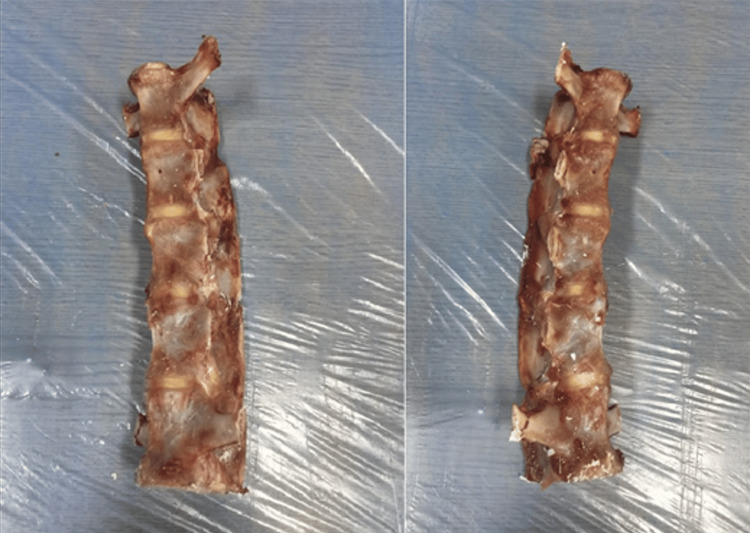
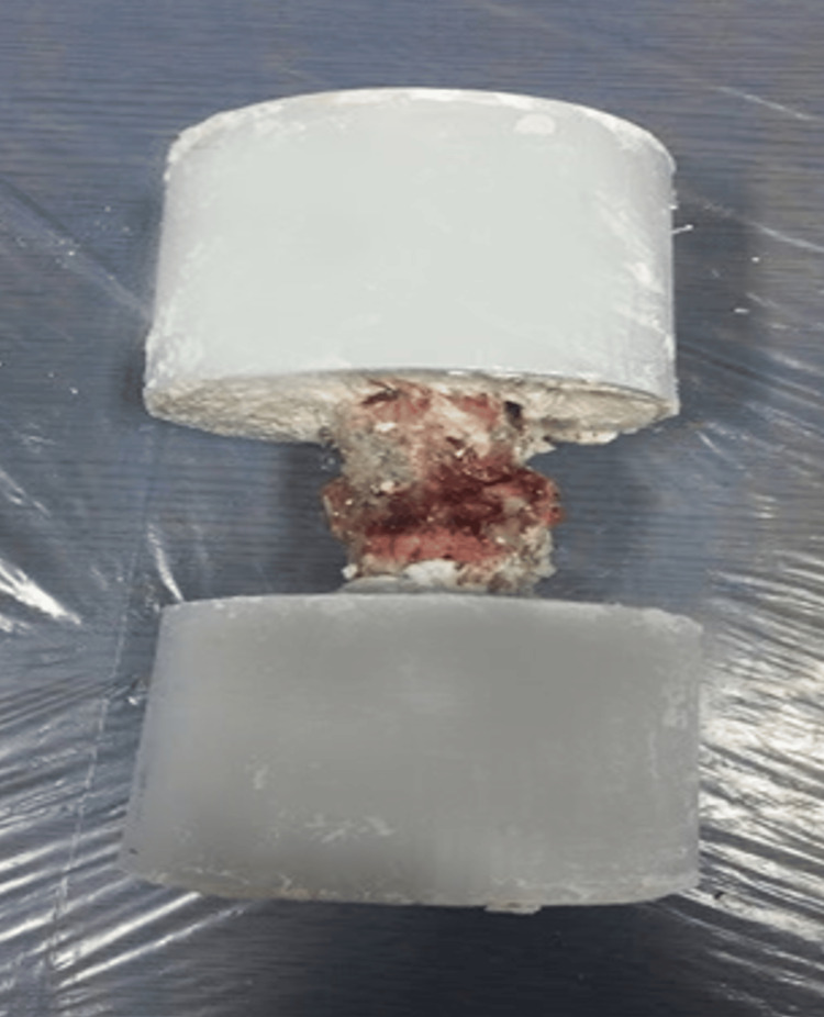
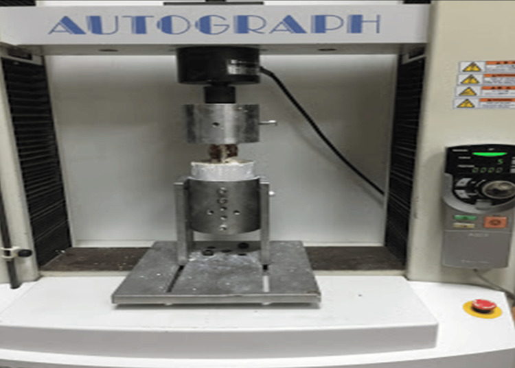
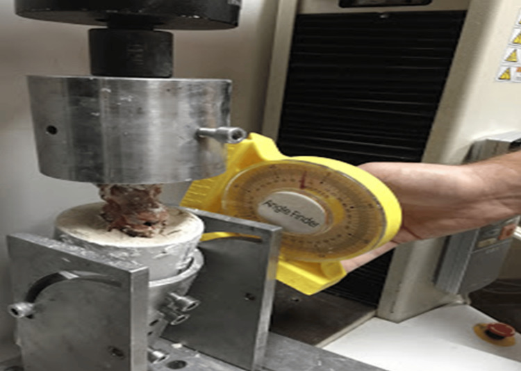
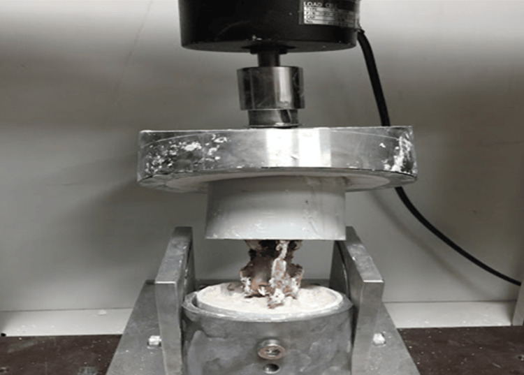
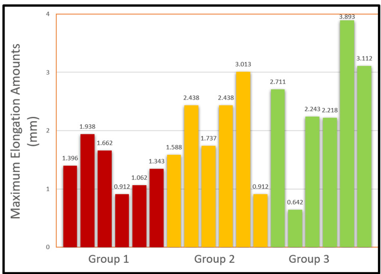
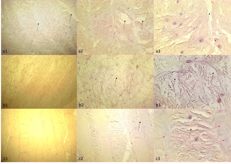
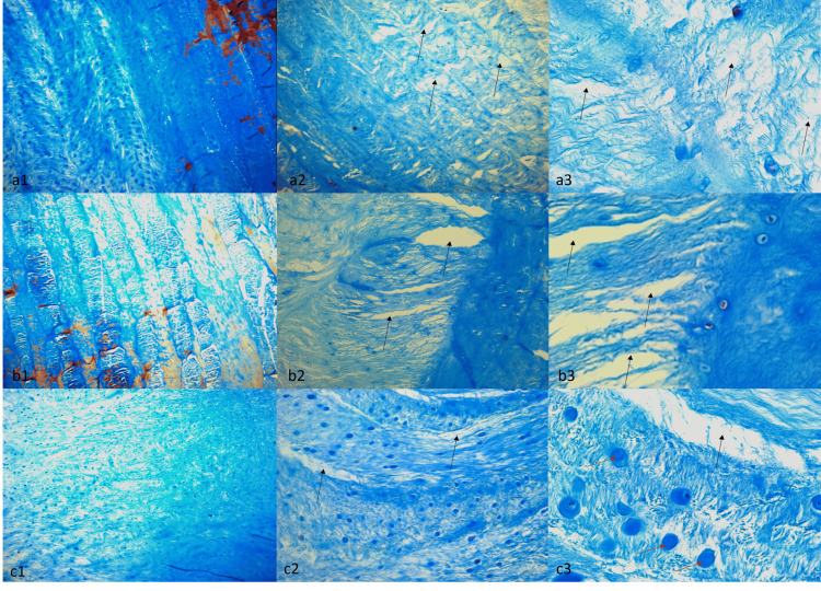
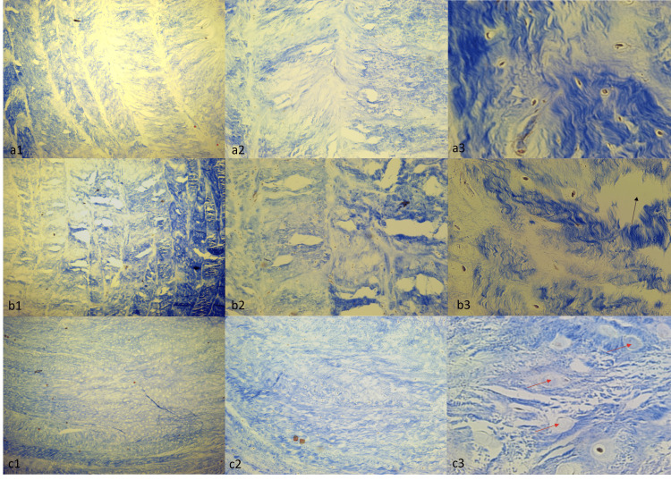
Background The human vertebral column generates movements under versatile, dynamic loads. Understanding how the spine reacts to these movements and loads is crucial for developing new spine implants and surgical treatments for intervertebral disc injuries. Mechanically uni-axial compression models have been extensively studied. However, the spine's daily loading is not limited to compression, so it is crucial to measure its behavior in all movements (flexion-extension, rotation, and axial compression). Methods This study utilized L1-L5 segments from 19 healthy adult sheep spines. The L2-L3 disc of the first spine underwent only histological evaluation without biomechanical testing to define basic histological parameters. The remaining 18 were divided into three groups of six and subjected to biomechanical tests. Different mechanisms for three groups of spinal segments were prepared, and tests were performed on Shimadzu AG-IS 10 KN (Universal Drawing Press, Kyoto, Japan). An axial load (800 N) was applied to the first group, an axial load with 15 degrees of flexion to the second group, and an axial load with 10 degrees of rotation plus 15 degrees of flexion to the third group. A biomechanical evaluation of the maximum elongation amounts (MEAs) was performed and compared between the groups. Then, the L2-L3 discs were removed from the sheep spines, and a histological examination of the discs was conducted using Hematoxylin-Eosin (HE), Alcian Blue (AB), and Masson's Trichrome (MT) staining. Results The mean MEA ± Standard Deviation (Range) was 1.39 ± 0.38 (0.91-1.94) for Group 1, 2.02 ± 0.75 (0.91-3.01) for Group 2, and 2.47 ± 1.09 (0.64-3.9) for Group 3. Biomechanically, although MEAs increased from Group 1 to Group 3 (meaning that the mean MEAs increased as the number of types of applied force increased), there was no statistically significant difference between the groups regarding the MEAs (P = 0.092). Histologically, no significant differences were observed between all groups after HE staining. In all groups, hypercellularity, edema in the connective tissue, separation between tissue layers, delamination, and signs of swelling and necrosis in the cells were observed similarly. For the AB staining, there was a decrease in the glycosaminoglycan (GAG) structure in the tissue samples compared to the control tissue, but no significant differences were observed between the groups. However, it was observed that the stratification in Group 3 was slightly more deteriorated than in the other groups. For the MT staining, collagen structure deterioration was observed in all groups. It was observed that the amount of collagen was significantly reduced compared to the control tissue. Conclusion As a result, when the axial load is applied biomechanically, there is more displacement of the vertebral discs in Group 3 with multidimensional movements. Furthermore, histological studies revealed deterioration between tissue layers when exposed to complex movements, and the degradation of stratification in group 3 compared to other loading combinations in groups 2 and 3 may indicate the role of complex loads in the formation of disc herniation.
Keywords: biomechanical study; cadaver model; complex loadings; histological study; intervertebral disc; lumbar disc hernia; multidisciplinary study; orthopedics; sheep spine; spine.
Copyright © 2024, Öztürk et al.
Conflict of interest statement
The authors have declared that no competing interests exist.
Figures
References
-
- Epidemiological features of chronic low-back pain. Andersson GBJ. Lancet (London, England) 1999;354:581–585. - PubMed
-
- A systematic review of the global prevalence of low back pain. Hoy D, Bain C, Williams G, et al. Arthritis Rheum. 2012;64:2028–2037. - PubMed
-
- Degenerative lumbar spinal stenosis: evaluation and management. Issack PS, Cunningham ME, Pumberger M, Hughes AP, Cammisa FP Jr. J Am Acad Orthop Surg. 2012;20:527–535. - PubMed
-
- Failure within one year following subtotal lumbar discectomy. Wera GD, Marcus RE, Ghanayem AJ, Bohlman HH. J Bone Joint Surg Am. 2008;90:10–15. - PubMed
-
- Physician office visits for low back pain. Frequency, clinical evaluation, and treatment patterns from a U.S. national survey. Hart LG, Deyo RA, Cherkin DC. Spine (Phila Pa 1976) 1995;20:11–19. - PubMed
LinkOut - more resources
Full Text Sources
Research Materials