. 2024 Sep;34(5):e13232.
doi: 10.1111/bpa.13232. Epub 2024 Jan 10.

The post-stroke young adult brain has limited capacity to re-express the gene expression patterns seen during early postnatal brain development

Affiliations

The post-stroke young adult brain has limited capacity to re-express the gene expression patterns seen during early postnatal brain development

Mihai Ruscu et al. Brain Pathol. 2024 Sep.

Abstract

The developmental origins of the brain's response to injury can play an important role in recovery after a brain lesion. In this study, we investigated whether the ischemic young adult brain can re-express brain plasticity genes that were active during early postnatal development. Differentially expressed genes in the cortex of juvenile post-natal day 3 and the peri-infarcted cortical areas of young, 3-month-old post-stroke rats were identified using fixed-effects modeling within an empirical Bayes framework through condition-specific comparison. To further analyze potential biological processes, upregulated and downregulated genes were assessed for enrichment using GSEA software. The genes showing the highest expression changes were subsequently verified through RT-PCR. Our findings indicate that the adult brain partially recapitulates the gene expression profile observed in the juvenile brain but fails to upregulate many genes and pathways necessary for brain plasticity. Of the upregulated genes in post-stroke brains, specific roles have not been assigned to Apobec1, Cenpf, Ect2, Folr2, Glipr1, Myo1f, and Pttg1. New genes that failed to upregulate in the adult post-stroke brain include Bex4, Cd24, Klhl1/Mrp2, Trim67, and St8sia2. Among the upregulated pathways, the largest change was observed in the KEGG pathway "One carbon pool of folate," which is necessary for cellular proliferation, followed by the KEGG pathway "Antifolate resistance," whose genes mainly encode the family of ABC transporters responsible for the efflux of drugs that have entered the brain. We also noted three less-described downregulated KEGG pathways in experimental models: glycolipid biosynthesis, oxytocin, and cortisol pathways, which could be relevant as therapeutic targets. The limited brain plasticity of the adult brain is illustrated through molecular and histological analysis of the axonal growth factor, KIF4. Collectively, these results strongly suggest that further research is needed to decipher the complex genetic mechanisms that prevent the re-expression of brain plasticity-associated genes in the adult brain.

Keywords: KIF4; adult brain; axon growth; brain plasticity; juvenile brain; kinesins; stroke; transcriptomics.


Conflict of interest statement

The authors declare that they have no conflicts of interest.

Figures
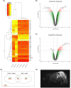
FIGURE 1
FIGURE 1
Transcriptomics analysis was conducted on the frontal and parietal cortex of postnatal, adult naïve rats, and perilesional cortex of rats. (A) The heatmap shows the differentially expressed genes and the relative expression values clearly distinguish naïve rats from both juvenile brains and their post‐stroke littermates. (B), (C) The volcano plot illustrates the differential expression analysis, and it is noteworthy that there is a highly symmetrical distribution of upregulated and downregulated genes in juvenile brains (B) compared to young naïve rats. In contrast, the distribution of DEGs in post‐stroke animals is highly asymmetrical, with a disproportionate ratio in favor of upregulated genes over downregulated ones (C). The The Venn diagram reveals that the number of common upregulated genes outnumbered that of downregulated genes by 256. However, the number of upregulated genes in the adult poststroke brain was just 33%. (D) The MRI image shows the infarct (arrow) (E).
FIGURE 2
FIGURE 2
KIF4 immunohistochemistry on the brains of 3‐day‐old postnatal mice and unlesioned young adult mice. Strong KIF4 expression was observed in neuronal precursor cells (NPCs) in the subventricular zone (A) and in glial cells of the third ventricle (B). In the cortical layer I, neurogenic radial glia‐like cells in the leptomeninges expressed KIF4 (C), inset C1). Occasionally, KIF4 was localized to dividing neuronal‐like cells (inset C2). KIF4 immunostaining was also observed lining juvenile blood vessels throughout the cortex (D), albeit sporadically. In the young adult brain, KIF4 immunostaining was expressed only occasionally by blood vessels (E). BV, blood vessel; CX‐l1, cortical layer I; SVZ, subventricular zone; third VT, third ventricle.
FIGURE 3
FIGURE 3
KIF4 immunohistochemistry in the 3 days post‐stroke young adult rats. KIF4 was robustly re‐expressed along with nestin, a marker of migratory neuroepithelial cells in NPCs of the SVZ of the 3 days post‐stroke young adult rats (A), (B). Of note, many newly born neurons originating from the SVZ began migrating toward the lesion (C). At this stage, post‐stroke adult animals co‐expressed KIF4 and nestin in migrating cells in close vicinity to the corpus callosum (D, arrows). In the periinfarcted area, KIF4 is expressed in endothelial cells of surviving blood vessels, as well as in astrocytes that are GFAP‐positive (E, arrows, and inset). Double‐labeled blood vessels (KIF4, green, and GFAP, red) are detected in remote areas relative to the lesion (F, arrowheads). PI, periinfarcted area; RA, remote area; SVZ, subventricular zone.
FIGURE 4
FIGURE 4
KIF4/NeuN immunohistochemistry was performed on young adult rats 3 days after stroke. At this time point, numerous KIF4/NeuN co‐labeled cells were observed in the peri‐infarcted region of adult rats (panel A, arrows). Co‐labeled KIF4/NeuN cells were also detected in remote cortical areas that were unaffected by the stroke (panel B, arrow). However, at this time point, BrdU was mainly incorporated by endothelial cells (panel C, blue, arrows). In the unlesioned, contralateral cortex, KIF4 was expressed only in blood vessels (panel D, green, arrows) surrounded with NeuN‐positive neuronal nuclei in their vicinity (panel D, red).

References

    1. Tamnes CK, Walhovd KB, Dale AM, Østby Y, Grydeland H, Richardson G, et al. Alzheimer's disease neuroimaging initiative. Brain development and aging: overlapping and unique patterns of change. Neuroimage. 2013;68:63–74. - PMC - PubMed
    1. Grandjean P, Landrigan PJ. Neurobehavioural effects of developmental toxicity. Lancet Neurol. 2014;13(3):330–338. - PMC - PubMed
    1. Poplawski GHD, Kawaguchi R, Van Niekerk E, Lu P, Mehta N, Canete P, et al. Injured adult neurons regress to an embryonic transcriptional growth state. Nature. 2020;581(7806):77–82. - PubMed
    1. Murphy TH, Corbett D. Plasticity during stroke recovery: from synapse to behaviour. Nat Rev Neurosci. 2009;10(12):861–872. - PubMed
    1. Joy MT, Ben Assayag E, Shabashov‐Stone D, Liraz‐Zaltsman S, Mazzitelli J, Arenas M, et al. CCR5 is a therapeutic target for recovery after stroke and traumatic brain injury. Cell. 2019;176(5):1143–1157.e13. - PMC - PubMed

Publication types