. 2024 Mar:151:106348.
doi: 10.1016/j.jmbbm.2023.106348. Epub 2023 Dec 28.

Material properties of nonpregnant and pregnant human uterine layers

Affiliations

Material properties of nonpregnant and pregnant human uterine layers

Daniella M Fodera et al. J Mech Behav Biomed Mater. 2024 Mar.

Abstract

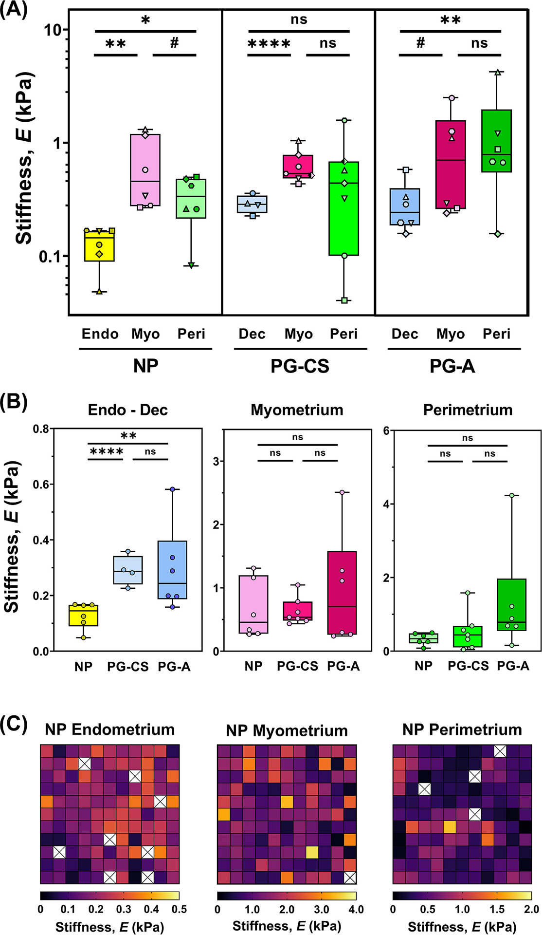
The uterus has critical biomechanical functions in pregnancy and undergoes dramatic material growth and remodeling from implantation to parturition. The intrinsic material properties of the human uterus and how they evolve in pregnancy are poorly understood. To address this knowledge gap and assess the heterogeneity of these tissues, the time-dependent material properties of all human uterine layers were measured with nanoindentation. The endometrium-decidua layer was found to be the least stiff, most viscous, and least permeable layer of the human uterus in nonpregnant and third-trimester pregnant tissues. In pregnancy, the endometrium-decidua becomes stiffer and less viscous with no material property changes observed in the myometrium or perimetrium. Additionally, uterine material properties did not significantly differ between third-trimester pregnant tissues with and without placenta accreta. The foundational data generated by this study will facilitate the development of physiologically accurate models of the human uterus to investigate gynecologic and obstetric disorders.

Keywords: Nanoindentation; Poro-viscoelasticity; Pregnancy; Reproductive biomechanics; Uterus.


Conflict of interest statement

Declaration of competing interest The authors declare that they have no known competing financial interests or personal relationships that could have appeared to influence the work reported in this paper.

Figures

Fig. 1.
Fig. 1.. Anatomy of the Human Uterus.
(A) Diagram of the (i) female reproductive tract in the frontal plane identifying all uterine layers: NP endometrium, PG decidua, myometrium, and perimetrium, (ii) human uterus in the sagittal plane indicating the anterior region from which samples were collected, (iii) cross-section of the uterine wall containing three uterine layers. Arrows indicate the orientation in which tissues were tested with nanoindentation. (B) Representative images of the three uterine layers from NP, cesarean section pregnancies (PG-CS), and placenta accreta pregnancies (PG-A) patient groups stained with Masson’s Trichrome (blue = collagen; red = muscle fibers, cytoplasm; black = nuclei). Images were taken at 10× magnification (scale bar = 100 μm). Note that the relative lengths of the tissue layer figure panels do not reflect actual layer proportions. (C) Relative proportion of collagen and smooth muscle in the myometrium. Bars indicate standard deviation. (D) Thickness of the perimetrium measured for all three patient groups. Data presented as min-max plots. Statistical significance is denoted by the following symbols: ns: p > 0.05; #p ≤ 0.1; *p ≤ 0.05.
Fig. 2.
Fig. 2.. Experimental Approach.
(A) Diagram of nanoindentation testing (Adapted from Optics11 Life). A spherical probe with radius (R) is attached at the end of a cantilever and indented into the sample at a fixed depth (h), recording load (F) over time. (B) Representative load relaxation plot generated from nanoindentation testing in displacement control and fitted with the combined poroelastic-viscoelastic (PVE) material model (solid red line = combined PVE model fit, dotted blue line = viscoelastic material response, solid green line = poroelastic material response).
Fig. 3.
Fig. 3.. Stiffness of Human Uterine Layers.
(A) Summary of stiffness values for each tissue layer at the anterior region organized by patient groups: nonpregnant (NP), cesarean section pregnancy (PG-CS), and placenta accreta pregnancy (PG-A). Each distinct symbol represents the median value of all indentation points measured for a single patient. Data are presented as box and whisker plots on a log10 scale. (B) Stiffness values separated by tissue layer, compared across all three patient groups, plotted on a linear scale. Statistical significance is denoted by the following symbols: ns: p > 0.05; #p ≤ 0.1; *p ≤ 0.05; **p ≤ 0.01; ****p ≤ 0.0001). (C) Representative stiffness heatmaps for NP uterine layers. Grid of 11 × 11 indentation points corresponds to a 2 mm × 2 mm region. [X] indicates points removed due to exclusion criteria.
Fig. 4.
Fig. 4.. Viscoelasticity of Human Uterine Layers.
(A) Summary of viscoelastic ratio values for each tissue layer at the anterior region organized by patient group (NP, PG-CS, PG-A). Each distinct symbol represents the median value of all indentation points measured for a single patient. Data are presented as box and whisker plots on a log10 scale. (B) Viscoelastic ratio values separated by tissue layer, compared across all three patient groups, plotted on a linear scale. Statistical significance is denoted by the following symbols: ns: p > 0.05; *p ≤ 0.05; **p ≤ 0.01; ****p ≤ 0.0001).
Fig. 5.
Fig. 5.. Permeability of Human Uterine Layers.
Summary of permeability values across uterine layers at the anterior region for each patient group (NP, PG-CS, PG-A). Each distinct symbol represents a single patient for which tens to hundreds of indentation points have been averaged. Data are presented as box and whisker plots on a log10 scale. Statistical significance is denoted by the following symbols: ns: p > 0.05; **p ≤ 0.01; ****p ≤ 0.0001).

Update of

References

    1. Abbas Y, Carnicer-Lombarte A, Gardner L, et al., 2019. Tissue stiffness at the human maternal–fetal interface. Hum Reprod Oxf Engl 34 (10), 1999–2008. 10.1093/humrep/dez139. - DOI - PMC - PubMed
    1. Aguilar HN, Mitchell BF, 2010. Physiological pathways and molecular mechanisms regulating uterine contractility. Hum. Reprod. Update 16 (6), 725–744. 10.1093/humupd/dmq016. - DOI - PubMed
    1. Almutairi WM, 2021. Literature review: physiological management for preventing postpartum hemorrhage. Healthcare 9 (6), 658. 10.3390/healthcare9060658. - DOI - PMC - PubMed
    1. Ameer MA, Fagan SE, Sosa-Stanley JN, Peterson DC, 2021. Anatomy, abdomen and pelvis, uterus. In: StatPearls. StatPearls Publishing. http://www.ncbi.nlm.nih.gov/books/NBK470297/. (Accessed 21 December 2021). - PubMed
    1. Aplin JD, 2003. Implantation. In: Henry HL, Norman AW (Eds.), Encyclopedia of Hormones. Academic Press, pp. 289–297. 10.1016/B0-12-341103-3/00165-0. - DOI

Publication types

LinkOut - more resources