. 2024 Feb;300(2):105631.
doi: 10.1016/j.jbc.2024.105631. Epub 2024 Jan 8.

PTP1B mediates the inhibitory effect of MFGE8 on insulin signaling through the β5 integrin

Affiliations

PTP1B mediates the inhibitory effect of MFGE8 on insulin signaling through the β5 integrin

Ritwik Datta et al. J Biol Chem. 2024 Feb.

Abstract

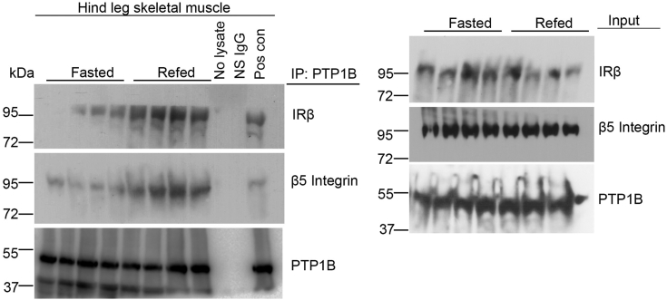
Integrins are cell adhesion receptors that dimerize to mediate cell-cell interactions and regulate processes, including proliferation, inflammation, and tissue repair. The role of integrins in regulating insulin signaling is incompletely understood. We have previously shown that binding of the integrin ligand milk fat globule epidermal growth factor like 8 (MFGE8) to the αvβ5 integrin promotes termination of insulin receptor signaling in mice. Upon ligation of MFGE8, integrin β5 complexes with the insulin receptor beta (IRβ) in skeletal muscle, resulting in dephosphorylation of IRβ and reduction of insulin-stimulated glucose uptake. Here, we investigate the mechanism by which the interaction between β5 and IRβ impacts IRβ phosphorylation status. We show in in vitro and in vivo in skeletal muscle in mice that antibody-mediated blockade of the β5 integrin inhibits and recombinant MFGE8 promotes PTP1B binding to and dephosphorylation of IRβ resulting in increased or reduced insulin-stimulated glucose uptake, respectively. The β5-PTP1B complex is recruited by MFGE8 to IRβ leading to termination of canonical insulin signaling. β5 blockade enhances insulin-stimulated glucose uptake in wildtype but not Ptp1b KO mice indicating that PTP1B functions downstream of MFGE8 in modulating insulin receptor signaling. Furthermore, in a human cohort, we report serum MFGE8 levels correlate with indices of insulin resistance. These data provide mechanistic insights into the role of MFGE8 and β5 in regulating insulin signaling.

Keywords: MFGE8; insulin recpetor; insulin resistance; insulin signaling; integrins.


Conflict of interest statement

Conflict of interest The authors declare no conflict of interest with the contents of this article.

Figures

Figure 1
Figure 1
Antibody-mediated blockade of β5 attenuates the effect of insulin on PTP1B phosphatase activity. A–C, PTP1B phosphatase activity in the presence of β5 blocking or isotype control antibody in (A) skeletal muscle lysates harvested from WT mice 15 and 60 min after intraperitoneal insulin (1U/kg) administration, (B) in differentiated C2C12 myotubes 5, 15, and 30 min after insulin treatment, and (C) in HeLa cells 30 min after EGF or insulin (100 nm) treatment. N = 4 male mice in each group for panel A. Data merged from two independent experiments. N = 5 and 3 independent experiments for panel B and C, respectively. Data are expressed as mean ± SEM; ∗p < 0.05, ∗∗p < 0.01, ∗∗∗p < 0.001 and analyzed by One-way ANOVA followed by Bonferroni’s post-test. EGF, epidermal growth factor; PTP1B, protein-tyrosine phosphatase 1B.
Figure 2
Figure 2
β5 complexes with PTP1B and IRβ.A, co-immunoprecipitation studies showing interaction of PTP1B with β5 and IRβ in hind leg skeletal muscle lysates from mice in presence of β5 blocking or isotype control antibody 60 min after IP insulin (1U/Kg) treatment. Blots represent two independent experiments with N = 4 male mice for insulin-treated groups and two mice without insulin treatment. B and C, co-immunoprecipitation studies showing interaction of PTP1B with β5 and IRβ in C2C12 myotubes treated with (B) β5 blocking (5 μg/ml) or isotype control antibody or (C) rMFGE8 (10 μg/ml) or BSA control in the presence and absence of insulin for 30 min. N = 4 male mice in total. Panel B represents two independent experiments. For panel C, cell culture, treatment, and protein isolation performed on three different days. After immunoprecipitation, protein samples were run on the same gel for Western blot. D, immunostaining of PTP1B (green) and β5 integrin (red) in skeletal muscle cross sections from mice treated with insulin for 60 min in presence of β5 blocking or isotype control antibody. The right-most panel shows the magnified portions (delineated with the dotted lines) of the merged images. The arrows in the magnified images point to the colocalization signals (yellow). Images are representative from two independent experiments. E, cell fractionation of insulin-treated skeletal muscle tissue samples from mice pretreated with β5 blocking or isotype control antibody and insulin for 60 min, followed by Western blot for PTP1B and β5 in the cytoplasmic and membrane fraction. Western blotting for CAVEOLIN-1 (CAV-1) confirmed enrichment of the membrane fractions. N = 1 per group per experiment. Western blots are representative of three independent experiments. IP, intraperitoneal; IRβ, insulin receptor β subunit; MFGE8, milk fat globule epidermal growth factor like 8; PTP1B, protein-tyrosine phosphatase 1B; rMFGE8, recombinant MFGE8.
Figure 3
Figure 3
Antibody-mediated blockade of β5 activates canonical insulin signaling. A, representative Western blot showing phosphorylated (serine 473 residue, S473; Threonine 308 residue, T308) AKT, total AKT, phosphorylated p70 S6Kinase, and total p70 S6K levels in differentiated C2C12 myotubes in the presence of β5 blocking or isotype control antibody treated with insulin for 30 min. N = 3 independent experiments. B, densitometric analysis of p-AKT (S473) Western blots including panel A. C–D, representative Western blot showing p-AKT (S473), p-AKT(T308), total AKT, p-p70S6K, and total p70S6K levels in differentiated C2C12 myotubes treated with rMFGE8 (10 μg/ml) and insulin for 20 min. N=3 to 6 independent samples from 1 to 2 independent experiments. D, densitometric analysis of Western blot of p-AKT (S473) including panel C. E, representative Western blot of phosphorylated and total AKT levels in skeletal muscle lysates from mice treated with insulin for 5, 15, and 30 min in presence of β5 blocking or isotype control antibody. The Western blot shown is representative of three independent experiments. F, densitometric analysis of Western blot of p-AKT (S473) including panel E. G, effect of GLUT-1 inhibitor (1 μM) or control peptide on 2NBDG uptake in C2C12 myotubes treated with β5 blocking or isotype control antibody after insulin stimulation. N = 3 independent experiments. Data expressed as relative fold change to untreated cells (NT). H, effect of antibody-mediated blockade of β5 (5 μg/ml) or rMFGE8 treatment (10 μg/ml) on 2NBDG uptake in 3T3 fibroblasts in the presence or absence of insulin. N = 3 independent experiments. I, Western blot showing GLUT-1 and GLUT-4 levels in cytosolic and membrane fractions isolated from C2C12 myotubes treated with insulin for 30 min in presence of β5 blocking or isotype control antibody. Na+K+ATPase and GAPDH were used as loading controls for membrane and cytosolic fractions, respectively. The Western blot represents three independent experiments. Data are expressed as mean ± SEM; ∗p < 0.05, ∗∗p < 0.01, ∗∗∗p < 0.001, and analyzed by One-way ANOVA followed by Bonferroni’s post-test. GLUT-1, glucose transporter 1; GLUT-4, glucose transporter type 4; MFGE8, milk fat globule epidermal growth factor like 8; rMFGE8, recombinant MFGE8.
Figure 4
Figure 4
Antibody-mediated blockade of β5 impacts insulin signaling through PTP1B. A and B, 2NBDG uptake assay in WT and Ptp1b KO myotubes in presence of (A) β5 blocking or isotype control antibody or (B) rMFGE8 or BSA with and without insulin treatment. N = 6 independent experiments for panel A, and N = 4 independent experiments for panel B. C, glucose tolerance test in 7- to 8-week-old male WT and Ptp1b KO mice after intraperitoneal injection of β5 blocking or isotype control antibody. N = 6 male mice per group from two independent experiments are presented. Data are expressed as mean ± SEM. Data in panel (A and B) were analyzed by one-way ANOVA followed by Bonferroni’s post-test. ∗p < 0.05, ∗∗p < 0.01 and ∗∗∗p < 0.001. Data in panel (C) were analyzed by 2-way ANOVA followed by Tukey’s post-test. ∗∗p < 0.01, ∗p < 0.05, when comparing WT+Con ab versus WT+ antibody-mediated blockade of β5 (β5 block) groups; #p < 0.05 when comparing WT+Con ab versus Ptp1bKO+Con ab groups. MFGE8, milk fat globule epidermal growth factor like 8; PTP1B, protein-tyrosine phosphatase 1B; rMFGE8, recombinant MFGE8.
Figure 5
Figure 5
Model depicting how MFGE8 impacts insulin signaling through PTP1B. MFGE8 binding of the β5 integrin on the outer cell membrane leads to recruitment of the β5-PTP1B complexes to IRβ. PTP1B subsequently dephosphorylates IRβ leading to reduced translocation of GLUT4 to the cell surface and reduced glucose uptake in response to insulin. GLUT-4, glucose transporter type 4; IRβ, insulin receptor β subunit; MFGE8, milk fat globule epidermal growth factor like 8; PTP1B, protein-tyrosine phosphatase 1B.
Figure 6
Figure 6
Physiological regulation of PTP1B/β5 interaction. Co-immunoprecipitation studies showing interaction of PTP1B with β5 and IRβ in hind leg skeletal muscle lysates from mice fasted for 16 h or mice refed for 1 h after a 16 h fast. N = 4 mice in each group. IRβ, insulin receptor β subunit; PTP1B, protein-tyrosine phosphatase 1B.
Supplementary figure 1
Dose-dependent inhibition of insulin-stimulated glucose uptake by rMFGE8. 2NBDG uptake assay in differentiated C2C12 myotubes treated with different doses of rMFGE8 (1, 3, 10 and 30μg/mL) with and without insulin for 20 minutes. N = 4 independent experiments. Data expressed as relative fold change to untreated cells (NT). Data are expressed as mean ± SEM; ∗p < 0.05, ∗∗∗p < 0.001, and analyzed by One-way ANOVA followed by Bonferroni’s post-test.
Supplementary figure 2
Densitometric analysis showing no effect of MFGE8/ β5 signaling on phosphorylation of AKT (T308) and p70S6 Kinase.A and B, Densitometric analysis of western blots including figure 3A for (A) phosphorylated AKT (Threonine 308 residue, T308) and (B) phosphorylated p70 S6Kinase in differentiated C2C12 myotubes in the presence of β5 blocking or isotype control antibody treated with insulin for 30 minutes. N = 3 independent experiments. C and D, Densitometric analysis of western blots in Figure 3B showing (C) phosphorylated AKT T308 and (D) phosphorylated p70 S6Kinase levels in differentiated C2C12 myotubes in the presence of rMFGE8 and insulin. N = 3 independent samples. E and F, Densitometric analysis of western blots including Figure 3C showing (E) phosphorylated AKT T308 and (F) phosphorylated p70S6K levels in skeletal muscle lysates from mice treated with insulin for 5, 15 and 30 minutes in presence of β5 blocking or isotype control antibody. N = 3 independent experiments. Data are expressed as mean ± SEM; and analyzed by One-way ANOVA followed by Bonferroni’s post-test.

Update of

Similar articles

Cited by

References

    1. Khalifeh-Soltani A., Ha A., Podolsky M.J., McCarthy D.A., McKleroy W., Azary S., et al. alpha8beta1 integrin regulates nutrient absorption through an Mfge8-PTEN dependent mechanism. Elife. 2016;5 doi: 10.7554/eLife.13063. - DOI - PMC - PubMed
    1. Khalifeh-Soltani A., McKleroy W., Sakuma S., Cheung Y.Y., Tharp K., Qiu Y., et al. Mfge8 promotes obesity by mediating the uptake of dietary fats and serum fatty acids. Nat. Med. 2014;20:175–183. - PMC - PubMed
    1. Datta R., Gholampour M.A., Yang C.D., Volk R., Lin S., Podolsky M.J., et al. MFGE8 links absorption of dietary fatty acids with catabolism of enterocyte lipid stores through HNF4gamma-dependent transcription of CES enzymes. Cell Rep. 2023;42 - PMC - PubMed
    1. Khalifeh-Soltani A., Gupta D., Ha A., Iqbal J., Hussain M., Podolsky M.J., et al. Mfge8 regulates enterocyte lipid storage by promoting enterocyte triglyceride hydrolase activity. JCI Insight. 2016;1 doi: 10.1172/jci.insight.87418. - DOI - PMC - PubMed
    1. Datta R., Lizama C.O., Soltani A.K., McKleroy W., Podolsky M.J., Yang C.D., et al. Autoregulation of insulin receptor signaling through MFGE8 and the alphavbeta5 integrin. Proc. Natl. Acad. Sci. U. S. A. 2021;118 doi: 10.1073/pnas.2102171118. - DOI - PMC - PubMed

Publication types